. 2007 Sep 15;21(18):2312-25.
doi: 10.1101/gad.1580507.

Ntr1 activates the Prp43 helicase to trigger release of lariat-intron from the spliceosome

Affiliations

Ntr1 activates the Prp43 helicase to trigger release of lariat-intron from the spliceosome

Naoko Tanaka et al. Genes Dev. .

Abstract

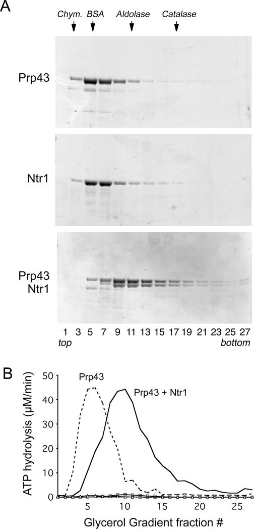
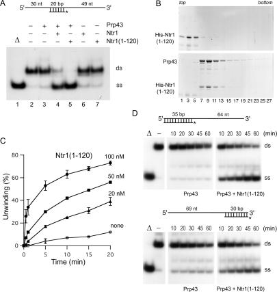
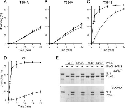
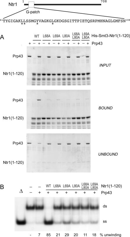
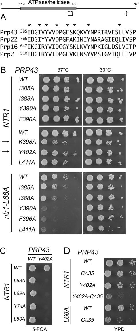
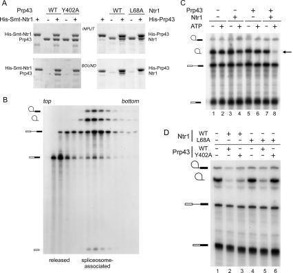
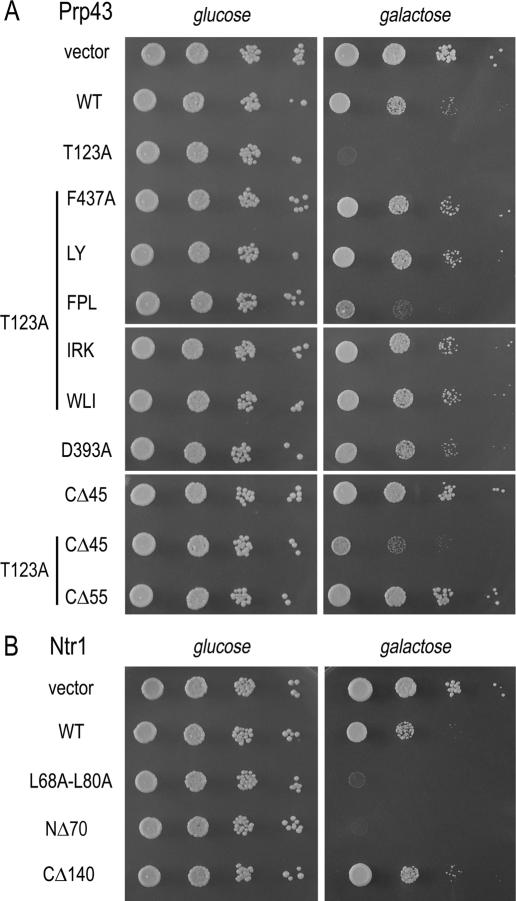
DEAD/H-box NTPases remodel the spliceosome at multiple steps during the pre-mRNA splicing cycle. The RNA-dependent NTPase Prp43 catalyzes dissociation of excised lariat-intron from the spliceosome, but it is unclear how Prp43 couples the energy of ATP hydrolysis to intron release. Here, we report that activation of Prp43's inherently feeble helicase activity by the splicing factor Ntr1 is required for lariat-intron release. Lethal Prp43 mutants T384A and T384V, which are active for ATP hydrolysis and fail to dissociate lariat-intron from spliceosomes, are refractory to stimulation of RNA unwinding by Ntr1. An N-terminal 120-amino-acid segment of Ntr1 suffices for binding to Prp43 and for stimulating its helicase activity. We identify missense mutations in Prp43 and Ntr1 that disrupt protein-protein interaction and impair Ntr1 enhancement of Prp43 RNA unwinding. Our results demonstrate for the first time that regulating the motor activity of a DEAH-box protein by an accessory factor is critical for mRNA splicing.


Figures

Figure 1.
Figure 1.
Prp43–Ntr1 interaction. (A) Glycerol gradient sedimentation of Prp43 and Ntr1 proteins. Native Ntr1 (83 kDa) and His10-Prp43 (90 kDa) proteins (200 μg) were sedimented in 15%–30% glycerol gradients, and aliquots (10 μL) of the odd-numbered gradient fractions were analyzed by 8% SDS-PAGE. The Coomassie blue-stained gels are shown. The positions of Prp43 and Ntr1 are indicated at the left. The peak positions of marker proteins chymotrypsinogen (Chym.) (25 kDa), BSA (67 kDa), aldolase (156 kDa), and catalase (232 kDa), sedimented in a parallel gradient, are indicated at the top. (B) ATPase activity. ATP hydrolysis was measured in the absence and presence of poly(A) RNA. ATPase activities (micromolar per minute) are plotted for all fractions from gradients in which Prp43 (dashed line, with RNA cofactor; Δ, without RNA) and Prp43 + Ntr1 (solid line, with RNA; ◽, activity without RNA) were analyzed.
Figure 2.
Figure 2.
Ntr1 stimulates Prp43 helicase. (A) Unwinding of a 5′/3′-tailed 20-bp RNA/DNA duplex. A schematic drawing of the substrate is shown at the top. The 99-nt top strand is RNA and the 32P-labeled 20-nt bottom strand is DNA. The positions of the duplex substrate (ds) and of the single-strand product of unwinding (ss) are indicated at the right of the panel. The 5′/3′-tailed substrate was incubated with the proteins as specified for 30 min at 37°C. The fraction of the single-stranded 20-mer DNA in the absence of protein (lane 2) constituted 10% of the total 32P-labeled nucleic acid in the reaction (background); Δ (lane 1) marks a reaction that was heated for 3 min at 95°C. (B) Glycerol gradient sedimentation. His10-Smt3-Ntr1(1–120) alone (top panel) and together with Prp43 (bottom panel) was sedimented in 15%–30% glycerol gradients. Aliquots (10 μL) of the odd-numbered gradient fractions were analyzed by 8% SDS-PAGE and the polypeptides were visualized by staining the gels with Coomassie blue. The positions of Prp43 and His10-Smt3-Ntr1(1–120) proteins are indicated at the left. (C) Reaction mixtures containing the 20-bp 5′/3′-tailed substrate (2.5 nM) and 100 nM Prp43 were incubated in the absence (○) or in the presence of 20 nM (▴), 50 nM (◼), and 100 nM (●) of His10-Smt3-Ntr1(1–120) protein. Aliquots were withdrawn after the indicated times and analyzed by native PAGE. The amount of displaced labeled 20-mer was determined as the percentage of total nucleic acid at each time point and adjusted for the background of labeled 20-mer in a reaction carried out without added protein. The values are averages of duplicate measurements; the error bars represent the deviation from the mean. (D) Unwinding of a 3′-tailed 35-bp RNA/DNA duplex (top panel) and a 5′-tailed 30-bp RNA/DNA duplex (bottom panel) as a function of time. Reaction mixtures contained the substrate (2.5 nM) depicted above the panel, 100 nM of the indicated proteins, and 1 mM ATP-Mg2+. Aliquots were withdrawn at the indicated times and analyzed by PAGE. Autoradiographs of the dried gels are shown.
Figure 3.
Figure 3.
(A–D) Influence of Ntr1 on unwinding by Prp43 mutants. The 5′/3′-tailed 20-bp nucleic acid duplex (2.5 nM), 1 mM ATP-Mg2+, and Prp43 proteins (500 nM) were incubated without Ntr1 (○), or with 500 nM His10-Smt3-Ntr1(1–120) (●). The Prp43 proteins were T384A (A), T384V (B), T384S (C), and wild-type Prp43 (D). Aliquots were withdrawn after the indicated times and analyzed by native PAGE. The amounts of the displaced 20-mer as percentage of total labeled nucleic acid were determined and the values were adjusted for background. Helicase activities (percentage unwinding) are plotted as a function of time. The values are averages from duplicate experiments. (E) Interaction of Prp43 mutant proteins with Ntr1. His10-Smt3-Ntr1 and various Prp43 proteins (10 μg each) were mixed as indicated above the panel prior to adsorption to Ni-agarose. Aliquots of the INPUT mixtures (10%) and the BOUND (20%) material, eluted with 500 mM imidazole, were analyzed by 8% SDS-PAGE. The polypeptides were visualized by staining the gels with Coomassie blue.
Figure 4.
Figure 4.
Binding of Ntr1(1–120) to Prp43 is necessary for stimulation. Ntr1 is drawn as a black line, with the rectangle indicating the G-patch motif. The amino acid sequence of the G-patch motif is shown in the expanded view. The stars below the sequence highlight positions that were replaced by alanine. (A) Purified Prp43 (10 μg) and partially purified His10-Smt3-Ntr1(1–120) (30 μg) proteins were mixed as indicated. Aliquots of the input mixtures (INPUT), the proteins eluted from the Ni-NTA agarose (BOUND), and the supernatants after binding (UNBOUND) were analyzed by 8% SDS-PAGE. Polypeptides were visualized by staining with Coomassie blue. The positions of Prp43 and His10-Smt3-Ntr1(1–120) proteins are indicated at the left. (B) Effect of His10-Smt3-Ntr1(1–120) mutant proteins on RNA helicase activity. Reaction mixtures containing 2.5 nM 5′/3′-tailed 20-bp RNA/DNA duplex and 1 mM ATP-Mg2+ were supplemented with proteins (100 nM) as indicated (+), incubated for 30 min at 37°C, and analyzed by native PAGE. (−) indicates that buffer instead of protein was added. The position of duplex substrate (ds) and displaced labeled 20-mer (ss) are indicated at the right. In Δ, the reaction mixture was heated for 3 min at 95°C.
Figure 5.
Figure 5.
Genetic interactions between Prp43 and Ntr1. Prp43 is drawn as a horizontal line with the gray rectangle highlighting the ATPase/helicase domain. The numbers refer to the first and last amino acids of Prp43 or of the ATPase/helicase domain. The wide and narrow arrows indicate the positions of the peptide shown in A, and of the C terminus in Prp43(1–722), respectively. (A) Alignment of conserved segments between motifs V and VI in DEAH splicing factors. The stars indicate positions that were replaced by alanine in Prp43. (B) Growth phenotypes of strains containing various PRP43 and NTR1 mutants. prp43Δ ntr1Δ cells harboring the indicated PRP43 and NTR1 alleles were grown in liquid cultures. Cultures were adjusted to A600 of 10−1 and 10-fold serial dilutions (3 μL) were spotted to YPD agar plates. Plates were photographed after 2.5 d at 30°C and 37°C. The arrows at the left indicate prp43 alleles that are lethal in ntr1-L68A. (C) prp43Δ ntr1Δ cells harboring an NTR1 PRP43 URA3 plasmid were transformed with the indicated NTR1 plus PRP43 alleles. His+Trp+ transformants were selected and grown in liquid culture in synthetic medium lacking tryptophan and histidine. Cultures were adjusted to A600 of 0.2 and spotted (5 μL) to 5-FOA-containing agar plates. Photographs of the plates after 4 d of incubation at 30°C are shown. (D) Tenfold serial dilutions of prp43Δ ntr1Δ cells harboring the indicated PRP43 and NTR1 alleles were spotted to YPD medium. Photographs were taken after 2 d of incubation at 30°C.
Figure 6.
Figure 6.
(A, left panel) Interaction of Prp43 and Ntr1. His10-Smt3-Ntr1 and wild-type Prp43 or the Y402A proteins (10 μg each) were mixed as indicated above the panel prior to adsorption to Ni-agarose. Aliquots of the INPUT mixtures (10%) and the BOUND (20%) material, eluted with 500 mM imidazole were analyzed by 8% SDS-PAGE. The polypeptides were visualized by staining the gels with Coomassie blue. The right panel shows a similar experiment using His10-Prp43 and tag-free Ntr1 proteins as indicated. (B) Lariat-intron RNA is retained in the Δntr1Δprp43 spliceosomes. Splicing was carried out in Δntr1Δprp43 extract for 20 min at 28°C. The reaction mixture (100 μL) was analyzed by sedimentation in a 15%–40% glycerol gradient. Fractions were collected from the top of the gradient and aliquots of odd-numbered fractions (3–29) were analyzed by denaturing PAGE. An autoradiogram of the gel is shown. (C) Reconstitution of intron release activity. 32P-labeled actin pre-mRNA was incubated in Δntr1Δprp43 extract in the presence of ATP (2 mM) for 20 min at 28°C. Then ATP was depleted from the reaction mixture by endogenous hexokinase upon addition of glucose (2.5 mM) during a 10-min incubation. Aliquots were then supplemented with proteins (±ATP) as indicated at the top of the panel. The reactions were halted after 10 min and analyzed by denaturing PAGE. An autoradiogram of the dried gel is shown. The symbols at the left indicate the positions of the following RNA species (from top to bottom): lariat-intermediate, lariat-intron, pre-mRNA, and mRNA. (D) Lariat-intron release activity of Ntr1 and Prp43 mutant proteins. 32P-labeled actin pre-mRNA was reacted in Δntr1Δprp43m extract (prepared from cell that express Prp43-Y402A-TAP and Ntr1TAP) in the presence of ATP (2 mM) for 20 min at 28°C. Aliquots of the mixture were then supplemented with proteins as indicated at the top of the panel. The reactions were halted after 10 min and analyzed by denaturing PAGE. An autoradiogram of the dried gel is shown.
Figure 7.
Figure 7.
Dominant-negative effects in vivo. Wild-type strains harboring an empty vector or plasmids (TRP1 CEN) with the indicated prp43 alleles (A), and ntr1 mutants (B) under the transcriptional control of the GAL1 promoter were grown in liquid raffinose-containing medium. The cultures were adjusted to A600 of 0.1 and 10-fold serial dilutions were spotted to synthetic agar medium lacking tryptophan and containing glucose or galactose. The plates were photographed after incubation for 2.5 d (glucose) or 4 d (galactose) at 30°C.

Similar articles

Cited by

References

    1. Ansari A., Schwer B., Schwer B. SLU7 and a novel activity, SSF1, act during the PRP16-dependent step of yeast pre-mRNA splicing. EMBO J. 1995;14:4001–4009. - PMC - PubMed
    1. Aravind L., Koonin E.V., Koonin E.V. G-patch: A new conserved domain in eukaryotic RNA-processing proteins and type D retroviral polyproteins. Trends Biochem. Sci. 1999;24:342–344. - PubMed
    1. Arenas J.E., Abelson J.N., Abelson J.N. Prp43: An RNA helicase-like factor involved in spliceosome disassembly. Proc. Natl. Acad. Sci. 1997;94:11798–11802. - PMC - PubMed
    1. Behzadnia N., Golas M.M., Hartmuth K., Sander B., Kastner B., Deckert J., Dube P., Will C.L., Urlaub H., Stark H., Golas M.M., Hartmuth K., Sander B., Kastner B., Deckert J., Dube P., Will C.L., Urlaub H., Stark H., Hartmuth K., Sander B., Kastner B., Deckert J., Dube P., Will C.L., Urlaub H., Stark H., Sander B., Kastner B., Deckert J., Dube P., Will C.L., Urlaub H., Stark H., Kastner B., Deckert J., Dube P., Will C.L., Urlaub H., Stark H., Deckert J., Dube P., Will C.L., Urlaub H., Stark H., Dube P., Will C.L., Urlaub H., Stark H., Will C.L., Urlaub H., Stark H., Urlaub H., Stark H., Stark H., et al. Composition and three-dimensional EM structure of double affinity-purified, human prespliceosomal A complexes. EMBO J. 2007;26:1737–1748. - PMC - PubMed
    1. Boon K.L., Auchynnikava T., Edwalds-Gilbert G., Barrass J.D., Droop A.P., Dez C., Beggs J.D., Auchynnikava T., Edwalds-Gilbert G., Barrass J.D., Droop A.P., Dez C., Beggs J.D., Edwalds-Gilbert G., Barrass J.D., Droop A.P., Dez C., Beggs J.D., Barrass J.D., Droop A.P., Dez C., Beggs J.D., Droop A.P., Dez C., Beggs J.D., Dez C., Beggs J.D., Beggs J.D. Yeast ntr1/spp382 mediates prp43 function in postspliceosomes. Mol. Cell. Biol. 2006;26:6016–6023. - PMC - PubMed

Publication types

MeSH terms

Substances