. 2007 Sep 15;21(18):2347-57.
doi: 10.1101/gad.434307.

N-cofilin is associated with neuronal migration disorders and cell cycle control in the cerebral cortex

Affiliations

N-cofilin is associated with neuronal migration disorders and cell cycle control in the cerebral cortex

Gian Carlo Bellenchi et al. Genes Dev. .

Abstract

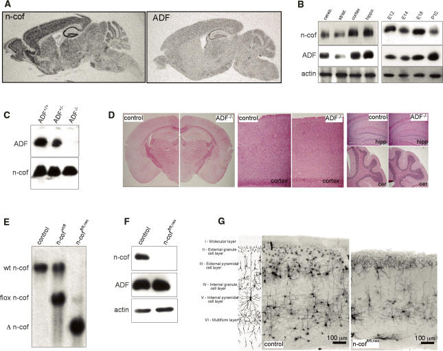
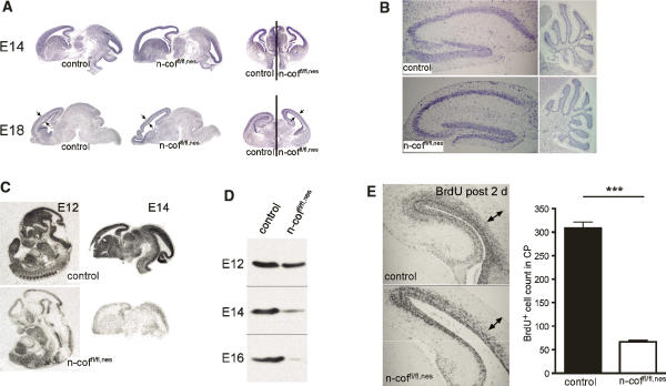
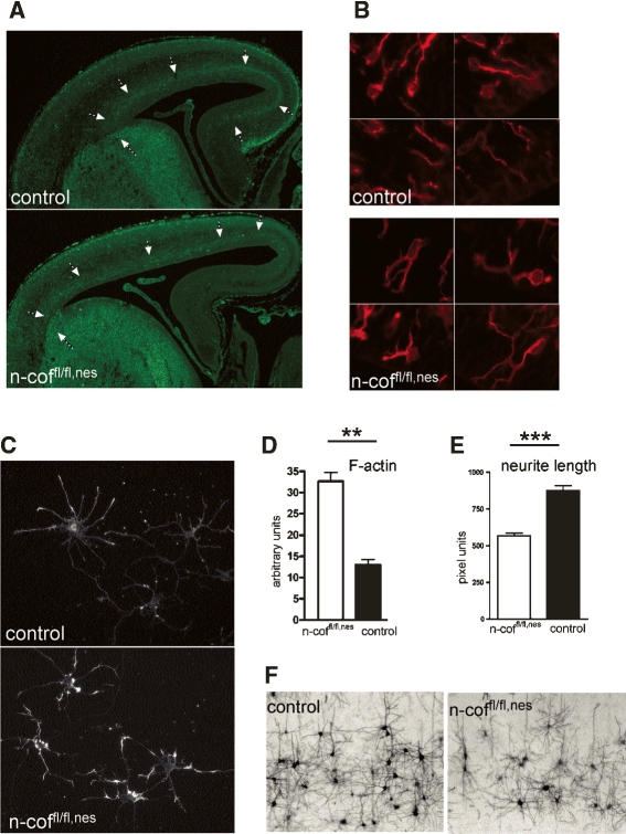
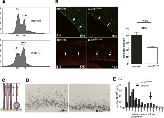
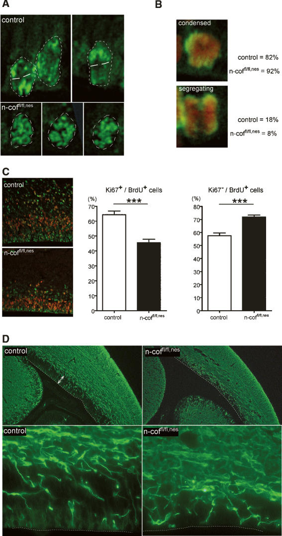
Many neuronal disorders such as lissencephaly, epilepsy, and schizophrenia are caused by the abnormal migration of neurons in the developing brain. The role of the actin cytoskeleton in neuronal migration disorders has in large part remained elusive. Here we show that the F-actin depolymerizing factor n-cofilin controls cell migration and cell cycle progression in the cerebral cortex. Loss of n-cofilin impairs radial migration, resulting in the lack of intermediate cortical layers. Neuronal progenitors in the ventricular zone show increased cell cycle exit and exaggerated neuronal differentiation, leading to the depletion of the neuronal progenitor pool. These results demonstrate that mutations affecting regulators of the actin cytoskeleton contribute to the pathology of cortex development.


Figures

Figure 1.
Figure 1.
Distinct roles of n-cofilin and ADF in cerebral cortex formation. (A) Expression of n-cofilin and ADF shown by radioactive in situ hybridization. The expression patterns of n-cofilin and ADF are comparable, although the ADF signal is weaker. (B, left panel) Protein levels of n-cofilin and ADF in different brain regions (cerebellum, striatum, cortex, hippocampus). (Right panel) Total brain lysates from E12 to postnatal day 10 show coexpression of n-cofilin and ADF in the developing brain. Actin serves as a loading control. (C) ADF is absent from the brain of ADF−/− mice. Western blots of total brain lysates were probed for ADF and n-cofilin. (D) Brain development and morphology is not affected in ADF−/− mice. Nissl staining confirmed normal organization of the cortex, hippocampus (hipp), and cerebellum (cer). (E) Southern blot analysis shows efficient deletion (Δ n-cof) of the conditional n-cofilin allele (flox n-cof, referred to as “fl”) in homozygous conditional mutants carrying the nestin-cre transgene (n-coffl/fl,nes). (F) N-cofilin is absent in the brain of n-coffl/fl,nes mice. Western blot of total brain extracts from postnatal n-coffl/fl,nes mice. ADF levels are not altered. (G) Golgi staining of coronal sections from the cortex of control and n-coffl/fl,nes mice. Cortical layers II–IV and parts of layer V are missing in mutant brains. The cartoon illustrates regular cortical layer organization in the mammalian brain.
Figure 2.
Figure 2.
Radial migration in the cortex is impaired. (A) Brain anatomy in n-coffl/fl,nes embryos at E14 and E18 shows enlarged ventricles and reduced thickness of the CP (see arrows). (B) In postnatal mutant brains (P20) the structure of the hippocampus and cerebellum is comparable with control littermates. (C) Specific deletion of n-cofilin mRNA in the developing brain is shown by in situ hybridization of E12 whole-embryo sections and E14 brain sections. (D) N-cofilin protein levels in total brain lysates of E12, E14, and E16 n-coffl/fl,nes embryos. At E12, n-cofilin levels start decreasing, while at E14–E16, n-cofilin is efficiently deleted. (E) BrdU pulse labeling of E16 embryos and analysis at E18 indicates a cell migration defect in the CP (see arrows). The number of BrdU+ cells translocating into the CP is reduced about sixfold in n-coffl/fl,nes embryos (P < 0.0001).
Figure 3.
Figure 3.
Tangential migration, cell shape, and actin remodeling in n-coffl/fl,nes neurons. (A) Tangential migration is partially impaired in the cortex of E16 n-cofilin mutants. In control embryos, GABAergic neurons labeled with an anti-VIAAT antibody emerge from the ganglionic eminence and migrate tangentially through the cortex. In n-coffl/fl,nes embryos, tangential migration through the cortex occurs, but GABAergic neurons do not reach the medial pallium (see arrows). (B) Cell shape of differentiating neurons (βIII-tubulin positive) in the VZ of n-coffl/fl,nes embryos is characterized by multiple cell protrusions, while in control embryos most migrating neurons have acquired an elongated bipolar shape. Four different representative fields from the VZ of mutant and control embryos (E16) are shown. (C) Cultured cortical neurons from control and n-coffl/fl,nes embryos were stained with phalloidin for F-actin after 3 d in culture. (D) In mutant cells, F-actin is increased about twofold (P = 0.0012). (E) Reduced average neurite length and outgrowth defects in n-coffl/fl,nes mice (P < 0.0001). (F) In vivo connectivity and outgrowth of pyramidal neurons is impaired, as seen in the cortex of postnatal n-coffl/fl,nes mice. Neurons are visualized by silver staining.
Figure 4.
Figure 4.
Cell cycle block and impaired interkinetic nuclear migration in the VZ of n-coffl/fl,nes embryos. (A) DNA content of total embryonic cells harvested from n-cof−/− embryos (Gurniak et al. 2005) suggests that n-cofilin is required for G2/M transition, as indicated by the increase in cells with a 4n DNA content. (B) Cell proliferation is impaired in the cortex of n-coffl/fl,nes embryos, as shown by the decreased number of pH3-positive and Ki67-positive cells in the VZ of E14 and E16 embryos, respectively. Quantitation of pH3-positive cells in the VZ of E14 embryos is depicted in the right panel (P = 0.0094). (C) Cartoon of interkinetic nuclear migration (adapted by permission from Macmillan Publishers Ltd.: Nature Reviews Molecular Cell Biology, Gotz and Huttner 2005, © 2005) to indicate the correlation with cell cycle. (D) Short-term pulse labeling with BrdU (60 min) in the VZ allows us to follow interkinetic nuclear movement. Note that in n-coffl/fl,nes embryos vertical translocation of nuclei is impaired. (E) Quantitation of nuclear translocation in the VZ of control embryos shows a second peak of S-phase nuclei distal from the ventricles (see arrow), while in n-coffl/fl,nes embryos this peak is absent.
Figure 5.
Figure 5.
Chromosome segregation is impaired and cell cycle exit is increased in the VZ of n-coffl/fl,nes embryos. (A) pH3 staining of dividing cells visualizes chromosome organization. Nuclei are marked by a dashed line. Control nuclei showed polarized chromosome figures with a defined cleavage plane (white lines), while in n-coffl/fl,nes nuclei chromosomes showed a mostly random organization. (B) Mitotic figures were classified according to the images shown, and “condensed” chromatin versus “segregating” figures were scored (pH3 in green, DNA in red). Eighteen percent of control cells, but only 8% of mutant cells, showed segregating chromosomes. (C) Cell cycle profiles of progenitor cells in the VZ. After pulse labeling with BrdU at E16, brain sections were double-stained for Ki67 (red) and BrdU (green). The numbers of Ki67+/BrdU+ and Ki67/BrdU+ cells are presented as percentages of BrdU+ cells (see histograms). In mutants, fewer cells enter the cell cycle, as indicated by the smaller Ki67+/BrdU+ population (P < 0.0001), while more cells exit the cell cycle as shown by the increased Ki67/BrdU+ population (P = 0.004). (D) Neuronal differentiation is increased in the VZ of n-coffl/fl,nes mice. E16 brains were stained using a neuronal differentiation marker (βIII-tubulin). The VZ in control embryos (white arrows) contains few differentiating neurons, while in n-coffl/fl,nes brains the VZ is depleted of progenitors and is instead populated with differentiated βIII-tubulin-positive neurons. For better orientation, the ventricle border is marked by a white dashed line.

Similar articles

Cited by

References

    1. Abe H., Obinata T., Minamide L.S., Bamburg J.R., Obinata T., Minamide L.S., Bamburg J.R., Minamide L.S., Bamburg J.R., Bamburg J.R. Xenopus laevis actin-depolymerizing factor/cofilin: A phosphorylation-regulated protein essential for development. J. Cell Biol. 1996;132:871–885. - PMC - PubMed
    1. Adams M.E., Minamide L.S., Duester G., Bamburg J.R., Minamide L.S., Duester G., Bamburg J.R., Duester G., Bamburg J.R., Bamburg J.R. Nucleotide sequence and expression of a cDNA encoding chick brain actin depolymerizing factor. Biochemistry. 1990;29:7414–7420. - PubMed
    1. Anderson S.A., Eisenstat D.D., Shi L., Rubenstein J.L., Eisenstat D.D., Shi L., Rubenstein J.L., Shi L., Rubenstein J.L., Rubenstein J.L. Interneuron migration from basal forebrain to neocortex: Dependence on Dlx genes. Science. 1997;278:474–476. - PubMed
    1. Ayala R., Shu T., Tsai L.H., Shu T., Tsai L.H., Tsai L.H. Trekking across the brain: The journey of neuronal migration. Cell. 2007;128:29–43. - PubMed
    1. Cappello S., Attardo A., Wu X., Iwasato T., Itohara S., Wilsch-Brauninger M., Eilken H.M., Rieger M.A., Schroeder T.T., Huttner W.B., Attardo A., Wu X., Iwasato T., Itohara S., Wilsch-Brauninger M., Eilken H.M., Rieger M.A., Schroeder T.T., Huttner W.B., Wu X., Iwasato T., Itohara S., Wilsch-Brauninger M., Eilken H.M., Rieger M.A., Schroeder T.T., Huttner W.B., Iwasato T., Itohara S., Wilsch-Brauninger M., Eilken H.M., Rieger M.A., Schroeder T.T., Huttner W.B., Itohara S., Wilsch-Brauninger M., Eilken H.M., Rieger M.A., Schroeder T.T., Huttner W.B., Wilsch-Brauninger M., Eilken H.M., Rieger M.A., Schroeder T.T., Huttner W.B., Eilken H.M., Rieger M.A., Schroeder T.T., Huttner W.B., Rieger M.A., Schroeder T.T., Huttner W.B., Schroeder T.T., Huttner W.B., Huttner W.B., et al. The Rho-GTPase cdc42 regulates neural progenitor fate at the apical surface. Nat. Neurosci. 2006;9:1099–1107. - PubMed

Publication types