. 2007 Sep 15;21(18):2358-70.
doi: 10.1101/gad.450707.

The prepattern transcription factor Irx3 directs nephron segment identity

Affiliations

The prepattern transcription factor Irx3 directs nephron segment identity

Luca Reggiani et al. Genes Dev. .

Abstract

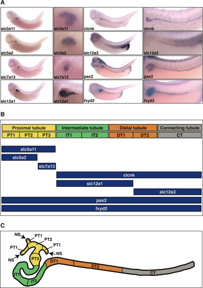
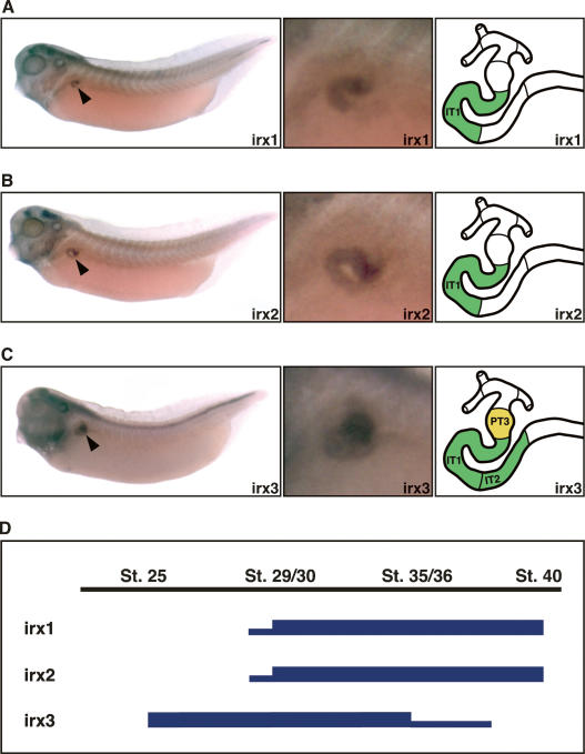
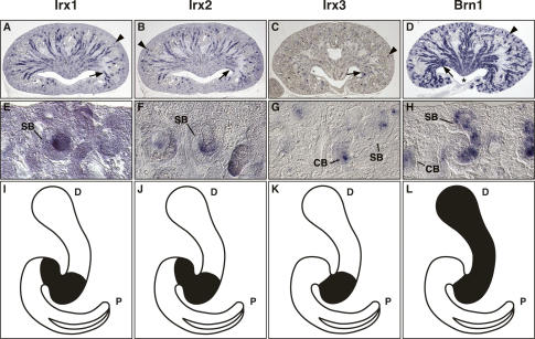
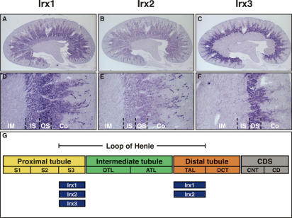
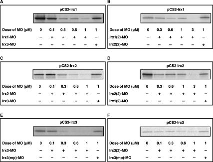
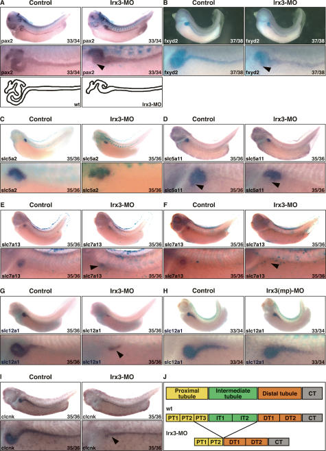
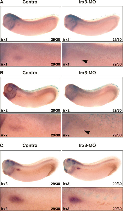
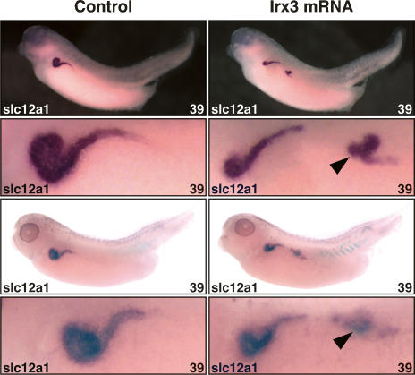
The nephron, the basic structural and functional unit of the vertebrate kidney, is organized into discrete segments, which are composed of distinct renal epithelial cell types. Each cell type carries out highly specific physiological functions to regulate fluid balance, osmolarity, and metabolic waste excretion. To date, the genetic basis of regionalization of the nephron has remained largely unknown. Here we show that Irx3, a member of the Iroquois (Irx) gene family, acts as a master regulator of intermediate tubule fate. Comparative studies in Xenopus and mouse have identified Irx1, Irx2, and Irx3 as an evolutionary conserved subset of Irx genes, whose expression represents the earliest manifestation of intermediate compartment patterning in the developing vertebrate nephron discovered to date. Intermediate tubule progenitors will give rise to epithelia of Henle's loop in mammals. Loss-of-function studies indicate that irx1 and irx2 are dispensable, whereas irx3 is necessary for intermediate tubule formation in Xenopus. Furthermore, we demonstrate that misexpression of irx3 is sufficient to direct ectopic development of intermediate tubules in the Xenopus mesoderm. Taken together, irx3 is the first gene known to be necessary and sufficient to specify nephron segment fate in vivo.


Figures

Figure 1.
Figure 1.
Segmental organization of the Xenopus pronephric nephron. (A) Spatial expression patterns of selected pronephric marker genes. Xenopus embryos (stage 35/36) were stained by whole-mount in situ hybridization for expression of clcnk (ClC-K), fxyd2 (Na, K-ATPase γ subunit), pax2, slc5a2 (SGLT2), slc5a11 (SGLT1L), slc7a13, slc12a1 (NKCC2), and slc12a3 (NCC). Lateral views are shown with accompanying enlargements of the pronephric kidney region. (B) Summary of marker gene expression along the proximodistal axis of the pronephric nephron. The localization of the expression domains is shown below the corresponding segments. (C) Schematic representation of the tubular portion of the Xenopus pronephric kidney. A stage 35/36 pronephric kidney is shown with the four tubular compartments color-coded. Each tubule may be further subdivided into distinct segments: proximal tubule (yellow; PT1, PT2, PT3), intermediate tubule (green; IT1, IT2, IT3), distal tubule (orange; DT1, DT2), and connecting tubule (gray; CT). The nephrostomes (NS) are ciliated peritoneal funnels that connect the coelomic cavity to the nephron.
Figure 2.
Figure 2.
Expression of irx genes is highly regionalized in the developing pronephric kidney. (A–C) Expression patterns of irx1 (A), irx2 (B), and irx3 (C) in the pronephric kidneys of stage 35/36 Xenopus embryos as determined by whole-mount in situ hybridization. Lateral views of whole embryos (left panels; arrowheads indicate pronephric expression), enlargements of the pronephric region (middle panels), and color-coded schematic representations of the segment-restricted expression domains (right panels) are shown. Note the sharp boundaries that limit the expression domains of irx genes in the developing nephron. (D) Summary of the temporal expression profiles of irx genes during pronephric kidney development. The embryonic stages of X. laevis development are indicated. High and low levels of irx gene expression are illustrated with thick and thin lines, respectively. The embryonic expression patterns in early embryos are shown in Supplementary Figure 1.
Figure 3.
Figure 3.
Irx gene expression marks an intermediate region of the S-shaped body. In situ hybridizations were performed on paraffin sections of E18.5 kidneys. Whole sagittal sections (A–D) and magnifications (E–H) of the renal cortex are shown to illustrate gene expression in developing nephrons. (I–L) The corresponding gene expression domains in the S-shaped body are indicated in the schematic drawings. (A–C) Irx1 and Irx2 are expressed in newly forming nephrons in the cortex (arrows) and the developing intermediate tubule epithelia (arrowheads). Irx3 expression is similar to Irx1 and Irx2, but fainter and more restricted. (D) Brn1 expression in the newly forming nephrons (arrowhead) and developing distal tubule epithelia (arrow). Note that in contrast to Irx genes, Brn1 is also highly expressed in epithelia of the renal papilla (asterisk). (I–K) Irx transcripts are confined to intermediate regions of S-shaped bodies. (L) Brn1 transcripts are detected in intermediate and distal domains of the S-shaped body. (CB) Comma-shaped body; (SB) S-shaped body; (P) proximal pole of the nephron; (D) distal pole of the nephron.
Figure 4.
Figure 4.
Irx gene expression is confined to distinct segments of Henle’s loop in the adult metanephric kidney. (A–F) Expression of Irx genes in the adult kidney. In situ hybridizations were performed on paraffin sections. Whole sagittal sections (A–C) and magnifications (D–F) are shown to illustrate Irx gene expression in detail. (Co) Cortex; (OS) outer stripe of outer medulla; (IS) inner stripe of the outer medulla; (IM) inner medulla. (A,B,D,E) Irx1 and Irx2 transcripts are detected in S3 and TAL. (C,F) Irx3 transcripts are confined to S3 only. (G) Summary of Irx gene expression in the adult metanephric kidney. The segmental organization of the adult metanephric nephron is shown schematically. The expression domains of each Irx gene are indicated below the scheme. (ATL) Ascending thin limb; (CD) collecting duct; (CDS) collecting duct system; (CNT) connecting tubule; (DCT) distal convoluted tubule; (DTL) descending thin limb; (S1, S2, S3) segments of the proximal tubule; (TAL) thick ascending limb.
Figure 5.
Figure 5.
Inhibition of irx1, irx2, and irx3 translation in vitro by antisense MOs. Plasmids (500 ng) encoding the ORF of irx1, irx2, or irx3 were used as templates in cell-free coupled transcription–translation reactions. MOs were tested for inhibition of translation at the doses indicated. Cell-free transcription–translation reactions were performed in the presence of [35S]methionine and analyzed by SDS-PAGE/autoradiography. (A,B) Dose-response analysis of inhibition of irx1 translation by Irx1-MO (A) and Irx1(2)-MO (B). (C,D) Dose-response analysis of inhibition of irx2 translation by Irx2-MO (C) and Irx2(2)-MO (D). (E,F) Dose-response analysis of inhibition of irx3 translation by Irx3-MO (E) and Irx3(2)-MO (F).
Figure 6.
Figure 6.
Irx3 is required for intermediate tubule formation in the Xenopus pronephric kidney. (A–I) Irx3-MO (5 ng; A–G, I) or Irx3(mp)-MO (5 ng; H) and mRNA (0.25 ng) for the lineage tracer nuclear β-galactosidase were coinjected into single V2 blastomeres of eight-cell-stage embryos. Injected embryos were raised to the embryonic stage indicated, fixed, and processed for β-gal activity. Expression of marker genes was subsequently visualized by in situ hybridization. Control and injected sides are shown as lateral views with accompanying enlargements of the pronephric kidney region. (A,B) Irx3 knockdown affects pronephric morphogenesis. Arrowheads indicate the central looped region that is abnormal. Schematic representations show the outline of the nephron in normal and Irx3-MO-injected embryos. (C) Irx3 knockdown does not affect PT1 and PT2. (D–F) Irx3 knockdown causes a loss of the proximal tubule segment PT3. The arrowhead indicates the position of the PT3 segment. Examples of strong reduction (E) and complete loss (F) of slc7a13 expression are shown. (G–I) Irx3 knockdown causes loss of IT1 and IT2 but not of DT1. Arrowheads indicate the location of DT1. Note that slc12a1 expression remains unaffected in the presence of the control Irx3(mp)-MO (H). (J) Summary illustrating the nephron segmentation defects seen in irx3 knockdown embryos. See Figure 1C for the nomenclature of pronephric nephron segments and their abbreviations.
Figure 7.
Figure 7.
Pronephric expression of irx1 and irx2 but not irx3 requires irx3 gene function. Irx3-MO (5 ng) and mRNA (0.25 ng) for the lineage tracer nuclear β-galactosidase were coinjected into single V2 blastomeres of eight-cell-stage embryos. Injected embryos were raised to the embryonic stage indicated, fixed, and processed for β-gal activity. Expression of marker genes was subsequently visualized by in situ hybridization. Control and injected sides are shown as lateral views with accompanying enlargements of the pronephric kidney region. (A,B) Irx3 knockdown disrupts irx1 and irx2 expression in the developing pronephric kidney. Arrowheads indicate the pronephric area devoid of irx1 (A) and irx2 (B) expression. (C) Irx3 knockdown does not affect irx3 expression.
Figure 8.
Figure 8.
Irx3 is sufficient for intermediate tubule formation in the Xenopus pronephric kidney. Single V2 blastomeres of eight-cell-stage embryos were injected with irx3 mRNA (0.15 ng). Injected embryos were raised to the embryonic stage indicated, fixed, and processed for β-gal activity. Expression of the slc12a1 marker gene was subsequently visualized by in situ hybridization. Lateral views of control and injected sides of two representative embryos displaying the gain-of-function phenotype are shown with accompanying enlargements of the pronephric kidney region. The arrowheads indicate the ectopic intermediate tubule tissues expressing slc12a1.

Similar articles

Cited by

References

    1. Aldaz S., Morata G., Azpiazu N., Morata G., Azpiazu N., Azpiazu N. The Pax-homeobox gene eyegone is involved in the subdivision of the thorax of Drosophila. Development. 2003;130:4473–4482. - PubMed
    1. Allanson J.E., Hunter A.G., Mettler G.S., Jimenez C., Hunter A.G., Mettler G.S., Jimenez C., Mettler G.S., Jimenez C., Jimenez C. Renal tubular dysgenesis: A not uncommon autosomal recessive syndrome: A review. Am. J. Med. Genet. 1992;43:811–814. - PubMed
    1. Bao Z.Z., Bruneau B.G., Seidman J.G., Seidman C.E., Cepko C.L., Bruneau B.G., Seidman J.G., Seidman C.E., Cepko C.L., Seidman J.G., Seidman C.E., Cepko C.L., Seidman C.E., Cepko C.L., Cepko C.L. Regulation of chamber-specific gene expression in the developing heart by Irx4. Science. 1999;283:1161–1164. - PubMed
    1. Bellefroid E.J., Kobbe A., Gruss P., Pieler T., Gurdon J.B., Papalopulu N., Kobbe A., Gruss P., Pieler T., Gurdon J.B., Papalopulu N., Gruss P., Pieler T., Gurdon J.B., Papalopulu N., Pieler T., Gurdon J.B., Papalopulu N., Gurdon J.B., Papalopulu N., Papalopulu N. Xiro3 encodes a Xenopus homolog of the Drosophila Iroquois genes and functions in neural specification. EMBO J. 1998;17:191–203. - PMC - PubMed
    1. Bilioni A., Craig G., Hill C., McNeill H., Craig G., Hill C., McNeill H., Hill C., McNeill H., McNeill H. Iroquois transcription factors recognize a unique motif to mediate transcriptional repression in vivo. Proc. Natl. Acad. Sci. 2005;102:14671–14676. - PMC - PubMed

Publication types