. 2007 Oct 1;204(10):2285-91.
doi: 10.1084/jem.20071143. Epub 2007 Sep 17.

Myosin IIA is required for cytolytic granule exocytosis in human NK cells

Affiliations

Myosin IIA is required for cytolytic granule exocytosis in human NK cells

Milena M Andzelm et al. J Exp Med. .

Abstract

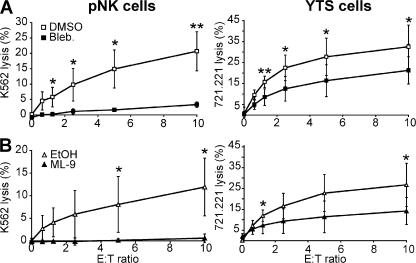
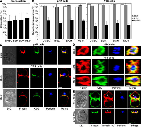
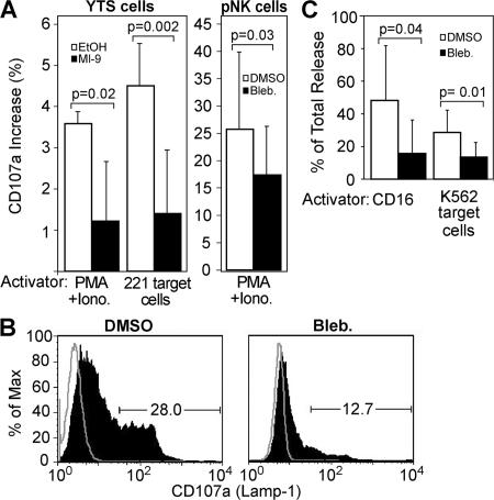
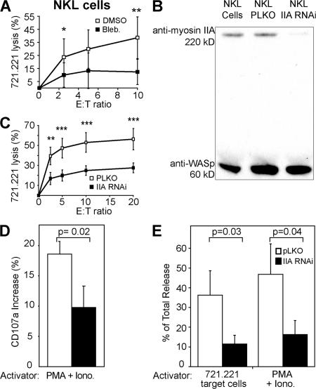
Natural killer (NK) cell cytotoxicity involves the formation of an activating immunological synapse (IS) between the effector and target cell through which granzymes and perforin contained in lytic granules are delivered to the target cell via exocytosis. Inhibition of nonmuscle myosin II in human NK cells with blebbistatin or ML-9 impaired neither effector-target cell conjugation nor formation of a mature activating NK cell IS (NKIS; formation of an actin ring and polarization of the microtubule-organizing center and cytolytic granules to the center of the ring). However, membrane fusion of lytic granules, granzyme secretion, and NK cell cytotoxicity were all effectively blocked. Specific knockdown of the myosin IIA heavy chain by RNA interference impaired cytotoxicity, membrane fusion of lytic granules, and granzyme secretion. Thus, myosin IIA is required for a critical step between NKIS formation and granule exocytosis.


Figures

Figure 1.
Figure 1.
Inhibition of myosin II blocks NK cell cytotoxicity. pNK cells and YTS cells were pretreated with inhibitors, and their killing ability was assayed using 51Cr release from K562 or 721.221 target cells, respectively. DMSO was used as a solvent for blebbistatin (Bleb; A) and EtOH was used as a solvent for ML-9 (B), and they are the control curves for their respective inhibitors. Both solvents showed nonspecific effects on cytotoxicity. Specifically, at an E/T ratio of 10:1 DMSO at the concentration used reduced killing of pNK cells by 18% and of YTS cells by 0%, whereas EtOH at the concentration used reduced killing of pNK cells by 52% and of YTS cells by 15%. Each curve represents the mean ± the SD of three independent experiments. *, P < 0.05 and **, P < 0.01 by Student's t test.
Figure 2.
Figure 2.
Myosin II inhibition does not affect conjugation or mature lytic synapse formation. (A) YTS-GFP cells were incubated with 721.221-RFP cells, and conjugation was quantified by FACS. Conjugates were defined as the percentage of GFP, RFP double-positive events measured by FACS compared with those seen with YTS-GFP cells alone. (B) The accumulation of F-actin, perforin, and CD2-GFP (YTS cells) or CD2-FITC (pNK cells) at the IS of an effector–target conjugate pair was evaluated for 50 conjugates in each experiment. Values are expressed as the percentage of synapses at which fluorescence was accumulated. Bar graphs represent the mean ± the SD of three independent experiments. (C) pNK cells are shown conjugated with K562 cells (top), and YTS-CD2-GFP cells are shown conjugated with 721.221 cells (bottom). NK cells are the top cells and target cells are the bottom cells in the conjugate pair. Differential interference contrast images are in the left column and fluorescent images are in the right columns. A merged overlay of all fluorescent channels is in the fifth column. (D) Representative lytic synapses were evaluated throughout their volume, and the z, x plane was reconstructed. The first row shows a lytic synapse in a pNK cell conjugated with a K562 cell. The bottom two rows show lytic synapses between YTS-CD2-GFP cells and 721.221 cells. The final column is a merged overlay of the fluorescent channels from the first three columns. The different inhibitor treatment for each row is indicated on the left of both C and D. In the case of both C and D, vehicle control images were identical in each case to those where NK cells were treated with inhibitors (solvent control for C and D are found in Fig. S1). (E) Myosin IIA colocalizes with F-actin and perforin. Representative conjugate pairs of both a YTS cell and a 721.221 target cell (top), as well as a pNK cell with a K562 cell (bottom) are shown, in the same format as C. Myosin II is found to associate with F-actin, as well as granules (white arrowhead). Bars, 5 μm.
Figure 3.
Figure 3.
Myosin II inhibitors block lytic granule exocytosis. (A) pNK and YTS cells were activated with either their respective target cells or PMA and ionomycin (Iono). The percentage of increase in CD107a surface expression was evaluated by FACS and expressed as the increase over identically treated (including with appropriate inhibitors) unstimulated cells. Thus, unstimulated cells were used as a baseline to account for any nonspecific stimulation of antibody endocytosis or granule exocytosis over the incubation time, as well as any antibody endocytosis nonspecifically induced by cell treatment. Unstimulated cells also accounted for any potential shifts in fluorescence caused by drug color. Bar graphs represent the mean ± the SD of three independent experiments. (B) A representative histogram of CD107a surface expression from pNK cells stimulated with PMA/ionomycin ± blebbistatin. The percentage of pNK cells expressing a high level of CD107a was gated, and values are shown on the graph. The percentage of Max indicates the number of events at each given fluorescence intensity, where the intensity with the greatest number of cells has been normalized to be 100%. (C) pNK cells were stimulated by plate-bound anti-CD16 or K562 target cells, and their supernatant was used in a serine esterase assay. Results are expressed as a percentage of total release from cells pretreated with inhibitors or appropriate controls to account for possible background effects on the colorimetric assay caused by either intrinsic compound color or direct effects on serine esterase activity. Bar graphs represent the mean ± the SD of four to five independent experiments. Paired and unpaired Student's t tests were performed to obtain P values.
Figure 4.
Figure 4.
Knockdown of myosin IIA in NKL cells impairs cytotoxicity and granule exocytosis. (A) NKL cells were pretreated with blebbistatin, and killing ability was assayed using 51Cr release from 721.221 target cells. (B) Relative protein levels were assessed after RNAi of myosin IIA by Western blot with WASp content as the control. (C) Killing ability of NKL cells with myosin IIA knockdown was assayed using the same 51Cr release assay. NKL cells infected with the pLKO vector only were used as a control. *, P < 0.1, **, P < 0.04, and **, P < 0.02 by Student's t test for both graphs. (D) The percentage of increase in CD107a surface expression was evaluated by FACS and expressed as the increase over identically treated cells, but with no stimulation by PMA and ionomycin (Iono). (E) NKL cells were stimulated by PMA and ionomycin or 721.221 target cells, and their supernatant was used in a serine esterase assay. Results are expressed as a percentage of total release. All data represent the mean ± the SD of three independent experiments. Unpaired Student's t tests were performed to obtain P values.

References

    1. Wülfing, C., B. Purtic, J. Klem, and J.D. Schatzle. 2003. Stepwise cytoskeletal polarization as a series of checkpoints in innate but not adaptive cytolytic killing. Proc. Natl. Acad. Sci. USA. 100:7767–7772. - PMC - PubMed
    1. Orange, J.S., K.E. Harris, M.M. Andzelm, M.M. Valter, R.S. Geha, and J.L. Strominger. 2003. The mature activating natural killer cell immunologic synapse is formed in distinct stages. Proc. Natl. Acad. Sci. USA. 100:14151–14156. - PMC - PubMed
    1. Kopcow, H.D., D.S. Allan, X. Chen, B. Rybalov, M.M. Andzelm, B. Ge, and J.L. Strominger. 2005. Human decidual NK cells form immature activating synapses and are not cytotoxic. Proc. Natl. Acad. Sci. USA. 102:15563–15568. - PMC - PubMed
    1. Sellers, J.R. 2000. Myosins: a diverse superfamily. Biochim. Biophys. Acta. 1496:3–22. - PubMed
    1. Wülfing, C., and M.M. Davis. 1998. A receptor/cytoskeletal movement triggered by costimulation during T cell activation. Science. 282:2266–2269. - PubMed

Publication types

MeSH terms

Substances