. 2007 Sep 15;67(18):8818-27.
doi: 10.1158/0008-5472.CAN-07-1214.

Targeting human medulloblastoma: oncolytic virotherapy with myxoma virus is enhanced by rapamycin

Affiliations

Targeting human medulloblastoma: oncolytic virotherapy with myxoma virus is enhanced by rapamycin

Xue Qing Lun et al. Cancer Res. .

Abstract

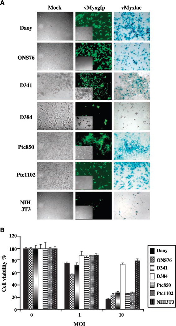
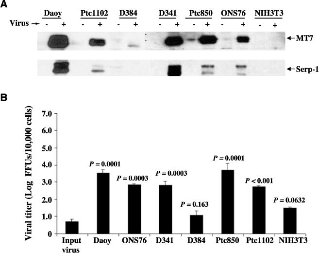
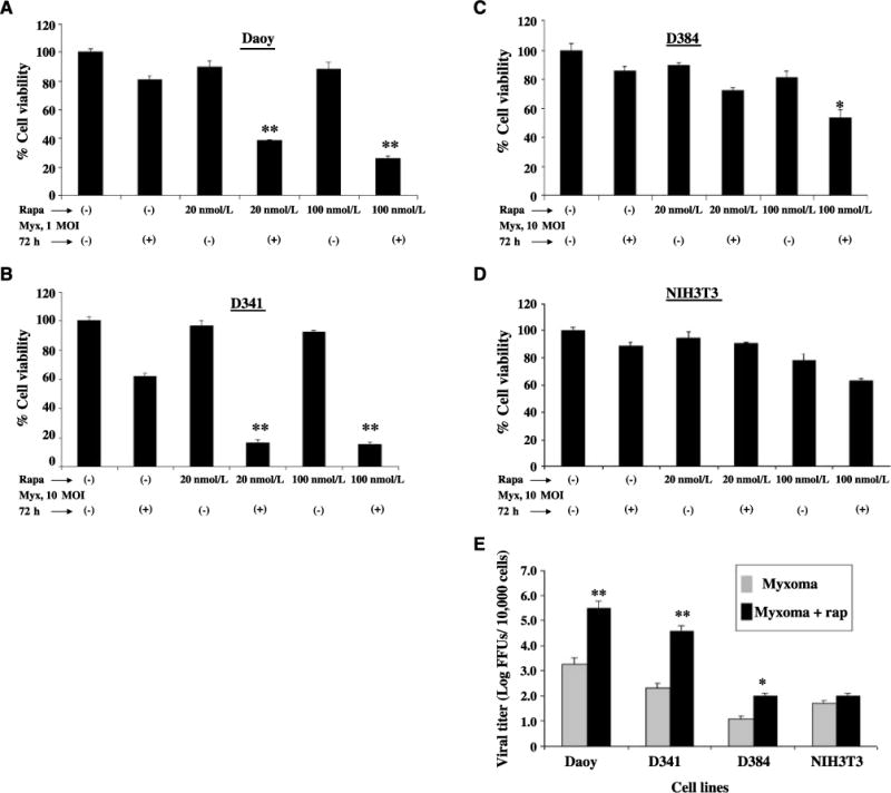
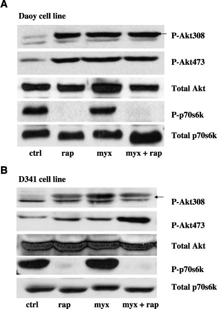
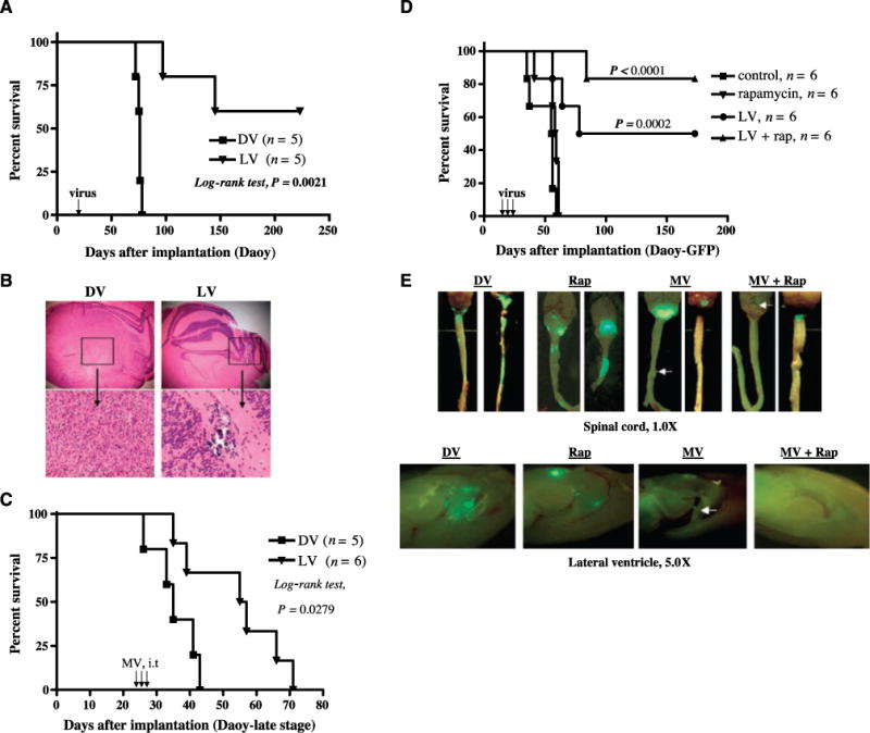
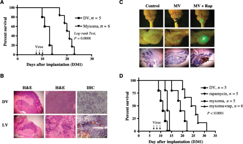
We have shown previously the oncolytic potential of myxoma virus in a murine xenograft model of human glioma. Here, we show that myxoma virus used alone or in combination with rapamycin is effective and safe when used in experimental models of medulloblastoma in vitro and in vivo. Nine of 10 medulloblastoma cell lines tested were susceptible to lethal myxoma virus infection, and pretreatment of cells with rapamycin increased the extent of in vitro oncolysis. Intratumoral injection of live myxoma virus when compared with control inactivated virus prolonged survival in D341 and Daoy orthotopic human medulloblastoma xenograft mouse models [D341 median survival: 21 versus 12.5 days; P = 0.0008; Daoy median survival: not reached (three of five mice apparently "cured" after 223 days) versus 75 days; P = 0.0021]. Rapamycin increased the extent of viral oncolysis, "curing" most Daoy tumor-bearing mice and reducing or eliminating spinal cord and ventricle metastases. Rapamycin enhanced tumor-specific myxoma virus replication in vivo and prolonged survival of D341 tumor-bearing mice (median survival of mice treated with live virus (LV) and rapamycin, versus LV alone, versus rapamycin alone, versus inactivated virus: 25 days versus 19, 13, and 11 days, respectively; P < 0.0001). Rapamycin increased the levels of constitutively activated Akt in Daoy and D341 cells, which may explain its ability to enhance myxoma virus oncolysis. These observations suggest that myxoma virus may be an effective oncolytic agent against medulloblastoma and that combination therapy with signaling inhibitors that modulate activity of the phosphatidylinositol 3-kinase/Akt pathway will further enhance the oncolytic potential of myxoma virus.


Figures

Figure 1
Figure 1
Effects of myxoma virus on established medulloblastoma cell lines in vitro. A, medulloblastoma (Daoy, ONS76, D341, D384, Ptc850, and Ptc1102) and murine fibroblast (NIH3T3) cell lines were infected with vMyxgfp or vMyxlac at a MOI of 5 and photographed 48h after infection. Middle, early viral gene expression (GFP) visualized by fluorescence microscopy. Inset, phase-contrast view of the same field. Right, late viral gene expression (β-galactosidase, blue foci) detected by X-gal staining. Magnification, ×100. Left, DV control. B, WST-1 assay comparing the effects of vMyxgfp on cell viability.
Figure 2
Figure 2
Myxoma virus productively infects medulloblastoma cell lines in vitro. A, Western blotting of whole-cell lysates (50 μg) for early (MT7) and late (Serp-1) myxoma virus gene expression following exposure to vMyxgfp (+) or no virus (−). B, viral titers were obtained in medulloblastoma and NIH3T3 cell lines after infection. Cells were infected with vMyxgfp (MOI of 0.1), incubated for 72 h, and lysed using three rounds of freeze-thawing to extract viral particles. The viral titers of samples were determined using a standard plaque titration assay on BGMK cells. Columns, mean focus forming units (FFU) of triplicate wells; bars, SD.
Figure 3
Figure 3
Effect of rapamycin combined with myxoma virus on medulloblastoma cell lines in vitro. A to D, the viability of medulloblastoma cell lines and murine fibroblast cell line (as a negative control) was measured using the WST-1 assay 72 h after infection with vMyxgfp in the presence (+) or absence (−) of rapamycin (Rapa). Myx, myxoma. E, cells were incubated for 1 h in medium alone or in medium containing 20 nmol/L rapamycin (rap) and then infected with 0.1 MOI of vMyxgfp as described in Materials and Methods. Viral titers were quantitated 72 h after infection using a standard plaque titration assay on BGMK cells. Columns, mean FFU of triplicate wells; bars, SD.
Figure 4
Figure 4
Rapamycin increases activation of Akt in human susceptible medulloblastoma cells. Daoy (A) and D341 (B) cells were pretreated with rapamycin (20 nmol/L) for 1 h, and then the cells were infected with vMyxgfp (5 MOI). Levels of phosphorylated p70S6K (P-p70S6K) and phosphorylated Akt (at Ser473 and Thr308; P-Akt473 and P-Akt308) in cell lysates were determined by Western blotting. Total Akt and mTOR levels were not changed.
Figure 5
Figure 5
Effects of i.t. injection of myxoma virus in the presence or absence of rapamycin on survival and metastasis in Daoy tumor-bearing CD-1 nude mice. A, Kaplan-Meier survival analysis of animals implanted in the cerebellum with Daoy (1 × 10⁵ cells per mouse) cells and treated with either DV or vMyxgfp (5 × 10⁶ PFUs per mouse, single injection). The LV-treated animals (n = 5) survived significantly longer than DV-treated controls (n = 5; P = 0.0021, log-rank test). B, representative histologic analysis in a live vMyxgfp- and a DV-treated mouse. Magnifications, ×25 (top) and ×100 (bottom). Left, H&E of the DV-treated mouse brain tumor; right, H&E of the vMyxgfp-treated mouse brain. C, Kaplan-Meier survival analysis of mice implanted with Daoy (2 × 10⁵ cells per mouse) and treated with either DV (n = 5) or vMyxgfp (n = 6; 5 × 10⁶ PFUs per mouse, three injections) at “late stage” (25 d after tumor implantation). All P values are two sided. Virus injections (arrows in A and C). D, Kaplan-Meier plot showing the survival of Daoy-GFP-bearing mice after treatment with DV (n = 6), vMyxgfp (n = 6), rapamycin (n = 6), or a combination of both vMyxgfp and rapamycin (n = 6). E, representative spinal cords and ventricle metastases (green fluorescence indicates GFP-expressing tumor cells) in animals treated with DV (n = 3), rapamycin (n = 3), vMyxgfp (n = 3), and vMyxgfp plus rapamycin (n = 3). Arrows, spinal cord or ventricle metastasis. Magnifications, ×10 (top) and ×50 (bottom).
Figure 6
Figure 6
Administration of myxoma virus inhibits growth of reovirus-resistant D341 medulloblastoma tumors in vivo and combined therapy with rapamycin synergistically increases myxoma virus oncolysis in D341 tumor-bearing mice. A, Kaplan-Meier survival curves of nude mice harboring human D341 tumor treated with DV (n = 5) or vMyxgfp (n = 6; 5 × 10⁶ PFUs per mouse). Arrows, day of virus administration. B, representative histologic analysis in a vMyxgfp- and a DV-treated mouse. Magnifications, ×25 (left), ×100 (middle), and ×400 (right). Top left and middle, H&E staining of the DV-treated mouse brain tumor; bottom left and middle, H&E staining of the LV-treated mouse brain. Right, immunohistochemistry (IHC) staining for myxoma virus protein. C, histologic characterization and GFP virus distribution of i.t. administered vMyxgfp in mice bearing D341 tumor. Mice bearing D341 tumor were treated i.p. with rapamycin 5 d after tumor cell implantation. The next day, animals were treated with vMyxgfp (i.t.) at a dose of 5 × 10⁶ PFUs per mouse. Animals were sacrificed 48h after virus infection. Top and middle, photomicrograph of GFP-labeled virus in cerebellum tumor (n = 3 mice per group. Magnification, top, ×10; middle, ×25. Arrows, GFP virus expression. Bottom, immunohistochemical staining for myxoma virus protein M-T7. Arrow, brown staining. Magnification, ×25. D, Kaplan-Meier plot showing the survival of D341 tumor-bearing mice after treatment with DV (n = 5), vMyxgfp (n = 5), rapamycin (n = 5), or a combination of both vMyxgfp and rapamycin (n = 6). All P values are two sided.

Similar articles

Cited by

References

    1. Walker DA, Wilne S. Treatment of medulloblastoma in young children. Lancet Oncol. 2005;6:541–2. - PubMed
    1. Ribi K, Relly C, Landolt MA, et al. Outcome of medulloblastoma in children: long-term complications and quality of life. Neuropediatrics. 2005;36:357–65. - PubMed
    1. Gilbertson RJ. Medulloblastoma: signalling a change in treatment. Lancet Oncol. 2004;5:209–18. - PubMed
    1. Louis DN, Pomeroy SL, Cairncross JG. Focus on central nervous system neoplasia. Cancer Cell. 2002;1:125–8. - PubMed
    1. Parato KA, Senger D, Forsyth PA, Bell JC. Recent progress in the battle between oncolytic viruses and tumours. Nat Rev Cancer. 2005;5:965–76. - PubMed

Publication types

MeSH terms