. 2007 Sep 24;178(7):1237-49.
doi: 10.1083/jcb.200702184. Epub 2007 Sep 17.

Proteolysis and membrane capture of F-spondin generates combinatorial guidance cues from a single molecule

Affiliations

Proteolysis and membrane capture of F-spondin generates combinatorial guidance cues from a single molecule

Sophie Zisman et al. J Cell Biol. .

Abstract

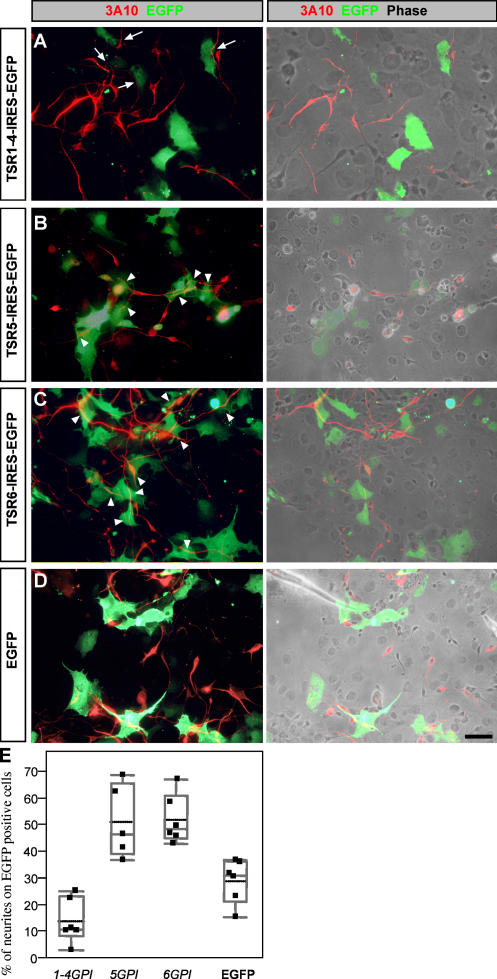
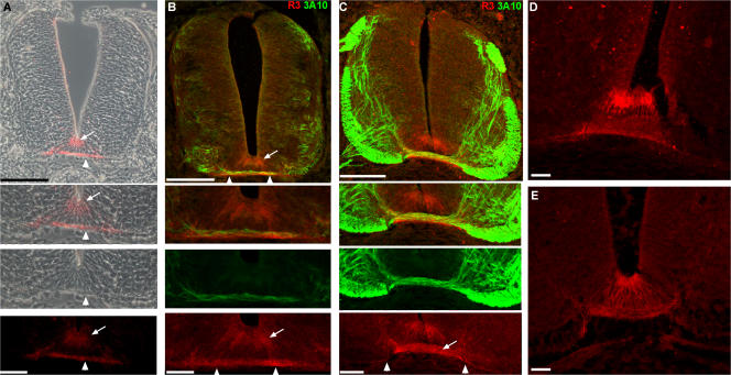
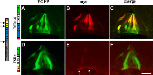
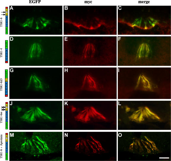
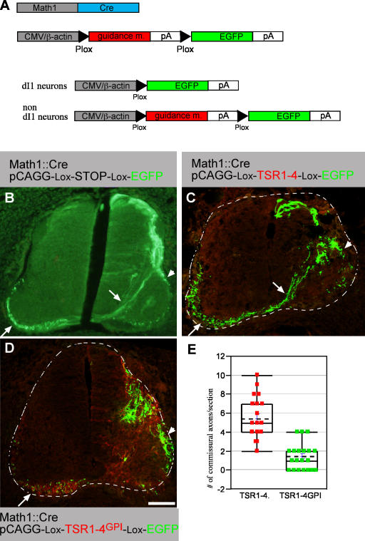
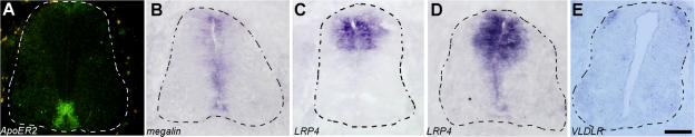
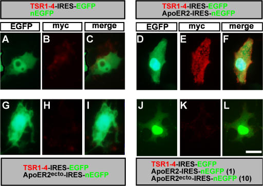
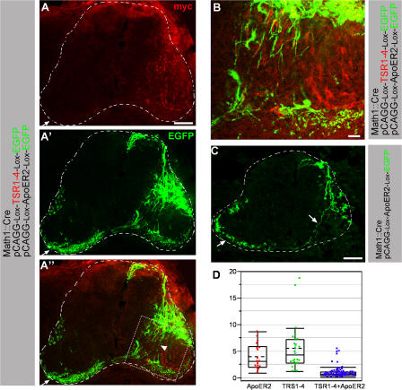
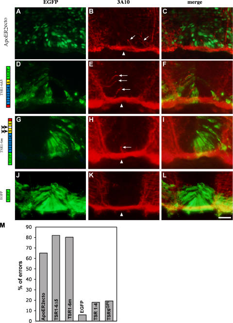
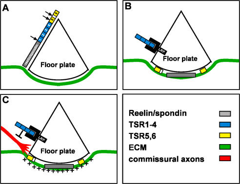
The formation of neuronal networks is governed by a limited number of guidance molecules, yet it is immensely complex. The complexity of guidance cues is augmented by posttranslational modification of guidance molecules and their receptors. We report here that cleavage of the floor plate guidance molecule F-spondin generates two functionally opposing fragments: a short-range repellent protein deposited in the membrane of floor plate cells and an adhesive protein that accumulates at the basement membrane. Their coordinated activity, acting respectively as a short-range repellant and a permissive short-range attractant, constricts commissural axons to the basement membrane beneath the floor plate cells. We further demonstrate that the repulsive activity of the inhibitory fragment of F-spondin requires its presentation by the lipoprotein receptor-related protein (LRP) receptors apolipoprotein E receptor 2, LRP2/megalin, and LRP4, which are expressed in the floor plate. Thus, proteolysis and membrane interaction coordinate combinatorial guidance signaling originating from a single guidance cue.


Figures

Figure 1.
Figure 1.
Differential outgrowth activity of TRSs of F-spondin. COS cells were transfected with pCAGG–TSR1-4GPI–IRES–EGFP (A), pCAGG–TSR5GPI–IRES–EGFP (B), pCAGG–TSR6GPI–IRES–EGFP (C), and pCAGG–EGFP (D). Dissociated E6 chick dorsal spinal cord was plated 24 h after transfection and cultured for an additional 48 h. Neurites are detected with 3A10 mAb. Fluorescence images are shown in the left, and combination of fluorescence and phase contrast images are shown in the right. Neurites are deflected from TSR1-4GPI–expressing cells (A, arrows) and grow preferentially on the adjacent nonexpressing cells. Neurites grow preferentially on TSR5GPI (B)- and TSR6GPI (C)-expressing cells (arrowheads) rather than on the neighboring nonexpressing cells. In the control experiment neurites grow uniformly on EGFP-expressing and nonexpressing cells (D). For quantification, images (n = 6 for each experiment) of the cultures were taken with a digital camera. The total neurite outgrowth was measured as a 3A10-positive area using ImageJ software. A total of 146 neurites were analyzed for TSR5GPI, 278 for TSR6GPI, 203 for TSR1-4GPI, and 455 for EGFP. The area occupied by neurites growing on EGFP-expressing cells was measured using the RG2B colocalization plugin of ImageJ software. The ratio between neurites extending on expressing cells versus total outgrowth is presented. Comparing each experimental group to the control using Dunnett's method (which takes into account multiple comparisons) shows a significant difference between the TSR5GPI (P = 0.004), TSR6GPI (P = 0.002), and TSR1-4GPI (P = 0.04) groups and the control EGFP group. Bar, 10 μm.
Figure 2.
Figure 2.
F-spondin protein is deposited in the apical floor plate and the basement membrane, and binds to commissural axons. F-spondin protein expression at stage 21 (A and B), 26 (C and E), and 27 (D) chick embryos as revealed by the R3 antibody. F-spondin accumulates at the apical floor plate (arrow) and the basement membrane (arrowhead). (B) The pioneer commissural axons (3A10 staining) extend on the F-spondin (R3 staining) that is deposited in the basement membrane as they cross the midline at stage 21. (C) At stage 26, the immunoreactivity of F-spondin is evident on the crossing fibers of the commissural axons (arrow) and basement membrane that flanks the midline ventral pia (arrowheads). Inhibition of serine proteases by in ovo aprotinin injection reduces the deposition of F-spondin at the basement membrane (E) and yields homogenous staining along the surface of floor plate cells. Fluorescence and phase-contrast images in A were taken with microscope using a digital camera. Fluorescence images in B–E were taken with a confocal microscope. Bar, 40 μm.
Figure 3.
Figure 3.
Expression and protein localization of F-spondin domains at the floor plate. In all the experiments, enhancer III of HoxA-1 gene (Li and Lufkin, 2000) was used to drive expression of Cre recombinants (HoxA-1–Cre). A TSR1-4 conditional plasmid (loxP–STOP–loxP–TSR1-4–IRES–EGFP; A–C) and a TSR6 conditional plasmid (loxP–STOP–loxP–TSR6–IRES–EGFP; D–F) cloned in a pCAGG vector were electroporated into the spinal cord of stage 12–14 chick embryos. The F-spondin proteins were tagged with 4× myc epitope at their amino end. Cross-sections of stage 22–24 electroporated embryos were stained with antibodies to myc (B and E) and EGFP (A and D). TSR1-4 protein (B and C) labels the margins of cytoplasmic EGFP (A and C), reflecting its deposition along the membrane of the expressing cells. (E and F) TSR6 is deposited at the basement membrane that underlies the floor plate (arrows). (D) Note that the expressing cells, EGFP-positive cells, do not present the protein. The gray box represents the reelin/spondin domain of F-spondin. The blue boxes represent the nonadhesive TSR1-4 of F-spondin. The yellow boxes represent the adhesive TSR5 and 6 of F-spondin. The red box represents a cassette of 4× myc epitope. The black arrows point to the sites of cleavage of F-spondin. The white arrows point to the site of deposition of TSR6. Images were taken with a microscope using a digital camera. Bar, 25 μm.
Figure 4.
Figure 4.
F-spondin is processed in vivo. In all the experiments the double-tagged F-spondin was cloned in a Cre-dependent plasmid and electroporated along with Hoxa-1–Cre. The myc epitope at the amino end contains four copies (E and H) and the myc epitope at the carboxyl end contains six copies (B, K, and N). (A–C) The TSR1-6 fragment of F-spondin is cleaved in vivo. The amino, EGFP-fused part of the protein stains the floor plate cells (A and C), whereas the carboxyl, myc-fused part of the protein stains the basement membrane (B and C). The TSR1-4 fragment of F-spondin (D–F), deletion of plasmin cleavage site (G–I), mutation of the plasmin cleavage sites (J–L), and inhibition of the endogenous serine proteases by aprotinin (M–O) resulted in the colocalization of the amino and carboxyl tags along the membrane of the floor plate cells, as revealed by the overlapping staining of the myc and EGFP epitopes. The black arrows point to the sites of cleavage of F-spondin. The crossed arrows indicate point mutations in the plasmin cleavage sites. Images in A–L were taken with a microscope using a digital camera. Images in M–O were taken with a confocal microscope. Bar, 50 μm.
Figure 5.
Figure 5.
Membrane-tethered TSR1-4 inhibits commissural outgrowth in vivo. (A, top) Two plasmids are coelectroporated into the chick neural tube. Math1 enhancer, which drives expression in the dI1 dorsal interneurons (Helms and Johnson, 1998), was used to drive expression of Cre recombinase and an alternate guidance molecule/EGFP plasmid. This plasmid contains a CMV enhancer followed by a guidance molecule (F-spondin or ApoER2) flanked with two Plox sites, followed by EGFP. (A, bottom) On electroporation into the chick spinal cord, the guidance molecule will be excised in dI1 cells that express Cre, leading to EGFP expression, whereas nondI1 cells present along the dI1 axonal pathway will express the guidance molecule. EGFP (B), TSR1-4 (C), and TSR1-4GPI (D) were electroporated using an EGFP alternating cassette along with a Math1-Cre plasmid. An alternating cassette contains a Plox-TSR-Plox-EGFP cloned in pCAGG vector. Electroporation was performed at stages 17–18 and embryos were analyzed with anti-myc (Cy3) and anti-EGFP (Cy2) antibodies at stage 26. (B) Expression of EGFP in dI1 cells. dI1 neurons project their axon toward and across the floor plate (arrow) and ipsilaterally (arrowhead). Commissural axons projecting diagonally toward the floor plate and across it to the contralateral side are observed (arrow). (C) dI1 EGFP-expressing cells are present at the dorsal, dorsal-lateral, and medial-lateral spinal cord. Commissural axons projecting diagonally toward the floor plate and across it to the contralateral side are observed (arrow). In addition, the medial-lateral dI1 subpopulation projects ipsilaterally (arrowhead). (D) Very few axons are extending diagonally toward the floor plate at the TSR1-4GPI domain (myc epitope in red). EGFP-expressing axons and TSR1-4GPI–expressing axons projecting longitudinally at the contralateral side are evident (arrow). (E) Quantification of the extent of commissural projection in B and C. Sections from four different embryos (for each group) with matching EGFP intensity were selected. 25 sections of the TSR1-4GPI and 20 sections of the TSR1-4 were analyzed. The number of diagonally crossing axons toward the floor plate was scored. Using a t test shows a significant difference between the TSR1-4 and TSR1-4GPI groups, under a significant level of α = 0.05. The dashed lines demarcate the spinal cord. The image in A was taken with a microscope using a digital camera. Images in C and D were taken with a confocal microscope. Bar, 100 μm.
Figure 6.
Figure 6.
Expression pattern of LRP receptors in the chick embryonic spinal cord. In situ hybridization of ApoER2 (A), LRP2/megalin (B), and LRP4 receptors at E5 chick spinal cord (D), and LRP4 (C) VLDLR (E) at E4 chick spinal cord. (A) ApoER2 expression is confined to the midline, the lateral floor plate cells, and the ventral ventricular zone. Lower levels are detected at the medial floor plate. (B) Megalin is expressed at the ventricular zone, including the floor plate. (C) LRP4 is expressed at E4 in the dorsal third neural tube, at the ventricular zone, and laterally to the ventricular zone. (D) At E5, LRP4 expression spreads to the ventricular zone and the floor plate. (E) VLDLR is expressed in subpopulation of dorsal interneurons. Colabeling with Lhx2 reveals that VLDLR is expressed in dI1 neurons (not depicted). The dashed lines demarcate the spinal cord. Bars: (A, B, and D) 75 μm; (C and E) 100 μm.
Figure 7.
Figure 7.
F-spondin is immobilized to the cell surface by the ApoER2 receptors. ApoER2 is required for the binding of myc-tagged TSR1-4 to the cell surface of COS cells. COS cells were transfected with TSR1-4–IRES–EGFP and nEGFP (A–C), TSR1-4–IRES–EGFP and ApoER2-IRES-nEGFP (D–F), a secreted form of ApoER2 (ApoER2ecto-IRES-nEGFP and TSR1-4–IRES–EGFP [G–I]), ApoER2ecto-IRES-nEGFP, ApoER2-IRES-EGFP, and TSR1-4–IRES–EGFP (J–L). At 48 h after the transfection, surface staining (without fixation) of the cells was preformed. TSR1-4 is bound to the cell surface, where it is coexpressed with ApoER2 (D–F). Bar, 5 μm.
Figure 8.
Figure 8.
ApoER2 is required for eliciting TSR1-4 repulsive activity. ApoER2 (A–C) was coelectroporated with TSR1-4 (A and B) using EGFP-alternating cassettes along with a Math1-Cre plasmid. An alternating cassette containing Plox-ApoER2-Plox-EGFP cloned in a pCAGG vector was used for ApoER2 expression. (A and B) The dotted square in A′ is presented as an enlargement in B. ApoER2 coexpressed with TSR1-4 in dI1-negative cells and EGFP in dI1 cells. dI1 axons are circumventing ApoER2 + TSR1-4–expressing domains (A and B). Many axons are deflected laterally (arrowhead). The number of diagonally crossing axons is reduced. (C) ApoER2 was expressed in dI1- negative cells and EGFP in dI1 cells. The dI1 cell projects axons toward the floor plate (arrow). The dashed lines demarcate the spinal cord. (D) Quantification of erroneous projection of dI1 axons. Sections with matching EGFP intensity were selected: ApoER2 18 (from two embryos), TSR1-4 25 (from two embryos), and ApoER2 + TSR1-4 73 (from five embryos). In each section, the number of axons projecting diagonally toward the floor plate versus the axons that are projecting laterally in the motor neurons domain was determined by quantifying EGFP brightness intensity using National Institutes of Health image software. The ordinate is the ratio between the normal diagonally crossing axons and the erroneous axon projecting at the ventral lateral spinal cord. The projection pattern of dI1 axons growing in a TSR1-4 milieu was compared with ApoER2 and ApoER2 + TRS1-4 milieus. Comparisonsfor all pairs using the Tukey-Kramer honestly significant difference test shows a significant difference between the ApoER2 + TRS1-4 group and the TSR1-4 and ApoER2 groups, and no difference between the TSR1-4 and ApoER2 groups, with a significance level of α = 0.05. Images in A and B were taken with a confocal microscope. The image in C was taken with a microscope using a digital camera. The ends of the box in D are the 25th and 75th percentiles. The line and the dashed line across the box identify the median and the mean, respectively. The horizontal line above the box represents the outermost data point that falls within the 75th percentile plus 1.5 the interquartile range (75th minus 25th percentile). The same applies for the line below the box. Bars, (A and C) 150 μm; (B) 20 μm.
Figure 9.
Figure 9.
Inhibiting LRPs/F-spondin binding enables growth of commissural axons into the floor plate cells. ApoER2ecto (A–C) or F-spondin EGFP-tagged isoforms (D–L) were electroporated into the ventral spinal cord at stages 12–14. Cross-sections of stage 22–24 embryos were stained with anti-EGFP (A, D, G, and J) or antineurofilament mAb 3A10 (B, E, H, and K). In the control experiment, expression of EGFP (J–L), commissural axons cross the midline as a tight bundle (arrowhead) under the floor plate cells whereas expression of ApoER2ecto (A–C) and the mutated F-spondin's isoforms (D–L) resulted in a dorsal erroneous neurites projection into the floor plate cells (B, E, and H, arrows). (M) Quantification of the extent of pathfinding errors at the midline. For each protein ∼100 sections were inspected. A cross-section with axons extending dorsally at the floor plate was scored as an error. The crossed arrows indicate point mutations in the plasmin cleavage sites. Bar, 50 μm.
Figure 10.
Figure 10.
The role of F-spondin in midline crossing. (A) F-spondin is expressed and secreted from the floor plate cells. F-spondin is subjected to proteolysis by serine proteases within the amino end of TSR5 and between TSR5 and 6 (arrows). A yet unidentified protease cleaves F-spondin between the spondin domain and TSR1 (arrow). (B) The reelin/spondin domain (Burstyn-Cohen et al., 1999) and the adhesive TSR5 and 6 bind to the ECM that underlies the floor plate. The TSR1-4 fragment binds to the apical floor plate cells via interaction with the LRP receptors ApoER2, megalin, and LRP4. (C) The cell surface–tethered TSR1-4 repels commissural axons and prevents their penetration into the floor plate cells. The adhesive TSRs provide an outgrowth supportive substrate for the commissural axons.

References

    1. Augsburger, A., A. Schuchardt, S. Hoskins, J. Dodd, and S. Butler. 1999. BMPs as mediators of roof plate repulsion of commissural neurons. Neuron. 24:127–141. - PubMed
    1. Bovolenta, P., and J. Dodd. 1990. Guidance of commissural growth cones at the floor plate in the embryonic rat spinal cord. Development. 109:435–447. - PubMed
    1. Brose, K., K.S. Bland, K.H. Wang, D. Arnott, W. Henzel, C.S. Goodman, M. Tessier-Lavigne, and T. Kidd. 1999. Slit proteins bind Robo receptors and have an evolutionarily conserved role in repulsive axon guidance. Cell. 96:795–806. - PubMed
    1. Burstyn-Cohen, T., V. Tzarfaty, A. Frumkin, Y. Feinstein, E. Stoeckli, and A. Klar. 1999. F-spondin is required for accurate pathfinding of commissural axons at the floor plate. Neuron. 23:233–246. - PubMed
    1. Charron, F., E. Stein, J. Jeong, A.P. McMahon, and M. Tessier-Lavigne. 2003. The morphogen sonic hedgehog is an axonal chemoattractant that collaborates with netrin-1 in midline axon guidance. Cell. 113:11–23. - PubMed

Publication types

MeSH terms

Substances