Review
. 2008 Feb;83(2):229-36.
doi: 10.1189/jlb.0707503. Epub 2007 Sep 17.

Mechanisms of eosinophil secretion: large vesiculotubular carriers mediate transport and release of granule-derived cytokines and other proteins

Affiliations
Review

Mechanisms of eosinophil secretion: large vesiculotubular carriers mediate transport and release of granule-derived cytokines and other proteins

Rossana C N Melo et al. J Leukoc Biol. 2008 Feb.

Abstract

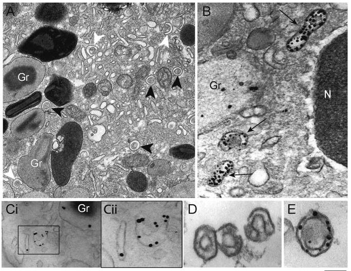
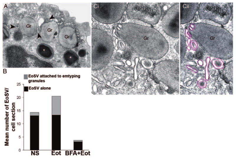
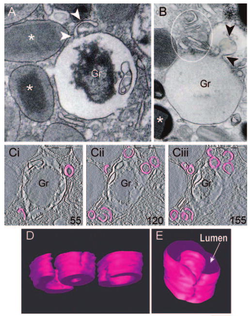
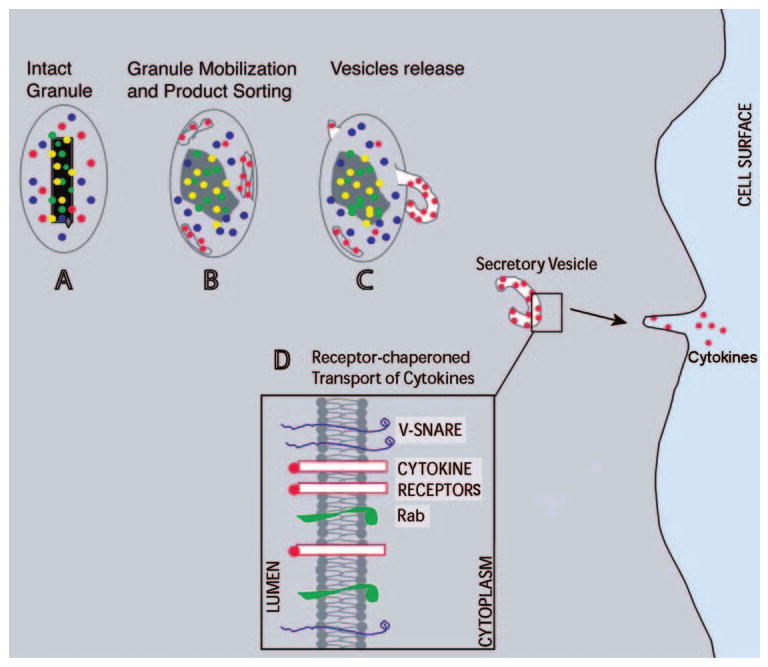
Eosinophils generate and store a battery of proteins, including classical cationic proteins, cytokines, chemokines, and growth factors. Rapid secretion of these active mediators by eosinophils is central to a range of inflammatory and immunoregulatory responses. Eosinophil products are packaged within a dominant population of cytoplasmic specific granules and generally secreted by piecemeal degranulation, a process mediated by transport vesicles. Large, pleiomorphic vesiculotubular carriers were identified recently as key players for moving eosinophil proteins from granules to the plasma membrane for extracellular release. During secretion, these specialized, morphologically distinct carriers, termed eosinophil sombrero vesicles, are actively formed and direct differential and rapid release of eosinophil proteins. This review highlights recent discoveries concerning the organization of the human eosinophil secretory pathway. These discoveries are defining a broader role for large vesiculotubular carriers in the intracellular trafficking and secretion of proteins, including selective receptor-mediated mobilization and transport of cytokines.


Figures

Fig. 1
Fig. 1
Multifunctional granule-stored products within human eosinophils. Ultrastructural image of a human eosinophil shows the cytoplasm packed with specific granules containing an internal, often electron-dense crystalline core, and cores are surrounded by an electron-lucent matrix. In response to a variety of stimuli, eosinophils secrete cytotoxic cationic proteins and an array of cytokines and chemokines. Eosinophil proteins documented within specific granules are listed. Original bar, 480 nm. Gr, Specific granules; N, nucleus; LB, lipid body. CCL5 (RANTES) [8]; CCL11 (eotaxin) [9]; ECP, eosinophil cationic protein [10]; EDN, eosinophil-derived neurotoxin [11]; ENA-78, epithelial cell-derived neutrophil-activating peptide/CXCL5 [12]; EPO, eosinophil peroxidase [10]; GM-CSF [13, 14]; GRO-α, growth-related oncogene-α [15]; IL-2 [16]; IL-3 [14]; IL-4 [17, 18]; IL-5 [14, 19]; IL-6 [20]; IL-10 (L. A. Spencer, P. F. Weller, unpublished data); IL-12 (L. A. Spencer, P. F. Weller, unpublished data); IL-13 [21]; INF-γ, (L. A. Spencer, P. F. Weller, unpublished data); MBP, major basic protein [22, 23]; NGF, nerve growth factor [24]; SCF, stem cell factor [25]; TGF-α [26]; TNF-α [27].
Fig. 2
Fig. 2
EoSVs are large, tubular carriers involved in the secretory pathway. (A) EoSVs (arrowheads) with typical morphology are observed in the cytoplasm by transmission electron microscopy (TEM). These vesicles show a “Mexican hat” (sombrero) appearance (black arrowheads) in conventional cross-thin sections (∼80 nm thickness) of eosinophils. EoSVs have a central area of cytoplasm and a brim of circular membrane-delimited vesicle. These tubular vesicles also exhibit a “C”-shaped morphology (white arrowheads). (B and C) EoSVs within activated eosinophils are immunolabeled for MBP and IL-4, respectively. Note a highly mobilized, electron lucent granule in B. (Cii) The boxed area of Ci in higher magnification. (D and E) EoSVs isolated by subcellular fractionation and visualized by, respectively, conventional TEM and after immunonanogold labeling for MBP. Note that although MBP is preferentially labeled within the vesicle lumen (B and E), IL-4 is clearly detected on vesicle membranes (C). Original bars, 400 nm (A); 230 nm (B); 250 nm (Ci); 200 nm (Cii); 180 nm (D); 150 nm (E). Gr, specific granules. (C) Reprinted from ref. [45]; (D and E) reprinted from ref. [44] with permission.
Fig. 3
Fig. 3
EoSVs associate with specific granules for protein transport. (A) EoSVs (arrowheads), within an eotaxin-stimulated eosinophil, are in contact with enlarged emptying granules (Gr) exhibiting reduced electron-density. Intact, nonemptying granules (*), with typical morphology, are seen close to emptying granules. (B) Quantification of EoSV numbers by TEM. Eotaxin (Eot) induced significant formation of EoSVs and association of these vesicular compartments with granules undergoing release of their products, as described in ref. [44]. Brefeldin-A (BFA) pretreatment suppressed all EoSV numbers dramatically (P<0.05). NS, Not stimulated. (C) Different profiles of EoSVs (highlighted in pink; Cii) in high magnification in contact and surrounding mobilized specific granules (Gr). Note the trilaminar unit membrane of vesicles and granules. Original bars, 400 nm (A); 250 nm (C).
Fig. 4
Fig. 4
EoSVs are formed from mobilized eosinophil granules. (A and B) Images from conventional TEM (80 nm thickness sections), showing progressive stages of granule emptying. (A) A clear, tubular extension (arrowheads) is seen on the granule surface. Note the disarranged core within the granule. (B) Several EoSV profiles (circle) are in close apposition to a virtually empty granule. Arrowheads point to a large vesicle, apparently formed from the granule surface. *, Full granules. (C) Representative serial tomographic slices (4 nm thickness), obtained from electron tomography of a mobilized granule, show substantial changes associated with EoSV formation. EoSVs, the lumens of which are highlighted in pink, are imaged as open, tubular-shaped structures [44]. Numbers on the lower right corners of the panels indicate slice numbers through the tomographic volume. Cells were stimulated with eotaxin as described [44]. Original bars, 300 nm (A, B); 500 nm (C); 150 nm (D); 100 nm (E). Gr, specific granules. C–E was reprinted from Traffic [44] with permission.
Fig. 5
Fig. 5
A model for mobilization and transport of cytokines from secretory granules to the plasma membrane in human eosinophils. (A) An intact granule with its electron-dense crystalline core and secretory products. (B and C) Sequential stages of structural changes related to granule mobilization and formation of tubular carriers (EoSVs), which transport a specific cytokine to the plasma membrane. (D) The differential sorting of cytokines is based on the presence of cytokine receptors in granule and vesicle membranes [45]. Rab-SNAREs mediate vesicle movement and plasma membrane fusion, resulting in cytokine secretion [31].

Similar articles

Cited by

References

    1. Rosenberg HF, Phipps S, Foster PS. Eosinophil trafficking in allergy and asthma. J Allergy Clin Immunol. 2007;119:1303–1310. - PubMed
    1. Trivedi SG, Lloyd CM. Eosinophils in the pathogenesis of allergic airways disease. Cell Mol Life Sci. 2007;64:1269–1289. - PMC - PubMed
    1. Jacobsen EA, Taranova AG, Lee NA, Lee JJ. Eosinophils: singularly destructive effector cells or purveyors of immunoregulation? J Allergy Clin Immunol. 2007;119:1313–1320. - PubMed
    1. Rothenberg ME, Hogan SP. The eosinophil. Annu Rev Immunol. 2006;24:147–174. - PubMed
    1. Gleich GJ. Mechanisms of eosinophil-associated inflammation. J Allergy Clin Immunol. 2000;105:651–663. - PubMed

Publication types