. 2007 Sep 18;69(12):1245-53.
doi: 10.1212/01.wnl.0000276947.59704.cf.

Clinical characterization of the HOXA1 syndrome BSAS variant

Affiliations

Clinical characterization of the HOXA1 syndrome BSAS variant

T M Bosley et al. Neurology. .

Abstract

Background: The Bosley-Salih-Alorainy syndrome (BSAS) variant of the congenital human HOXA1 syndrome results from autosomal recessive truncating HOXA1 mutations. We describe the currently recognized spectrum of ocular motility, inner ear malformations, cerebrovascular anomalies, and cognitive function.

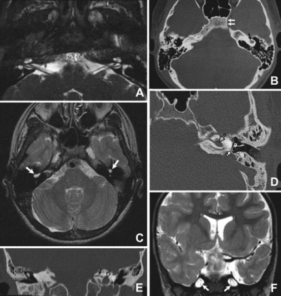
Methods: We examined nine affected individuals from five consanguineous Saudi Arabian families, all of whom harbored the same I75-I76insG homozygous mutation in the HOXA1 gene. Patients underwent complete neurologic, neuro-ophthalmologic, orthoptic, and neuropsychological examinations. Six individuals had CT, and six had MRI of the head.

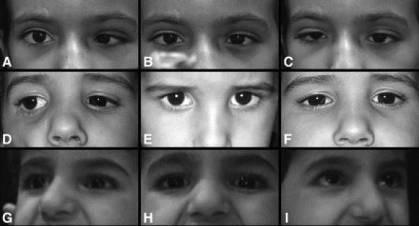
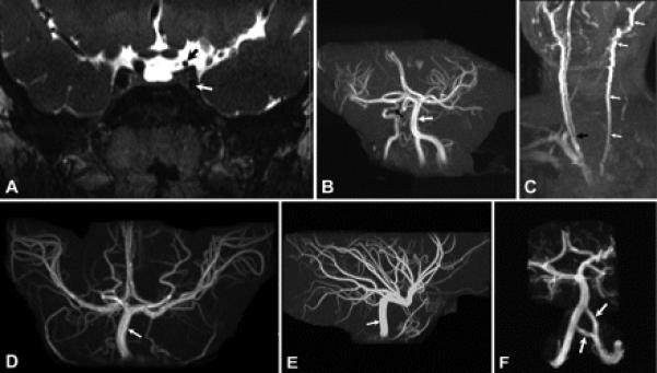
Results: All nine individuals had bilateral Duane retraction syndrome (DRS) type 3, but extent of abduction and adduction varied between eyes and individuals. Eight patients were deaf with the common cavity deformity of the inner ear, while one patient had normal hearing and skull base development. Six had delayed motor milestones, and two had cognitive and behavioral abnormalities meeting Diagnostic and Statistical Manual of Mental Disorders-IV criteria for autism spectrum disorder. MRI of the orbits, extraocular muscles, brainstem, and supratentorial brain appeared normal. All six appropriately studied patients had cerebrovascular malformations ranging from unilateral internal carotid artery hypoplasia to bilateral agenesis.

Conclusions: This report extends the Bosley-Salih-Alorainy syndrome phenotype and documents the clinical variability resulting from identical HOXA1 mutations within an isolated ethnic population. Similarities between this syndrome and thalidomide embryopathy suggest that the teratogenic effects of early thalidomide exposure in humans may be due to interaction with the HOX cascade.


Figures

Figure 1
Figure 1. Variability of ocular alignment and motility
Images A, D, and G are right gaze, while images B, E, and H are in primary position, and images C, F, and I are left gaze. Top row (A, B, and C) are of Patient 4 who had a primary position esotropia with no anomalous head position when fixing with the right eye. This patient was unable to abduct either eye beyond the midline but demonstrated modest adduction bilaterally with globe retraction and fissure narrowing of the adducting eye. Middle row (D, E, and F) are of Patient 8 who was orthotropic in primary position but adopted a small face turn left to maintain comfortable binocular fixation. He had no eye movements to the left but modest symmetric movement of both eyes into right gaze. Bottom row (G, H, and I) are of Patient 5, who was orthotropic in primary position and had severely limited eye movements bilaterally with only minimal adduction the right eye. No abnormal head posture was present in primary position but she used a face turn to view objects to the right or left.
Figure 2
Figure 2. Variability of skull base neuroimaging
(A) High resolution axial constructive interference in steady-state MR image of the inner ear and low brainstem of Patient 8 demonstrating normal inner ear structures and normal lower cranial nerves. (B, C) Axial CT (B) and T2-weighted MR images (C) at the level of the inner ear of Patient 6 demonstrating bilateral common cavity deformity (black arrow in B and white arrows in C). The medial part of the left carotid canal is absent (double arrows in B) while the right one is present. (D) Axial CT of the petrous bone of Patient 3 showing common cavity deformity of the left inner ear (open arrow) with normal stapes (double arrows) and lack of development of the oval window opposite to the footplate of stapes (solid arrow). (E) Reconstructed coronal CT image of the petrous bone of Patient 3 showing the common cavity deformity of the left inner ear (open arrow) and severe stenosis of the left internal auditory canal (black arrow). Inner ear structures on the right side are absent. (F) Coronal T2-weighted MR image of Patient 7 showing patulous Meckle's caves (arrows).
Figure 3
Figure 3. Variability of MR angiography
(A) Coronal T2-weighted MR image of Patient 8 demonstrating hypoplasia of the cavernous (white arrow) and supraclinoid (black arrow) portions of the left internal carotid artery. The cavernous and supraclinoid portions of the right internal carotid artery have normal caliber. (B) MR angiogram of the brain of Patient 6 showing absence of the left internal carotid artery and hypoplasia of the cavernous portion of the right internal carotid artery (black arrow). Vertebral and basilar arteries (white arrow) have larger caliber than the right internal carotid artery. (C) MR angiogram of the neck of Patient 6 demonstrating absence of the left internal carotid artery with the common carotid artery ending as a continuation of external carotid artery (white arrows). The right common carotid artery has a bifurcation (black arrow) at about the level of the C7 vertebra, while normal bifurcation of common carotid artery is about the level of C4. (D, E) Anterior (D) and lateral (E) projections of the MR angiogram of the brain of Patient 5 demonstrating absence of both internal carotid arteries and enlargement of the basilar (arrow) and posterior communicating arteries, which supply both the anterior and posterior cerebral circulations. (F) MR angiogram of the brain of Patient 5 showing duplication of the intracranial portion of the left vertebral artery (arrows).

References

    1. Tischfield MA, Bosley TM, Salih MA, et al. Homozygous HOXA1 mutations disrupt human brainstem, inner ear, cardiovascular and cognitive development. Nat Genet. 2005;37:1035–1037. - PubMed
    1. Chisaka O, Musci TS, Capecchi MR. Developmental defects of the ear, cranial nerves and hindbrain resulting from targeted disruption of the mouse homeobox gene Hox-1.6. Nature. 1992;355:516–520. - PubMed
    1. Lufkin T, Dierich A, LeMeur M, Mark M, Chambon P. Disruption of the Hox-1.6 homeobox gene results in defects in a region corresponding to its rostral domain of expression. Cell. 1991;66:1105–1119. - PubMed
    1. Holve S, Friedman B, Hoyme HE, et al. Athabascan brainstem dysgenesis syndrome. Am J Med Genet A. 2003;120:169–173. - PubMed
    1. Jen JC, Chan WM, Bosley TM, et al. Mutations in a human ROBO gene disrupt hindbrain axon pathway crossing and morphogenesis. Science. 2004;304:1509–1513. - PMC - PubMed

MeSH terms