. 2007 Dec;27(23):8113-26.
doi: 10.1128/MCB.00794-07. Epub 2007 Sep 17.

Identification of phosphotyrosine binding domain-containing proteins as novel downstream targets of the EphA8 signaling function

Affiliations

Identification of phosphotyrosine binding domain-containing proteins as novel downstream targets of the EphA8 signaling function

Jongdae Shin et al. Mol Cell Biol. 2007 Dec.

Abstract

Eph receptors and ephrins have been implicated in a variety of cellular processes, including morphology and motility, because of their ability to modulate intricate signaling networks. Here we show that the phosphotyrosine binding (PTB) domain-containing proteins AIDA-1b and Odin are tightly associated with the EphA8 receptor in response to ligand stimulation. Both AIDA-1b and Odin belong to the ankyrin repeat and sterile alpha motif domain-containing (Anks) protein family. The PTB domain of Anks family proteins is crucial for their association with the juxtamembrane domain of EphA8, whereas EphA8 tyrosine kinase activity is not required for this protein-protein interaction. In addition, we found that Odin is a more physiologically relevant partner of EphA8 in mammalian cells. Interestingly, overexpression of the Odin PTB domain alone attenuated EphA8-mediated inhibition of cell migration in HEK293 cells, suggesting that it acts as a dominant-negative mutant of the endogenous Odin protein. More importantly, small interfering RNA-mediated Odin silencing significantly diminished ephrinA5-induced EphA8 signaling effects, which inhibit cell migration in HEK293 cells and retract growing neurites of Neuro2a cells. Taken together, our findings support a possible function for Anks family proteins as scaffolding proteins of the EphA8 signaling pathway.


Figures
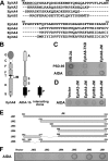
FIG. 1.
FIG. 1.
Yeast library screening identified AIDA-1 as an EphA8-interacting protein. (A) The JM amino acid sequences of the indicated Eph family members were aligned using the ClustalW program. Gaps are represented by dashes. The numbers above the two sets of sequences mark the locations of amino acids within the EphA8 JM region. The dotted underline indicates the specific amino acid residues of the EphA8 JM region that are critical for interaction with the AIDA-1b PTB domain. (B) Domain structures of EphA8, AIDA-1b, and the interacting clone of AIDA identified in the yeast two-hybrid screen. G, immunoglobulin-like domain; C, cysteine-rich motif; F, fibronectin type III repeat domain; T, transmembrane domain; JM, juxtamembrane domain; K, kinase domain; S, SAM domain; A, ankyrin repeat domain; P, PTB domain. (C) X-Gal staining analysis indicated that the PTB domain of AIDA-1 binds to the JM domain of EphA8. Yeast transformants were cultured on Trp Leu selective plates in the absence of 3-AT prior to X-Gal staining using a filter-lifting method. Yeast cells transformed with NR-2A and PSD-95 served as positive controls for these yeast two-hybrid systems. Yeast cells transformed with the tyrosine kinase domain (TKD) of EphA8 as bait served as a negative control. Yeast transformants showing negative X-Gal staining on Trp Leu selective plates did not grow on His Trp Leu selective plates in the presence of 1 mM 3-AT (data not shown). (D) AIDA-1 PTB domain interacts only with the JM domain of EphA8, not with those of EphA2, EphA4, and EphB2, in yeast. The JM sequences of the Eph family members shown in panel A were expressed as bait proteins fused to the LexA DNA binding domain in yeast. Yeast transformants were cultured on Trp Leu selective plates, and X-Gal staining was performed essentially as described for panel C. (E) Schematic diagram showing the various EphA8-JM deletion constructs that were subcloned into the bait construct described for panels C and D. (F) The AIDA PTB domain was coexpressed in the yeast two-hybrid assay with various deletion mutants of EphA8-JM, and X-Gal staining was performed essentially as described for panel C.
FIG. 2.
FIG. 2.
Evidence that the EphA8 JM domain associates directly with the PTB domain of Anks family proteins. (A) Demonstration of AIDA-1 PTB domain interaction with the EphA8 JM domain, using purified proteins. Purified GST or GST-JM fusion proteins were mixed with purified His-tagged AIDA-1 PTB protein. Bound proteins were precipitated using His-bind resin, fractionated by 10% sodium dodecyl sulfate-polyacrylamide gel electrophoresis (SDS-PAGE), and detected by Western blotting with anti-GST antibody (top panel). The same blot was stripped and reprobed with anti-AIDA antibody (middle panel). GST or GST-JM fusion proteins used for this experiment were directly detected by Coomassie staining (bottom panel). (B) Demonstration of AIDA-1 PTB domain interaction with the full-length EphA8 receptor. Purified proteins (GST or GST-PTB) were mixed with whole-cell lysates from HEK293 cells transfected with the control vector (lanes 1 and 2) or an EphA8 expression construct (lanes 3 and 4). Bound proteins were pulled down using glutathione beads, fractionated by 10% SDS-PAGE, and detected by Western blotting using anti-EphA8 antibody (top panel). The same blot was stripped and reprobed with anti-GST antibody (middle panel). A sample (2%) of each total cell lysate was analyzed directly by Western blotting using anti-EphA8 antibody (bottom panel). (C) Evidence that the Odin PTB domain interacts with the EphA8 JM domain, using purified proteins. Experiments were performed essentially as described for panel A, except that purified His-tagged Odin PTB protein and anti-Odin antibody were used. PD, pulldown; WB, Western blot.
FIG. 3.
FIG. 3.
Anks family proteins associate with EphA8 in HEK293 cells. (A) Coimmunoprecipitation of AIDA-1b and EphA8 in HEK293 cells. Cells were transfected with AIDA-1b and EphA8 as indicated in each lane, and then total cell lysates (100 μg) were immunoprecipitated (IP) with anti-AIDA antibody, followed by immunoblotting (WB) with anti-EphA8 antibody (top panel). The same blot was stripped and reprobed with anti-AIDA antibody (middle panel). A sample (1%) of each total cell lysate (WCL) was analyzed directly by Western blotting using anti-EphA8 antibody (bottom panel). (B) Reciprocal coimmunoprecipitation of AIDA-1b and EphA8 in HEK293 cells. Experiments were performed essentially as described for panel A, except that reciprocal antibodies were used, as indicated in each panel. (C) Coimmunoprecipitation of AIDA-1b and KD EphA8 in HEK293 cells. Experiments were performed essentially as described for panel B. (D) Coimmunoprecipitation of Odin and EphA8 in HEK293 cells. Experiments were performed essentially as described for panel A, except that AIDA-1b was replaced by Odin. (E) Reciprocal coimmunoprecipitation of Odin and EphA8 in HEK293 cells. Experiments were performed as described for panel D, except that reciprocal antibodies were used, as indicated in each panel. (F) Coimmunoprecipitation of Odin and KD EphA8 in HEK293 cells as described for panel E. (G) Binding of Odin-PTB to EphA8-JM is inhibited by a specific peptide (KKRHCGY), which contains the docking site in EphA8-JM for the PTB domain. Purified proteins (GST or GST-JM) were mixed with whole-cell lysates from HEK293 cells transfected with control vector (lanes 1 and 3) or an Odin-PTB expression construct (lanes 2, 4, and 5). In the peptide competition experiment, 50 μM of specific peptides was preincubated with cell lysates for 30 min on ice (lane 5). GST pull-down experiments using glutathione beads were performed essentially as described in the legend to Fig. 2B, and bound proteins were detected by Western blotting using anti-Odin antibodies (top panel). The same blot was stripped and reprobed with anti-GST antibodies (middle panel). A sample (2%) of each total cell lysate was analyzed directly by Western blotting using anti-Odin antibodies (bottom panel). (H and I) Evidence that both Odin-PTB and Odin-ΔPTB interact with EphA8. Experiments were performed essentially as described for panel D, except that the indicated constructs and antibodies were used for transfection and immunoprecipitation, respectively.
FIG. 4.
FIG. 4.
EphrinA5 stimulation enhances specific complex formation of EphA8 with AIDA-1 family proteins. (A) HEK293 cells were transiently transfected with AIDA-1b and WT or KD EphA8. Forty-eight hours after transfection, cells were stimulated with ephrinA2-expressing HEK293 cells for 15 min. Total cell lysates (100 μg) were immunoprecipitated with anti-AIDA antibody or control IgG and then immunoblotted with anti-EphA8 antibody (top panel). The same blot was stripped and reprobed with anti-AIDA antibody (middle panel). A sample (1%) of each total cell lysate was analyzed directly by Western blotting using anti-EphA8 antibody (bottom panel). (B) Experiments were performed essentially as described for panel A, except that AIDA-1b was replaced by Odin. Note that the coprecipitated EphA8 protein was barely detectable in the absence of ephrinA2 stimulation because of the very short exposure time (top panel, lanes 1 and 4). (C) Mouse brains at the indicated developmental stages were homogenized in lysis buffer, and the lysates were analyzed directly by Western blotting with the indicated antibodies. SC, superior colliculus; E, embryonic day; P, postnatal day. (D) Association of endogenous Odin and EphA8 in the mouse superior colliculus. Lysates from E14.5 mouse superior colliculi were immunoprecipitated with anti-Odin antibodies or with control IgG and then immunoblotted with anti-EphA8 antibodies (top panel). The same blot was stripped and reprobed with anti-Odin antibodies (middle panel). A sample (10%) of each lysate was analyzed directly by Western blotting using anti-EphA8 antibodies (bottom panel). (E) Odin colocalizes with EphA in superior collicular neurons. E14.5 mouse superior collicular neurons were treated with aggregated ephrinA5-Fc for 10 min, fixed, and then stained for Odin (red) and EphA (green). Bar = 20 μm. WB, Western blot; WCL, whole-cell lysate.
FIG. 5.
FIG. 5.
(A) EphrinA5 inhibits cell migration in an EphA8 kinase activity-dependent manner. The lower chambers of Transwell inserts were coated with fibronectin, and then 1 × 10⁵ cells were added to the top chamber and allowed to migrate toward the lower chamber, filled with medium containing 5 μg/ml ephrinA5-Fc or Fc, for 2 h. Cells were fixed and stained with eosin prior to being counted under an inverted microscope. WT-6, -7, and -9 and KD-3, -7, and -8 represent different clones of stably transfected cells. Values represent means plus standard deviations. Asterisks mark cell migration whose inhibition was significantly different (P < 0.01) from that of the vector-transfected cells. (B and C) A sample (10%) of each total cell lysate was analyzed directly by Western blotting using anti-EphA8 antibody (B) or anti-Odin antibody (C). (D) Overexpression of Odin-PTB reverses the inhibitory effect of cell migration by the ephrinA5-stimulated EphA8 receptor. A Boyden chamber migration assay was performed as described for panel A. The EphA8-expressing cells used for this experiment correspond to the WT-7 clone shown in panel A. Values represent means plus standard deviations. Asterisks mark cell migration whose inhibition was significantly different (P < 0.01) from that of the vector-transfected cells. (E to G) A sample (10%) of each total cell lysate was analyzed directly by Western blotting using anti-Odin antibody (E and F) or anti-EphA8 antibody (G). (H) Evidence that Odin-PTB competes with endogenous Odin for binding to EphA8. Cells were stimulated with preclustered ephrinA5-Fc (+) or Fc (−) for 15 min, and equal amounts of protein (500 μg) from each cell lysate were immunoprecipitated with anti-EphA8 antibodies and analyzed by immunoblotting with anti-Odin antibodies. The WT-7, EphA8/PTB-2, and EphA8/ΔPTB-2 clones shown in panels A and D were used for this experiment. (I) The same blot shown in panel H was stripped and reprobed with anti-EphA8 antibodies. (J to L) A sample (5%) of each total cell lysate was analyzed directly by Western blotting using anti-EphA8 (J) or anti-Odin (K and L) antibodies. WB, Western blot.
FIG. 6.
FIG. 6.
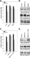
Inhibition of cell migration by ephrinA5-stimulated EphA8 requires Odin function in HEK293 cells. The EphA8-expressing cells used for this experiment correspond to the WT-7 clone shown in Fig. 5A. (A) HEK293 cells were transfected with control siRNA or human Odin siRNA, and 72 h later, cells were subjected to cell migration assay in the presence of Fc or ephrinA5-Fc as described in the legend to Fig. 5A. An asterisk marks cell migration whose inhibition was significantly different (P < 0.01) from that of Odin siRNA-transfected cells. (B) Seventy-two hours after siRNA transfection, cell lysates (10%) were probed by immunoblotting with anti-Odin antibodies (top panel), anti-EphA8 antibodies (middle panel), or anti-actin antibodies (bottom panel). (C) Experiments were performed essentially as described for panel A, except that the control vector (bars 1 to 4) or Odin expression construct was retransfected after 72 h of transfection with siRNA. An asterisk marks cell migration whose inhibition was significantly different (P < 0.01) from that of cells transfected with Odin siRNA and the control vector (bar 4). (D) Experiments were performed essentially as described for panel B. WB, Western blot.
FIG. 7.
FIG. 7.
(A) Coexpression of EphA8 and Odin in Neuro2a cells. One hundred micrograms of whole-cell extract (WCL) from PC12 or Neuro2a cells was resolved by SDS-PAGE and then subjected to Western blot analysis using the indicated antibodies. Note that Odin was detected as a smaller protein in PC12 cells than in Neuro2a cells. This may be because PC12 cells predominantly express a 1,125-amino-acid rat Odin protein, which is 25 amino acids shorter than mouse Odin in Neuro2a cells. (B) Coimmunoprecipitation of Odin and EphA8 in Neuro2a cells. Neuro2a cells stably expressing the WT EphA8 receptor were generated as described previously. Total cell lysates (500 μg) were immunoprecipitated with anti-Odin antibody and then immunoblotted with the anti-EphA8 antibody (top panel). The same blot was stripped and reprobed with anti-Odin antibody (middle panel). A sample (0.5%) of each total cell lysate was analyzed directly by Western blotting using anti-EphA8 antibody (bottom panel). (C) Odin colocalizes with EphA8 in the neurite tips of Neuro2a cells. Neuro2a cells stably expressing the EphA8 receptor were stained with either anti-EphA8 or anti-Odin antibody followed by Alex Fluor 594-anti-rabbit secondary antibody (top panels). Neuro2a cells transiently transfected with EphA8-EGFP were stained with anti-Odin antibody followed by Alex Fluor 594-anti-rabbit secondary antibody (bottom panels). WB, Western blot.
FIG. 8.
FIG. 8.
Neurite retraction is induced by ephrinA5-stimulated EphA8 in Neuro2a cells. Neuro2a cells stably transfected with the indicated constructs were cultured on a poly-l-lysine-coated cover glass for 2 to 3 days in the presence of dibutyryl cAMP to induce neurite outgrowth. (A) Neurites were imaged by time-lapse microscopy at 1 frame per min. The 0-min images show neuronal tips just prior to treatment with preclustered ephrinA5-Fc. (B) Neurite retraction was scored for Fc-treated and ephrinA5-Fc-treated cells by using time-lapse microscopy. The data represent three or more separate experiments in which at least 20 neurites were scored for each cell. Asterisks mark neurite retraction that was significantly different (*, P < 0.01; **, P < 0.02) from that of the vector transfectants. (C) A sample (10%) of each total cell lysate was analyzed directly by Western blotting (WB) using anti-EphA8 antibody.
FIG. 9.
FIG. 9.
Odin is required for neurite retraction induced by ephrinA5-stimulated EphA8 in Neuro2a cells. Experiments were performed essentially as described in the legend to Fig. 8A and B, except that FITC-labeled control siRNA was included in each transfection to visualize the siRNA-transfected cells. Once the transfectants were localized by fluorescence, neurite retraction was observed by time-lapse microscopy, using a differential interference contrast imaging system. The data represent three or more separate experiments in which at least 20 neurites were scored for each cell. *, P < 0.05 compared with cells transfected with the control vector; **, P < 0.02 compared with cells transfected with control siRNA. (B) Neuro2a cells were transfected with control siRNA or Odin siRNA, and 72 h later, cell lysates (10%) were probed by immunoblotting with anti-Odin antibodies (top panel), anti-EphA8 antibodies (middle panel), or antiactin antibodies (bottom panel). WB, Western blot.

Similar articles

Cited by

References

    1. Carter, N., T. Nakamoto, H. Hirai, and T. Hunter. 2002. EphrinA1-induced cytoskeletal re-organization requires FAK and p130(cas). Nat. Cell Biol. 4:565-573. - PubMed
    1. Chen, Z. Y., C. Sun, K. Reuhl, A. Bergemann, M. Henkemeyer, and R. Zhou. 2004. Abnormal hippocampal axon bundling in EphB receptor mutant mice. J. Neurosci. 24:2366-2374. - PMC - PubMed
    1. Choi, S., and S. Park. 1999. Phosphorylation at Tyr-838 in the kinase domain of EphA8 modulates Fyn binding to the Tyr-615 site by enhancing tyrosine kinase activity. Oncogene 18:5413-5422. - PubMed
    1. Dail, M., M. Richter, P. Godement, and E. B. Pasquale. 2006. Eph receptors inactivate R-Ras through different mechanisms to achieve cell repulsion. J. Cell Sci. 119:1244-1254. - PubMed
    1. Elowe, S., S. J. Holland, S. Kulkarni, and T. Pawson. 2001. Downregulation of the Ras-mitogen-activated protein kinase pathway by the EphB2 receptor tyrosine kinase is required for ephrin-induced neurite retraction. Mol. Cell. Biol. 21:7429-7441. - PMC - PubMed

Publication types

MeSH terms