. 2007 Dec;27(23):8401-8.
doi: 10.1128/MCB.01478-07. Epub 2007 Sep 17.

Complex formation of yeast Rev1 with DNA polymerase eta

Affiliations

Complex formation of yeast Rev1 with DNA polymerase eta

Narottam Acharya et al. Mol Cell Biol. 2007 Dec.

Abstract

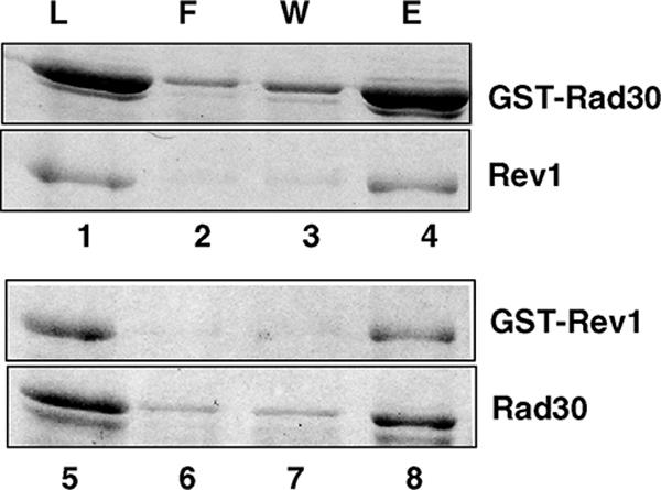
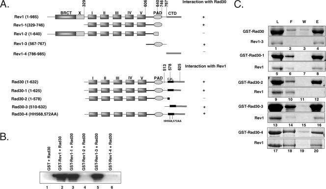
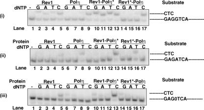
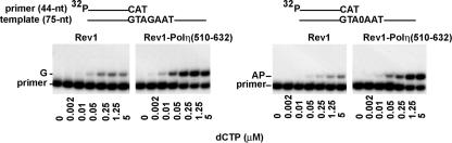
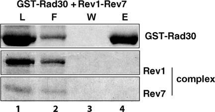
In Saccharomyces cerevisiae, Rev1 functions in translesion DNA synthesis (TLS) together with polymerase zeta (Pol zeta), comprised of the Rev3 catalytic and Rev7 accessory subunits. Rev1 plays an indispensable structural role in promoting Pol zeta function, and deletion of the Rev1-C terminal region that is involved in physical interactions with Rev3 inactivates Pol zeta function in TLS. In humans, however, Rev1 has been shown to physically interact with the Y-family polymerases Pol eta, Pol iota, and Pol kappa, and the Rev1 C terminus mediates these interactions. Since all the available genetic and biochemical evidence in yeast support the requirement of Rev1 as a structural element for Pol zeta and not for Pol eta, these observations have raised the possibility that in its structural role, Rev1 has diverged between yeast and humans. Here we show that although in yeast a stable Rev1-Pol eta complex can be formed, this complex formation involves the polymerase-associated domain of Rev1 and not the Rev1 C terminus as in humans. We also found that the DNA synthesis activity of Rev1 is enhanced in this complex. We discuss the implications of these and other observations for the possible divergence of Rev1's structural role between yeast and humans.


Figures

FIG. 1.
FIG. 1.
GST pull-down of Polη and Rev1 proteins. Yeast Rev1 or Rad30 (Polη) was mixed with GST-Rad30 (lanes 1 to 4) or GST-Rev1 (lanes 5 to 8), respectively. About 1 μg of each protein was used for this study. After being incubated, samples were bound to glutathione-Sepharose beads, after which they were washed and the bound proteins were eluted by SDS-sample buffer. Aliquots of each sample before being added to the beads (L) and of the flow-through fraction (F), the last washed fraction (W), and the eluted proteins (E) were analyzed on a SDS-12% polyacrylamide gel developed with silver staining.
FIG. 2.
FIG. 2.
Mapping of Rev1 and Polη regions involved in complex formation. (A) Schematic representation of the wild type and various mutants of Rev1 and Polη (Rad30). Based on amino acid sequence homology among the Y-family DNA polymerases, the conserved polymerase domains of yeast Rev1 (aa 1 to 985) and Rad30 (aa 1 to 632) are indicated as I and II (fingers), III and IV (palm), and V (thumb). Although not as conserved, the PAD is also shared among these polymerases. Also indicated are the carboxyl-terminal domain (CTD) and the N-terminal BRCT domain in Rev1. Following the PAD, Rad30 contains a C2H2 UBZ motif. The positions of various deletions or point mutations made in the two proteins are shown. (B) The PAD of Rev1 is necessary and sufficient for interaction with Polη. Yeast Rad30 was mixed with GST alone (lane 1), with GST-Rev1 (lane 2), or with GST-Rev1 proteins with different portions of Rev1 deleted (lanes 3 to 6). About 0.5 μg of each protein was used for this study. After being incubated, samples were bound to glutathione-Sepharose beads and extensively washed, and the bound proteins were eluted by SDS-sample buffer. Half of the eluted proteins were resolved on a SDS-12% polyacrylamide gel, and Western blot analysis was performed, using yeast anti-Polη antibodies. (C) Involvement of the Polη C terminus in mediating interactions with Rev1. Lanes 1 to 4, yeast Rev1-3 mixed with GST-Rad30; lanes 5 to 16, Rev1 mixed with GST-Rad30 with different portions of Rad30 deleted; lanes 17 to 20, Rev1 mixed with GST-Rad30 carrying the HH568,572AA mutations. After being incubated, samples were bound to glutathione-Sepharose beads and washed, and the bound proteins were eluted by SDS-sample buffer. Aliquots of each sample before being added to the beads (L) and of the flow-through fraction (F), the last washing fraction (W), and the eluted proteins (E) were analyzed on a SDS-12% polyacrylamide gel and developed with silver staining.
FIG. 3.
FIG. 3.
Comparison of the DNA polymerase activity of Rev1 and Rev1-Polη complexes. (i to iii) The complete standard reaction mixture contained 0.2 nM Rev1 or Polη or the Rev1-Polη complex, 10 μM of a single dNTP, and 10 nM of a DNA substrate. Reaction mixtures were incubated at 30°C for 10 min, and the reactions were stopped by the addition of loading buffer (30 μl) containing 20 mM EDTA, 95% formamide, 0.3% bromphenol blue, and 0.3% cyanol blue. The reaction products were resolved on 15% polyacrylamide gels containing 8 M urea. Lane 1 is the buffer control. Incorporation of each of the nucleotides by Rev1 is shown in lanes 2 to 5; by Polη, in lanes 6 to 9, by the Rev1-Polη* complex, in lanes 10 to 13, and by the Rev1*-Polη complex, in lanes 14 to 17. The DNA substrates used are indicated to the right of the panels. (iii) The abasic site in the substrate is indicated by 0. Polη* and Rev1* denote the catalytically inactive forms of the polymerases.
FIG. 4.
FIG. 4.
dCTP incorporation opposite template G and opposite an abasic (AP) site by Rev1 in the presence or absence of the 122-amino-acid C-terminal peptide of Polη. Rev1 (100 nM) alone or together with the Polη peptide (aa 510 to 632) (200 nM) was first preincubated overnight at 0°C. Rev1 and Rev1 plus the Polη peptide (each containing 0.2 nM Rev1) were incubated with a primer-template DNA substrate (10 nM) and increasing concentrations of dCTP for 5 min at 30°C. The nucleotide incorporation rate was plotted against the dCTP concentration, and the data were fit to the Michaelis-Menten equation describing a hyperbola. Apparent Km and kcat values were obtained from the fit and used to calculate the efficiency of deoxynucleotide incorporation (kcat/Km). The position of the abasic site in the DNA substrate is indicated by 0.
FIG. 5.
FIG. 5.
Inhibition of Rev1-Polη complex formation by Rev7. A purified Rev1-Rev7 complex was mixed with GST-Rad30 (lanes 1 to 4). About 1 μg of each protein was used for the study. After being incubated, samples were bound to glutathione-Sepharose beads and washed, and the bound proteins were eluted by SDS-sample buffer. Aliquots of each sample before being added to the beads (L) and of the flow-through fraction (F), the last washing fraction (W), and the eluted proteins (E) were analyzed on an SDS-12% polyacrylamide gel developed with silver stain.
FIG. 6.
FIG. 6.
Secondary structural elements in the Rev1 PAD. The distal region of the Rev1 PAD could mediate its binding to Rev7, whereas the proximal region could be involved in interactions with Polη.

References

    1. Acharya, N., L. Haracksa, R. E. Johnson, I. Unk, S. Prakash, and L. Prakash. 2005. Complex formation of yeast Rev1 and Rev7 proteins: a novel role for the polymerase-associated domain. Mol. Cell. Biol. 25:9734-9740. - PMC - PubMed
    1. Acharya, N., R. E. Johnson, S. Prakash, and L. Prakash. 2006. Complex formation with Rev1 enhances the proficiency of yeast DNA polymerase ζ for mismatch extension and for extension opposite from DNA lesions. Mol. Cell. Biol. 26:9555-9563. - PMC - PubMed
    1. Bailly, V., J. Lamb, P. Sung, S. Prakash, and L. Prakash. 1994. Specific complex formation between yeast RAD6 and RAD18 proteins: a potential mechanism for targeting RAD6 ubiquitin-conjugating activity to DNA damage sites. Genes Dev. 8:811-820. - PubMed
    1. Bailly, V., S. Lauder, S. Prakash, and L. Prakash. 1997. Yeast DNA repair proteins Rad6 and Rad18 form a heterodimer that has ubiquitin conjugating, DNA binding, and ATP hydrolytic activities. J. Biol. Chem. 272:23360-23365. - PubMed
    1. Baynton, K., A. Bresson-Roy, and R. P. P. Fuchs. 1999. Distinct roles for Rev1p and Rev7p during translesion synthesis in Saccharomyces cerevisiae. Mol. Microbiol. 34:124-133. - PubMed

Publication types

MeSH terms

LinkOut - more resources