. 2007 Dec;27(23):8228-42.
doi: 10.1128/MCB.00374-07. Epub 2007 Sep 17.

Mutant p53 attenuates the SMAD-dependent transforming growth factor beta1 (TGF-beta1) signaling pathway by repressing the expression of TGF-beta receptor type II

Affiliations

Mutant p53 attenuates the SMAD-dependent transforming growth factor beta1 (TGF-beta1) signaling pathway by repressing the expression of TGF-beta receptor type II

Eyal Kalo et al. Mol Cell Biol. 2007 Dec.

Abstract

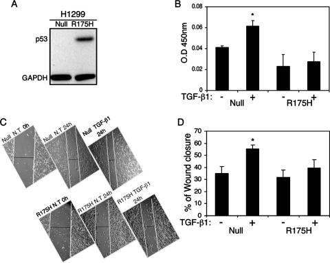
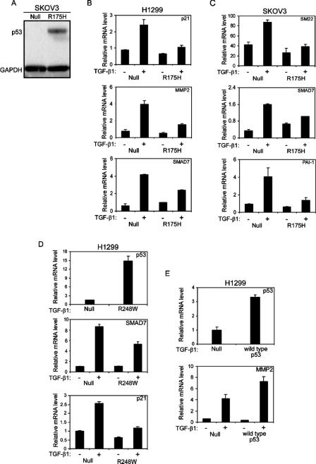
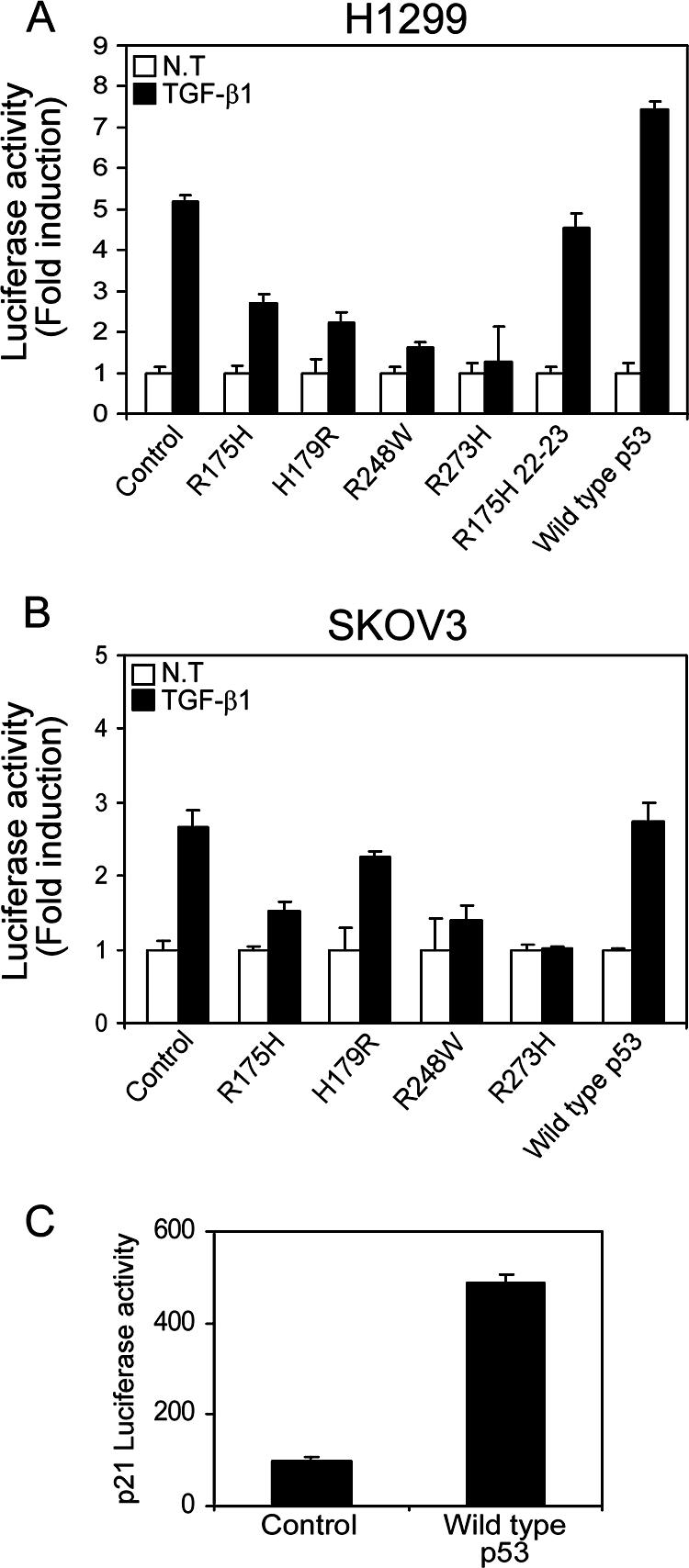
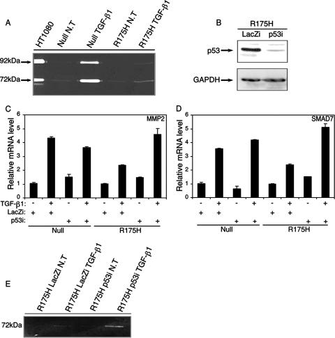
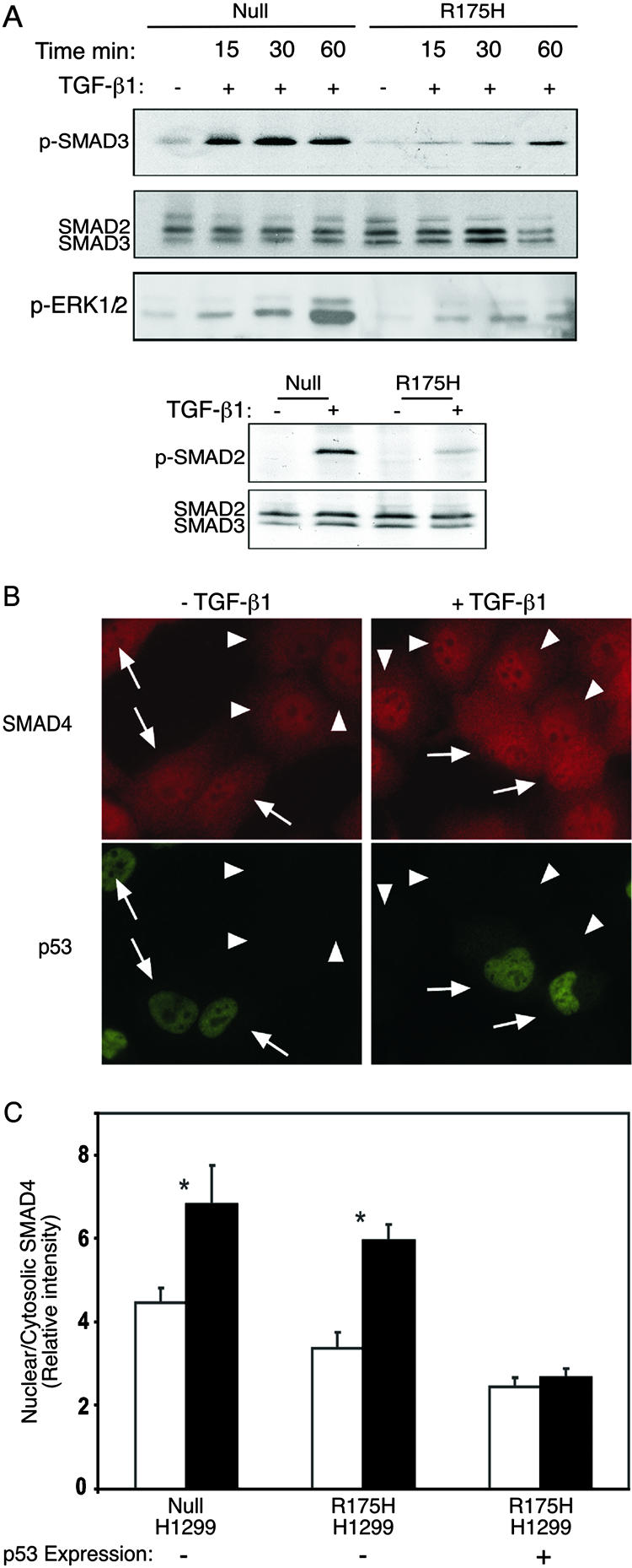
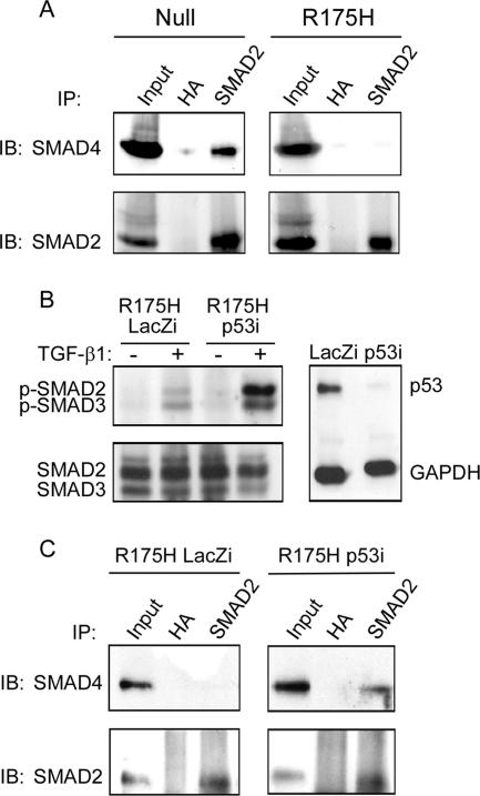
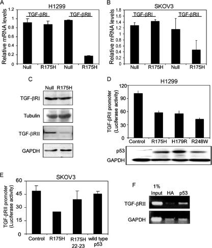
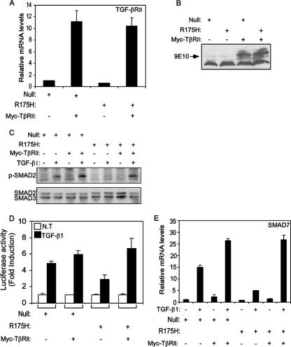
Both transforming growth factor beta (TGF-beta) and p53 have been shown to control normal cell growth. Acquired mutations either in the TGF-beta signaling pathway or in the p53 protein were shown to induce malignant transformation. Recently, cross talk between wild-type p53 and the TGF-beta pathway was observed. The notion that mutant p53 interferes with the wild-type p53-induced pathway and acts by a "gain-of-function" mechanism prompted us to investigate the effect of mutant p53 on the TGF-beta-induced pathway. In this study, we show that cells expressing mutant p53 lost their sensitivity to TGF-beta1, as observed by less cell migration and a reduction in wound healing. We found that mutant p53 attenuates TGF-beta1 signaling. This was exhibited by a reduction in SMAD2/3 phosphorylation and an inhibition of both the formation of SMAD2/SMAD4 complexes and the translocation of SMAD4 to the cell nucleus. Furthermore, we found that mutant p53 attenuates the TGF-beta1-induced transcription activity of SMAD2/3 proteins. In searching for the mechanism that underlies this attenuation, we found that mutant p53 reduces the expression of TGF-beta receptor type II. These data provide important insights into the molecular mechanisms that underlie mutant p53 "gain of function" pertaining to the TGF-beta signaling pathway.


Figures

FIG. 1.
FIG. 1.
Mutant p53 attenuates TGF-β1-induced cell migration and wound healing. (A) Western blot analysis depicting the protein levels of p53R175H. (B) p53-null and p53R175H-producing H1299 cells were treated with TGF-β1 and subjected to a cell migration assay. Migrated cells were fixed, stained with crystal violet, dissolved in acetic acid, and analyzed for OD by using an enzyme-linked immunosorbent assay reader. The OD values represent the amounts of migrated cells (P < 0.05). (C) p53-null and p53R175H-producing H1299 cells were seeded to confluence, scratched to create an artificial wound, and treated with TGF-β1 for 24 h. The photographs indicate wound closure. (D) Wound closure, analyzed by subtracting the distance measured between the edges of the wound at hour 24 from that at hour 0 and presented as a percentage (P < 0.001).
FIG. 2.
FIG. 2.
Mutant p53 attenuates TGF-β1-induced gene expression. (A) Western blot analysis depicting protein levels of p53R175H in SKOV3 cells. H1299 (B) and SKOV3 (C) cells expressing either the p53R175H mutant or empty vector were serum starved and treated with TGF-β1 for 12 h. Total RNA was extracted and analyzed for the expression of TGF-β1-induced specific genes by RT-PCR. (D) H1299 cells expressing either p53R248W or empty vector were treated with TGF-β1 and evaluated for the expression of p21 and SMAD7 by RT-PCR analysis. The expression of the p53R248W mutant was confirmed using RT-PCR analysis. (E) H1299 cells were transiently transfected with either wild-type p53 (30 ng) or a β-Gal expression plasmid (30 ng) as a control. Twenty-four hours following transfection, the cells were starved, treated with TGF-β1, and analyzed for the expression of MMP2. The expression of wild-type p53 was confirmed using RT-PCR analysis.
FIG. 3.
FIG. 3.
Mutant p53 attenuates the activation of TGF-β1-induced reporter gene. p53-null H1299 (A) and p53-null SKOV3 (B) cells were transfected with p3TP-Luc and p3TP-Luc(+), respectively, together with various p53 mutant or wild-type p53 expression plasmids, and luciferase activity was measured following TGF-β1 treatment. The amount of activation was calculated as the ratio of p3TP-Luc activity in TGF-β1-treated cells to p3TP-Luc activity in nontreated (NT) cells. (C) Wild-type p53-mediated induction of p21 promoter activity in H1299 cells.
FIG. 4.
FIG. 4.
Mutant p53R175H attenuates the production of MMP2 and MMP9 enzymes. (A) p53-null and p53R175H-producing H1299 cells were serum starved and treated with TGF-β1, and the activities of MMP2 and MMP9 were analyzed using a zymography assay. The electrophoretic positions of the 92-kDa MMP9 zymogen and the 72-kDa MMP2 zymogen are indicated. (B) siRNA for either p53 or LacZ was introduced into p53R175H-producing H1299 cells, and the expression of mutant p53 was detected by Western blot analysis using a monoclonal anti-DO1 antibody. (C) RT-PCR analysis of MMP2 in p53-null and p53R175H-producing cells that were exposed to siRNA for either p53 or LacZ and treated with TGF-β1. (D) RT-PCR analysis of SMAD7 in p53-null and p53R175H-producing cells that were exposed to siRNA for either p53 or LacZ and treated with TGF-β1. (E) Zymography analysis of MMP2 in p53R175H-producing cells expressing siRNA for either p53 or LacZ following TGF-β1 treatment. NT, not treated.
FIG. 5.
FIG. 5.
Mutant p53 attenuates TGF-β1-induced SMAD2/3 and ERK1/2 phosphorylation and abolishes the nuclear translocation of SMAD4. (A) p53-null and p53R175H-producing H1299 cells were serum starved and treated with TGF-β1 for 15, 30, and 60 min. The phosphorylated form of SMAD2 and SMAD3 was analyzed by Western blotting, using either rabbit anti-p-SMAD2 or rabbit anti-p-SMAD3 and total rabbit anti-SMAD2/3 antibody. The phosphorylation of ERK1/2 was monitored using mouse anti-p-ERK1/2. (B) p53-null or p53R175H-producing H1299 cells were serum starved and incubated with or without TGF-β1 (2 ng/ml) for 30 min at 37°C. Cells were then fixed/permeabilized and labeled for SMAD4 and p53 by successive incubations with (i) murine anti-SMAD4 (5 μg/ml) together with anti-p53 rabbit serum (1:1,000) and (ii) Cy3-GαM IgG together with Alexa 488-GαR IgG (2 μg/ml each). Arrows indicate cells expressing the p53R175H mutant, identified by fluorescence labeling for p53. Arrowheads point to cells devoid of mutant p53. (C) Quantitative measurements of the relative levels (nuclear/cytoplasmic ratio) of SMAD4 were taken using the point confocal method (see Materials and Methods). The laser beam (528.7 nm) was focused on well-defined spots within the nucleus and the cytoplasm of each cell, and the fluorescence intensity of Cy3-labeled SMAD4 was measured in both locations. The ratio of the nuclear to cytosolic intensities was recorded for each cell. Bars represent means plus standard errors of the means of 150 measurements in each case. Asterisks indicate a significant increase in the SMAD4 nuclear/cytosolic ratio when cells were stimulated with TGF-β1 (P < 0.001); the effect of TGF-β1 in p53R175H-expressing cells was not significant (P > 0.46).
FIG. 6.
FIG. 6.
Mutant p53 interferes with formation of the SMAD2-SMAD4 complex. (A) p53-null and p53R175H-producing H1299 cells were treated with TGF-β1 as described in Materials and Methods. Cell lysates were immunoprecipitated with rabbit anti-SMAD2 antibody, and the precipitates were analyzed with mouse anti-SMAD4 antibodies. (B) p53R175H-producing H1299 cells were transfected with siRNA for either p53 or LacZ, treated with TGF-β1 for 30 min, and tested for the presence of p-SMAD2/3 by means of Western blot analysis. (C) p53R175H-producing H1299 cells were transfected with siRNA for p53 or LacZ, immunoprecipitated with rabbit anti-SMAD2, and analyzed for the presence of SMAD4 using a mouse anti-SMAD4 antibody. IP, immunoprecipitation; IB, immunoblotting.
FIG. 7.
FIG. 7.
Mutant p53 represses the expression of TGF-βRΙΙ but not TGF-βRΙ. RT-PCR analysis of the expression of TGF-βRI/II was performed with p53-null and p53R175H-producing H1299 cells (A) or p53-null and p53R175H-producing SKOV3 cells (B). (C) Western blot analysis depicting the levels of TGF-βRI and TGF-βRΙΙ in p53-null and p53R175H-producing H1299 cells. (D) H1299 cells were transiently transfected with the TGF-βRII promoter (300 ng) along with plasmids encoding the indicated p53 mutants. A β-Gal expression plasmid was also included in all transfections, and luciferase activity was normalized to β-Gal activity. (E) SKOV3 cells were transiently transfected with the promoter of TGF-βRII together with either the p53R175H mutant, p53R175H 22-23 mutant, or wild-type p53 expression plasmid, and luciferase activity was measured and normalized to β-Gal. (F) Chromatin immunoprecipitation experiment performed on H1299 cells stably expressing the p53R175H mutant. After cross-linking of proteins to DNA, DNA was fragmented, and the p53 protein was immunoprecipitated with a specific antibody against p53 or with a nonspecific antibody against HA as a control. PCR analysis was performed on the immunoprecipitated DNA samples, using either TGF-βRII- or GAPDH-specific primers.
FIG. 8.
FIG. 8.
Overexpressing Myc-tagged TGF-βRII restores TGF-β1-mediated signaling pathway and gene expression. Both p53-null and mutant p53R175H-producing H1299 cells were infected with either empty vector or Myc-TβRII expression plasmid, and the expression of TGF-βRII was measured by RT-PCR analysis (A) and Western blot analysis using the 9E10 monoclonal anti-Myc antibody (B). (C) Western blot analysis depicting the levels of phosphorylated SMAD2 protein following treatment of H1299 cells with TGF-β1 for 30 min. (D) p53-null and mutant p53R175H-producing H1299 cells were transiently transfected with p3TP-Luc, together with either empty vector or Myc-TβRII expression plasmid, and luciferase activity was measured following TGF-β1 treatment. The amount of activation was calculated as the ratio of p3TP-Luc activity in TGF-β1-treated cells to p3TP-Luc activity in nontreated (NT) cells. (E) p53-null and mutant p53R175H-producing H1299 cells were infected with either a Myc-TβRII expression plasmid or empty vector, and the expression of TGF-β1-induced SMAD7 was analyzed using RT-PCR.

References

    1. Aas, T., A. L. Borresen, S. Geisler, B. Smith-Sorensen, H. Johnsen, J. E. Varhaug, L. A. Akslen, and P. E. Lonning. 1996. Specific P53 mutations are associated with de novo resistance to doxorubicin in breast cancer patients. Nat. Med. 2:811-814. - PubMed
    1. Abdollah, S., M. Macias-Silva, T. Tsukazaki, H. Hayashi, L. Attisano, and J. L. Wrana. 1997. TbetaRI phosphorylation of Smad2 on Ser465 and Ser467 is required for Smad2-Smad4 complex formation and signaling. J. Biol. Chem. 272:27678-27685. - PubMed
    1. Blandino, G., A. J. Levine, and M. Oren. 1999. Mutant p53 gain of function: differential effects of different p53 mutants on resistance of cultured cells to chemotherapy. Oncogene 18:477-485. - PubMed
    1. Buganim, Y., E. Kalo, R. Brosh, H. Besserglick, I. Nachmany, Y. Rais, P. Stambolsky, X. Tang, M. Milyavsky, I. Shats, M. Kalis, N. Goldfinger, and V. Rotter. 2006. Mutant p53 protects cells from 12-O-tetradecanoylphorbol-13-acetate-induced death by attenuating activating transcription factor 3 induction. Cancer Res. 66:10750-10759. - PubMed
    1. Cadwell, C., and G. P. Zambetti. 2001. The effects of wild-type p53 tumor suppressor activity and mutant p53 gain-of-function on cell growth. Gene 277:15-30. - PubMed

Publication types

MeSH terms

Substances