. 2007 Nov;27(22):7839-47.
doi: 10.1128/MCB.00438-07. Epub 2007 Sep 17.

The basic helix-loop-helix transcription factor NeuroD1 facilitates interaction of Sp1 with the secretin gene enhancer

Affiliations

The basic helix-loop-helix transcription factor NeuroD1 facilitates interaction of Sp1 with the secretin gene enhancer

Subir K Ray et al. Mol Cell Biol. 2007 Nov.

Abstract

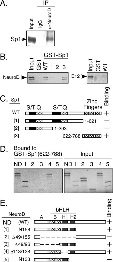
The basic helix-loop-helix transcription factor NeuroD1 is required for late events in neuronal differentiation, for maturation of pancreatic beta cells, and for terminal differentiation of enteroendocrine cells expressing the hormone secretin. NeuroD1-null mice demonstrated that this protein is essential for expression of the secretin gene in the murine intestine, and yet it is a relatively weak transcriptional activator by itself. The present study shows that Sp1 and NeuroD1 synergistically activate transcription of the secretin gene. NeuroD1, but not its widely expressed dimerization partner E12, physically interacts with the C-terminal 167 amino acids of Sp1, which include its DNA binding zinc fingers. NeuroD1 stabilizes Sp1 DNA binding to an adjacent Sp1 binding site on the promoter to generate a higher-order DNA-protein complex containing both proteins and facilitates Sp1 occupancy of the secretin promoter in vivo. NeuroD-dependent transcription of the genes encoding the hormones insulin and proopiomelanocortin is potentiated by lineage-specific homeodomain proteins. The stabilization of binding of the widely expressed transcription factor Sp1 to the secretin promoter by NeuroD represents a distinct mechanism from other NeuroD target genes for increasing NeuroD-dependent transcription.


Figures

FIG. 1.
FIG. 1.
Transcriptional synergism between NeuroD1 and Sp1 on the secretin gene. (A) Conserved alignment of the E-box and Sp1 binding sites in the rat, mouse, and human secretin gene promoters. (B) Loss of transcriptional activity of the secretin reporter in HIT and STC-1 cells, with mutations in the E-box or the adjacent Sp1 binding site. Luciferase activity of the E-box mutant (black bars) and GC1 mutant (striped bars) is expressed as a percentage of the wild-type (white bars) reporter gene activity. Results are shown as the means ± standard errors of the means of three separate experiments done in duplicate. *, P < 0.0001. (C) Secretin reporter gene activity in C33A cells or SL2 cells cotransfected with expression plasmids for NeuroD1, Sp1, or NeuroD1 plus Sp1 and the reporter plasmids indicated in panel B. Results are expressed as luciferase activity relative to the activity in the absence of NeuroD1 or Sp1. The dashed line indicates the sum of transcriptional activities by NeuroD1 and Sp1 expression vectors cotransfected individually with the respective reporter. Results are shown as the means ± standard errors of the means for at least five separate experiments. Significant differences from the additive activities of NeuroD1 and Sp1 are shown by asterisks (*, P < 0.0001; **, P < 0.05). WT, wild type.
FIG. 2.
FIG. 2.
NeuroD1 and Sp1 bind cooperatively to the secretin gene to form a ternary DNA-protein complex. (A) EMSA showing protein complexes formed by indicated combinations of in vitro-translated NeuroD1, E12, or Sp1 bound to a 32P-labeled probe containing the E-box and GC1 Sp1 site (lanes 1 to 3) or the same probes with mutations in either the E-box (lanes 4 to 6) or GC1 (lanes 7 to 9) or with the insertion of a 6-bp (Ins 6) or a 10-bp (Ins 10) sequence between the two sites (lanes 10 and 11). The arrowhead denotes a slow-migrating complex seen only in the presence of all three proteins (lane 2) but not seen with any of the mutant probes (lanes 4 to 11). (B) Failure to generate the ternary complex with E12 and Sp1 in the absence of NeuroD1 (lane 2). (C) Quantitation of Sp1 and NeuroD1/E12 binding to DNA. The fraction of probe present in complexes formed by NeuroD1 plus E12 alone (panel A, lane 1) or Sp1 alone (panel A, lane 3) or in the ternary complex formed by NeuroD1, E12, and Sp1 together (panel A, lane 2) was measured by phosphorimaging. Results are shown as the means ± standard errors of the means for at least three separate experiments. *, significantly different (P < 0.05) from the fraction predicted for independent binding events measured as described before (24). WT, wild type.
FIG. 3.
FIG. 3.
Presence of NeuroD1 and Sp1 in a higher-order DNA-protein complex formed from nuclear extracts. Shown are results of an EMSA of factors in HIT cell nuclear extract binding to the secretin promoter regions containing the E-box and adjacent Sp1 binding site. (A) Effect of competition with unlabeled oligonucleotides for an Sp1, mutant Sp1 site, or E-box on ternary complex formation. (B) Presence of Sp1 and NeuroD1 in the ternary complex. Antibodies to NeuroD1 (lane 2) or E12 (lane 5) reduced the slow-migrating complex. Sp1 antibody (lane 3) supershifted the ternary complex. (C) EMSA showing that probes with increased spacing between the E-box and Sp1 binding site cannot generate the ternary complex from nuclear proteins extracted from HIT cells (lanes 2 and 3). (D) Effect of spacing (6 or 10 bp) between the E-box and the adjacent Sp1 binding site on secretin gene transcription. Luciferase activity of the insertion mutants is expressed as a percentage of activity of the wild-type reporter gene transfected in HIT (white bars) or STC-1 (black bars) cells. Results are shown as the means ± standard errors of the means for at least four independent experiments. Significant differences from the wild type are shown by an asterisk (P < 0.001). WT, wild type.
FIG. 4.
FIG. 4.
Physical association between Sp1 and NeuroD1. (A) Coimmunoprecipitation of Sp1 with NeuroD1 in HIT cells. Lane 1 shows 4% of the input used for immunoprecipitation. (B) Sp1 directly interacts with NeuroD1 but not its dimerization partner, E12, through zinc finger domains. 35S-labeled NeuroD1 (left) or 35S-labeled E12 (right) was incubated with GST or the indicated GST-Sp1 fusion proteins as indicated in panel C. NeuroD1 bound to fusion proteins was affinity isolated, subjected to SDS-PAGE, and detected by autoradiography. Input lanes were loaded with 10% of the labeled proteins used in the binding assay. (C) Structure of the Sp1 deletion mutants and summary of the results. (D and E) Identification of the Sp1 binding domain of NeuroD1. (D) Wild type or different 35S-labeled NeuroD1 mutants bound to GST-Sp1 (amino acids 622 to 788) were affinity isolated and separated by SDS-PAGE (left panel). Input (right panel) lanes were loaded with 10% of the labeled proteins used in the binding assay. (E) Structures of the NeuroD1 deletion mutants. WT, wild type; ND, NeuroD1.
FIG. 5.
FIG. 5.
Stabilization of Sp1-DNA binding in the ternary DNA-protein complex with NeuroD1. (A) EMSA performed with indicated recombinant proteins and a probe containing the contiguous E-box and Sp1 binding site. Slower-migrating complex i represents the ternary DNA-protein complex (lane 3) whereas complexes ii (lanes 1 and 3) and iii (lanes 2 and 3) were generated by NeuroD1-E12 and Sp1, respectively. All three complexes were competed by a 100-fold molar excess of the unlabeled probe added to the probe prior to the binding reaction. (B) An EMSA where a 500-fold molar excess of unlabeled oligonucleotide competitor was added after the probe and proteins reached equilibrium. Aliquots were removed at 4-minute intervals and quickly loaded onto an acrylamide gel for electrophoresis. (C) Time course of dissociation of DNA-protein complexes. The percentage of probe remaining in complexes with Sp1 alone (complex iii), with NeuroD1-E12 (complex ii), or with NeuroD1-E12-Sp1 (complex i) was quantitated by phosphorimaging and plotted relative to the zero time point. Results are shown as the means ± standard errors of the means for three separate experiments. *, significantly different (complex iii in comparison to complex i or complex ii; P < 0.01).
FIG. 6.
FIG. 6.
NeuroD1 facilitates interaction of Sp1 with the secretin enhancer. (A) Recruitment of transiently expressed NeuroD1 to the endogenous secretin promoter in HeLa cells. ChIP experiments were performed using soluble chromatin fragments prepared from HeLa cells transfected with an expression plasmid for E12 in the absence (−) or the presence (+) of a NeuroD1 expression plasmid. DNA purified from the chromatin input or the immunoprecipitate (α-NeuroD1 or control IgG) was amplified by PCR for 32 cycles with primers covering the secretin promoter (−284 to −57) (top panel) or a control promoter (−310 to +17 of human DHFR) (bottom panel). (B) Effect of NeuroD1 on recruitment of Sp1 to the secretin promoter in vivo. ChIP experiments were performed with anti-Sp1 antibody (α-Sp1) as described for panel A. The human GAPDH gene served as a negative control (right).
FIG. 7.
FIG. 7.
Depletion of NeuroD1 in STC-1 cells reduces recruitment of Sp1 to the secretin promoter. Shown are results of transient ChIP assays for transfected secretin and hTERT promoters. (A) Sp1 and NeuroD1 occupancy of the wild-type secretin promoter (top panel), a secretin promoter with mutations in the Sp1 binding sites (MutGC; middle panel), and a mutant (MutE/GC) secretin promoter lacking Sp1 and NeuroD1 binding sites (bottom panel) in STC-1 cells. (B) Effect of NeuroD1 depletion on transient ChIP of Sp1 and NeuroD1 in STC-1 cells cotransfected with a plasmid containing the secretin promoter (left) or hTERT promoter (right). Cells were cotransfected with either shRNA plasmids for NeuroD1 or GFP as a control. The final DNA extractions were PCR amplified for detection of the transfected promoters using pairs of primers described in Materials and Methods. (C) Effect of NeuroD1 depletion on transcription of a secretin reporter gene in STC-1 cells cotransfected with shRNA plasmids for NeuroD1 and GFP as indicated. Results are shown as the means ± standard errors of the means of at least three separate experiments. *, significantly different from control or GFP shRNAs (P < 0.01).

Similar articles

Cited by

References

    1. Biesiada, E., Y. Hamamori, L. Kedes, and V. Sartorelli. 1999. Myogenic basic helix-loop-helix proteins and Sp1 interact as components of a multiprotein transcriptional complex required for activity of the human cardiac α-actin promoter. Mol. Cell. Biol. 19:2577-2584. - PMC - PubMed
    1. Cong, Y.-S., J. Wen, and S. Bacchetti. 1999. The human telomerase catalytic subunit hTERT: organization of the gene and characterization of the promoter. Hum. Mol. Genet. 8:137-142. - PubMed
    1. Courey, A. J., and R. Tjian. 1988. Analysis of Sp1 in vivo reveals multiple transactivational domains, including a novel glutamine-rich activation motif. Cell 55:887-898. - PubMed
    1. Dumonteil, E., B. Laser, I. Constant, and J. Philippe. 1998. Differential regulation of the glucagon and insulin I gene promoters by basic helix-loop-helix transcription factors E47 and Beta2. J. Biol. Chem. 273:19945-19954. - PubMed
    1. Edlund, T., M. D. Walker, P. J. Barr, and W. J. Rutter. 1985. Cell-specific expression of the rat insulin gene: evidence for role of two distinct 5′ flanking elements. Science 230:912-916. - PubMed

Publication types

MeSH terms

LinkOut - more resources