. 2007 Nov;27(22):7802-15.
doi: 10.1128/MCB.02179-06. Epub 2007 Sep 17.

Sox17 and Sox4 differentially regulate beta-catenin/T-cell factor activity and proliferation of colon carcinoma cells

Affiliations

Sox17 and Sox4 differentially regulate beta-catenin/T-cell factor activity and proliferation of colon carcinoma cells

Débora Sinner et al. Mol Cell Biol. 2007 Nov.

Abstract

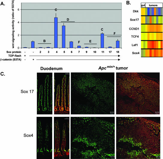
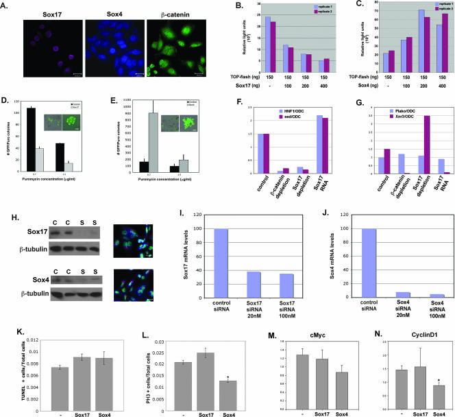
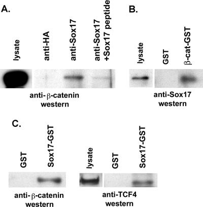
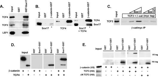
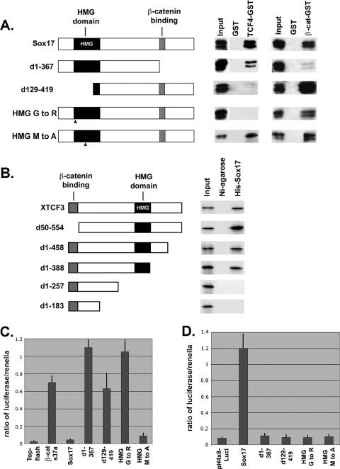
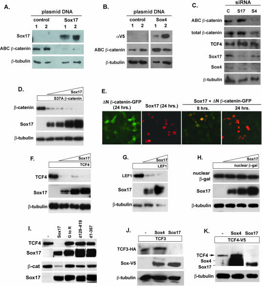
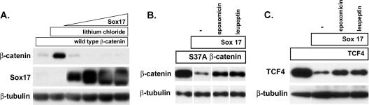
The canonical Wnt pathway is necessary for gut epithelial cell proliferation, and aberrant activation of this pathway causes intestinal neoplasia. We report a novel mechanism by which the Sox family of transcription factors regulate the canonical Wnt signaling pathway. We found that some Sox proteins antagonize while others enhance beta-catenin/T-cell factor (TCF) activity. Sox17, which is expressed in the normal gut epithelium but exhibits reduced expression in intestinal neoplasia, is antagonistic to Wnt signaling. When overexpressed in SW480 colon carcinoma cells, Sox17 represses beta-catenin/TCF activity in a dose-dependent manner and inhibits proliferation. Sox17 and Sox4 are expressed in mutually exclusive domains in normal and neoplastic gut tissues, and gain- and loss-of-function studies demonstrate that Sox4 enhances beta-catenin/TCF activity and the proliferation of SW480 cells. In addition to binding beta-catenin, both Sox17 and Sox4 physically interact with TCF/lymphoid enhancer factor (LEF) family members via their respective high-mobility-group box domains. Results from gain- and loss-of-function experiments suggest that the interaction of Sox proteins with beta-catenin and TCF/LEF proteins regulates the stability of beta-catenin and TCF/LEF. In particular, Sox17 promotes the degradation of both beta-catenin and TCF proteins via a noncanonical, glycogen synthase kinase 3beta-independent mechanism that can be blocked by proteasome inhibitors. In contrast, Sox4 may function to stabilize beta-catenin protein. These findings indicate that Sox proteins can act as both antagonists and agonists of beta-catenin/TCF activity, and this mechanism may regulate Wnt signaling responses in many developmental and disease contexts.


Figures

FIG. 1.
FIG. 1.
Different Sox factors are antagonists or agonists of the canonical Wnt pathway and are expressed in the normal and neoplastic gut. (A) Effect of different Sox family members on Wnt signaling activity. 293T cells (shown) and COS cells (not shown) were transfected with Sox2, Sox3, Sox4, Sox5, Sox6, Sox7, Sox9, Sox10, Sox11, Sox17, and Sox18 together with a TCF-luciferase reporter plasmid (TOP-flash) and an expression plasmid encoding a stabilized form of β-catenin (carrying the S37A mutation). Transfection with β-catenin(S37A) alone resulted in significant activation of the reporter plasmid. Sox2, Sox3, Sox7, Sox9, Sox10, and Sox17 repressed the β-catenin-mediated activation of TOP-flash, whereas Sox4, Sox5, and Sox11 enhanced β-catenin/Wnt activity. Data are shown as ratios of the activity in cotransfected cells to the activity in cells transfected with β-catenin(S37A) alone. Transfection with Sox factors without β-catenin(S37A) did not result in significant TOP-flash activity (data not shown). Letters above the bars denote the Sox subfamily. Error bars indicate standard errors of the means for replicate samples from two separate experiments. −, absent; +, present. (B) Analysis of published microarray data (8, 32, 38). The expression levels of the Wnt antagonists Dkk and Sox17 were reduced in seven of eight tumor samples relative to those in the normal gut samples. The levels of expression of several pro-Wnt signaling genes, Sox4, TCF4, and LEF1, and the Wnt target gene cyclin D1 (CCND1) were elevated in tumor samples relative to those in normal gut samples. The color scale for normalized transcript levels is as follows: red, high; yellow, moderate; and blue, low. (C) Sox4 and Sox17 proteins are expressed in the normal gut epithelium and in APCmin/+ tumors. In the small intestine, Sox17 and Sox4 proteins (green) are expressed in the nuclei of the epithelial cells (a duodenum sample is shown). The Sox4 expression domain is located predominantly in the crypt, whereas Sox17 protein is found throughout the crypt and villus. The right panels show adjacent sections from APCmin/+ tumors in which Sox17 and Sox4 proteins appear to be in mutually exclusive domains. Nuclei are stained with the red nuclear dye PoPro3. Scale bars in duodenum panels, 50 μm.
FIG. 2.
FIG. 2.
Sox17 and Sox4 have opposite effects on Wnt activity and proliferation of SW480 colon carcinoma cells. (A) Endogenous Sox17, Sox4, and β-catenin proteins are present in SW480 cells. (B) Sox17 represses endogenous Wnt signaling activity in colon carcinoma cells. Transfection with the TOP-flash reporter alone (lane 1) demonstrates that SW480 cells have high levels of endogenous Wnt/β-catenin/TCF activity. Cells cotransfected with increasing concentrations of a Sox17 expression plasmid (100 to 400 ng) showed dose-dependent reduction in endogenous Wnt activity (lanes 2 to 4). TOP-flash activity is expressed in relative light units that were normalized to the transfection efficiency by using a renilla plasmid. The results shown are representative of those from at least three independent experiments. −, absent. (C) Sox4 enhances endogenous Wnt signaling activity in colon carcinoma cells. SW480 cells that were cotransfected with increasing levels of a Sox4 expression plasmid exhibited a dose-dependent increase in TOP-flash activity. Results for replicate samples are shown, and experiments were done three times. −, absent. (D) Sox17 represses the proliferation of SW480 cells. The formation of puromycin-resistant SW480 cell colonies was used to measure the effect of Sox17 on cell proliferation. SW480 cells were cotransfected with the pBABE-puromycin resistance plasmid, the pmax-GFP plasmid, and either an empty vector or a Sox17 expression vector and grown in puromycin-containing medium for 5 days. Insets show GFP-positive cells before (left) and after (right) puromycin selection. Only colonies that had ≥10 GFP-positive cells were counted. Error bars indicate standard errors of the means of results for three replicate samples. Puro, puromycin. (E) Sox4 promotes the proliferation of SW480 cells. SW480 cells that expressed Sox4 formed more colonies than vector-alone controls. Insets show GFP-positive cells before (left) and after (right) puromycin selection. (F and G) The effects of the loss and gain of function of Sox17 on Wnt and Sox17 target gene expression in Xenopus embryos. MO (20 ng each) previously used to deplete Xenopus embryos of Sox17 (6) or β-catenin (13) were injected into two-cell embryos, and stage 11 embryos were harvested and pooled in groups of three for each condition. Real-time RT-PCR analysis was used to analyze the relative levels of expression of the Wnt target genes Xnr3 and Siamois (data not shown), the Sox17 target gene Hnf1β, or the ubiquitously expressed gene Plakoglobin (Plako). All gene expression levels are shown as a ratio of the expression of the indicated gene to that of the ubiquitously expressed ODC gene (44). These results demonstrate that Sox17 is required to restrict the activity of Wnt/β-catenin/TCF in Xenopus embryos. (H) siRNA-mediated knockdown of Sox17 and Sox4 proteins in SW480 cells. SW480 cells were transfected with 50 nM scrambled control siRNA (C) or the Sox siRNA (S). The left panels show Western blot analyses of Sox17 and Sox4 proteins, and the right panels are confocal images of transfected SW480 cells showing the typical perinuclear localization of the fluorescently labeled control siRNA (red). Nuclei are stained with SYTOX green. (I and J) siRNA knockdown of endogenous Sox17 and Sox4 mRNA levels. Transfection with Sox17 siRNAs resulted in a 70% reduction of endogenous mRNA, whereas Sox4 siRNAs effected >90% reduction relative to mRNA levels in cells transfected with a control siRNA. (K) Quantitative assessment of apoptosis in SW480 cells as measured by terminal deoxynucleotidyltransferase-mediated dUTP-biotin nick end labeling (TUNEL) staining 48 h after transfection with Sox4 and Sox17 siRNAs. −, control. (L) Quantitative assessment of proliferation of SW480 cells as measured by phosphohistone-H3 (PH3) staining 48 h after transfection with siRNAs. * indicates significance corresponding to a P value of 0.001 versus control samples as calculated using a t test analysis. −, control. (M and N) Quantitative RT-PCR analysis of the Wnt target genes c-myc (I) and cyclin D1 (J) in SW480 cells 48 h after transfection with Sox4 and Sox17 siRNAs. Quantitative PCR analyses of triplicate biological samples were performed. * indicates significance corresponding to a P value of 0.02 versus control samples as calculated using a t test analysis. −, control. Scale bars in panels A, D, E, and H, 20 μm.
FIG. 3.
FIG. 3.
Endogenous Sox17, β-catenin, and TCF4 proteins interact in SW480 colon carcinoma cell lysates. (A) Sox17 antibodies were used to coimmunoprecipitate interacting proteins from nuclear extracts of SW480 cells. Lysates were immunoprecipitated with a control antibody (lane 1; anti-HA) or with a Sox17 antibody alone (lane 2) or in the presence of a Sox17 peptide or a control peptide (lanes 3 and 4). The Sox17 peptide used to generate the antibody inhibited the immunoprecipitation. (B) β-catenin (β-cat)-GST-linked beads or glutathione beads alone (GST) were incubated with cell lysates from SW480 cells, and precipitates were analyzed by Western blotting with an anti-Sox17 antibody. The Sox17 antibody was raised in rabbits and recognizes both mouse and human Sox17. (C) Sox17-GST beads or GST beads alone (GST) were incubated with cell lysates from SW480 cells, and precipitates were analyzed by Western blotting with anti β-catenin or anti-TCF4 antibodies.
FIG. 4.
FIG. 4.
Sox17 forms a stable, trimeric complex with TCF/LEF and β-catenin proteins. (A) Sox17 interacts with TCF3, TCF4, and LEF proteins. Sox17-GST beads pulled down in vitro-translated, V5 epitope-tagged TCF4, TCF3, and LEF1, and GST beads did not. (B) The interaction between Sox17 and β-catenin is enhanced in the presence of TCF4. Sox17 or TCF4 independently bound to β-catenin-GST beads but not to GST beads alone (panels 1 and 2). The binding of Sox17 to β-catenin-GST beads was enhanced in the presence of TCF4 (compare the ratios of input to bound Sox17 in the presence and absence of TCF4 [lanes marked by *]). β-catenin-interacting proteins were analyzed by Western blotting with an anti-V5 antibody. (C) The interaction between β-catenin (β-cat) and TCF3 is stabilized by Sox17 and requires a Sox17-β-catenin interaction. In vitro-translated, S35-labeled TCF3 protein (input) was incubated with β-catenin (myc tagged) alone or with increasing concentrations of purified His-tagged Xenopus Sox17β or His-tagged Xenopus Sox17 consisting of amino acids 1 to 150, which removed the β-catenin interaction domain (ΔC Sox17). β-catenin and its associated proteins were precipitated with an anti-myc antibody, and TCF3 protein was analyzed by SDS-PAGE and autoradiography. IP, immunoprecipitate. (D and E) Under high-stringency conditions, Sox17-GST beads would not efficiently pull down β-catenin. However, in the presence of TCF4 (D) or TCF3 (E), Sox17 was able to pull down β-catenin. This interaction depends on the β-catenin interaction domain of TCF3, which is deleted in the N terminus-lacking TCF3 protein (ΔN TCF3). +, present; −, absent.
FIG. 5.
FIG. 5.
Sox4 and Sox17 have similar affinities for TCF4 and β-catenin, but Sox4 does not form a stable complex. (A) Sox4 has affinities for TCF4 and β-catenin (β-cat) similar to those of Sox17. The ratios of the input (I) and bound (B) protein fractions are similar for Sox17 and Sox4. (B) Sox17 formed a complex with β-catenin and TCF4. Also, more Sox17 protein was pulled down by TCF4-GST beads in the presence of β-catenin. (C) GST-TCF4 beads were able to pull down either β-catenin or Sox4 but did not efficiently pull down Sox4 in the presence of β-catenin. Moreover, Sox4 binding to TCF4 was somewhat inhibited in the presence of β-catenin, suggesting that Sox4 and β-catenin compete for binding.
FIG. 6.
FIG. 6.
Structure-function analyses of Sox17-TCF-β-catenin interactions. (A) Sox17 interacts with TCF4 protein via its HMG domain. The left panel shows a schematic diagram of Sox17 deletions and point mutations. HMG mutant 1 carries a stringent mutation (G to R at amino acid 103), and HMG mutant 2 carries a more conservative mutation (M to A at amino acid 76). d1-367, now contains amino acids 1 to 367; d129-419, now contains amino acids 129 to 419. The right panel shows the interaction of in vitro-translated Sox17 mutant proteins (V5 tagged) with GST alone, TCF4-GST, or β-catenin (β-cat)-GST. (B) Identification of protein domains of TCF3 that mediate its interaction with Sox17. The left panel shows a schematic diagram of deletions of Xenopus TCF3. The right panel shows the interaction of the in vitro-translated S35-labeled TCF3 mutant proteins with His-tagged Xenopus Sox17β or Ni-agarose alone. (C) Sox17 requires interactions with both β-catenin and TCF/LEF to antagonize Wnt activity. COS cells were transfected with β-catenin(S37A) and TOP-flash alone or together with the Sox17 mutant forms, and TOP-flash activity was measured after 48 h. Error bars indicate standard errors of the means of results for replicate samples from at least three separate experiments. (D) Sox17 does not require transcriptional activity to antagonize Wnt activity. 293T cells were cotransfected with Sox17 and derived mutant forms and a Sox17 reporter plasmid (pH4X8-Luci). The transcriptional activity of Sox17 requires a functional DNA binding domain (HMG domain) and a functional transactivation domain (at the C terminus). While the Sox17 HMG mutant form carrying an M-to-A mutation is transcriptionally inactive, it still is able to interact with both TCF4 and β-catenin and antagonize Wnt activity. Error bars indicate standard errors of the means of results for replicate samples from two separate experiments.
FIG. 7.
FIG. 7.
Sox proteins affect β-catenin and TCF/LEF protein levels. (A and B) Effects of Sox17 and Sox4 overexpression on endogenous β-catenin protein levels in SW480 cells. The overexpression of Sox17 in SW480 cells causes significant reduction in the levels of endogenous unphosphorylated β-catenin protein, which was detected using an antibody specific for unphosphorylated β-catenin (ABC β-catenin). The overexpression of Sox4 caused an increase in the levels of endogenous unphosphorylated β-catenin protein. αV5, anti-V5 antibody. (C) Impact of siRNA knockdown of Sox17 (S17) and Sox4 (S4) on β-catenin and TCF4 levels in SW480 cells. Reduced levels of endogenous Sox4 protein coincide with a significant decrease in unphosphorylated β-catenin protein. Reduced levels of endogenous Sox17 protein coincide with a slight increase in β-catenin and TCF4 protein levels. C, control. (D) Sox17 promotes the degradation of stabilized β-catenin protein in a dose-dependent manner. COS cells (or 293T cells [data not shown]) were transfected with a plasmid encoding stabilized β-catenin (carrying the S37A mutation and tagged with V5 epitope; 100 ng) alone or with increasing amounts of a Sox17 expression plasmid (50, 100, 200, and 400 ng). After 24 to 48 h, cell lysates were harvested and analyzed by Western blotting using anti-V5 (β-catenin), anti-Sox17, and anti-β-tubulin antibodies. (E) Time course of Sox17-mediated reduction in stabilized β-catenin protein. Cells were transfected with a Sox17 plasmid (100 ng) and a plasmid encoding stabilized, GFP-tagged β-catenin (ΔN β-catenin-GFP; 100 ng) alone or together. The expression levels of Sox17 and β-catenin were analyzed 8 and 24 h posttransfection by the analysis of GFP for β-catenin or by immunohistochemistry analysis with a Sox17 antibody. (F and G) Sox17 promotes the degradation of TCF4 (F) and LEF1 (G) proteins in a dose-dependent manner. (H) A Sox17 plasmid (50, 100, and 200 ng) had no effect on the levels of expression of nuclear β-galactosidase protein (β-gal), demonstrating that Sox17 did not generally affect the transcription or translation of plasmids in cotransfected cells. β-tubulin protein was used to indicate that each lane contained roughly equivalent amounts of total protein. (I) The interaction of Sox17 with either TCF4 or β-catenin (β-cat) is required for the degradation of these proteins. COS cells were cotransfected with plasmids encoding Sox17 mutant forms and TCF4 or β-catenin, and degradation was analyzed by Western blotting of cell extracts after 48 h. The relative levels of expression of the V5 epitope-tagged Sox17 mutant forms are shown in the bottom panel. Mutant forms of Sox17 that retained the ability to interact with TCF but failed to interact with β-catenin were unable to promote TCF4 degradation and vice versa, suggesting that interaction with both is required for degradation. −, control. (J and K) Effects of Sox4 versus Sox17 on TCF3 and TCF4 protein levels. Sox4, Sox17, and TCF4 proteins were detected by Western blotting with an anti-V5 antibody and closely comigrate, as indicated in panel K. TCF3 protein was detected using a HA epitope tag, and the Sox proteins were detected using a V5 epitope tag. β-Tubulin protein was used to indicate that each lane contained roughly equivalent amounts of total protein. −, control. For all panels, experiments were repeated at least three times and results from a representative experiment are shown.
FIG. 8.
FIG. 8.
Sox17-mediated degradation of β-catenin and TCF4 is independent of GSK3β activity and mediated by the proteasome. (A) LiCl does not affect Sox17-mediated degradation of β-catenin. COS cells were transfected with an expression plasmid encoding wild-type β-catenin (100 ng; myc tagged) in the absence (lane 1) or presence(lanes 2 to 6) of 40 mM LiCl. Cells were cotransfected with increasing amounts of Sox17 plasmid (50, 100, 200, and 400 ng) and the wild-type β-catenin gene in the presence of lithium chloride (lanes 3 to 6). LiCl effectively stabilized wild-type β-catenin protein in the absence of Sox17 but could not inhibit Sox17-mediated degradation of wild-type β-catenin protein. (B and C) The proteasome inhibitor epoxomicin inhibits Sox17-induced degradation of stabilized β-catenin (B) and TCF4 (C). Cells were transfected with the β-catenin(S37A) mutant form (100 ng) or TCF4 (100 ng) alone or together with Sox17 (100 ng). Culturing cotransfected cells with epoxomicin (0.1 μM) or leupeptin (25 μM) inhibited Sox17-mediated degradation of β-catenin and TCF4 proteins. The lysosomal acidification inhibitor cholorquine (25 μM) had no effect (data not shown). −, control.

Similar articles

Cited by

References

    1. Aberle, H., A. Bauer, J. Stappert, A. Kispert, and R. Kemler. 1997. β-Catenin is a target for the ubiquitin-proteasome pathway. EMBO J. 16:3797-3804. - PMC - PubMed
    1. Akiyama, H., J. P. Lyons, Y. Mori-Akiyama, X. Yang, R. Zhang, Z. Zhang, J. M. Deng, M. M. Taketo, T. Nakamura, R. R. Behringer, P. D. McCrea, and B. de Crombrugghe. 2004. Interactions between Sox9 and beta-catenin control chondrocyte differentiation. Genes Dev. 18:1072-1087. - PMC - PubMed
    1. Ambrosetti, D. C., C. Basilico, and L. Dailey. 1997. Synergistic activation of the fibroblast growth factor 4 enhancer by Sox2 and Oct-3 depends on protein-protein interactions facilitated by a specific spatial arrangement of factor binding sites. Mol. Cell. Biol. 17:6321-6329. - PMC - PubMed
    1. Cavallo, R. A., R. T. Cox, M. M. Moline, J. Roose, G. A. Polevoy, H. Clevers, M. Peifer, and A. Bejsovec. 1998. Drosophila Tcf and Groucho interact to repress Wingless signalling activity. Nature 395:604-608. - PubMed
    1. Chang, C. M., C. L. Kao, Y. L. Chang, M. J. Yang, Y. C. Chen, B. L. Sung, T. H. Tsai, K. C. Chao, S. H. Chiou, and H. H. Ku. 2007. Placenta-derived multipotent stem cells induced to differentiate into insulin-positive cells. Biochem. Biophys. Res. Commun. 357:414-420. - PubMed

Publication types

MeSH terms

Substances