. 2007 Oct;22(5):411-24.
doi: 10.1177/0748730407306043.

Melanopsin-dependent nonvisual responses: evidence for photopigment bistability in vivo

Affiliations

Melanopsin-dependent nonvisual responses: evidence for photopigment bistability in vivo

Ludovic S Mure et al. J Biol Rhythms. 2007 Oct.

Abstract

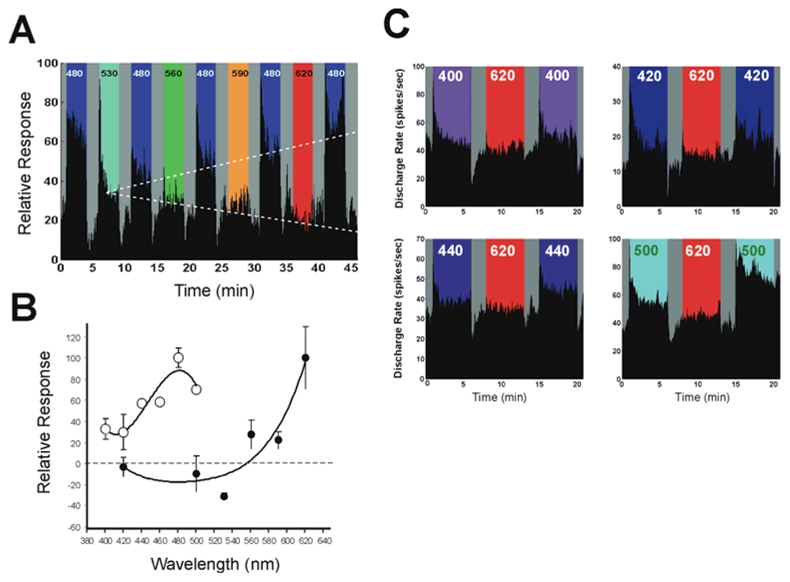
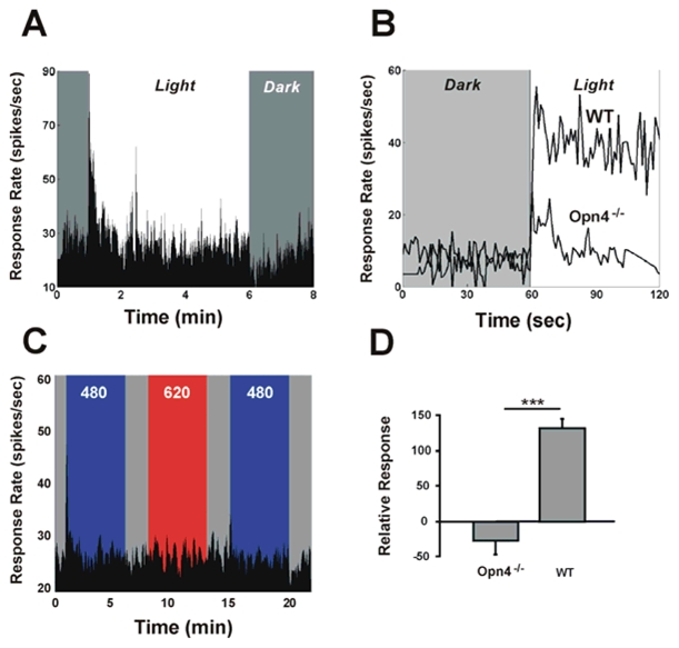
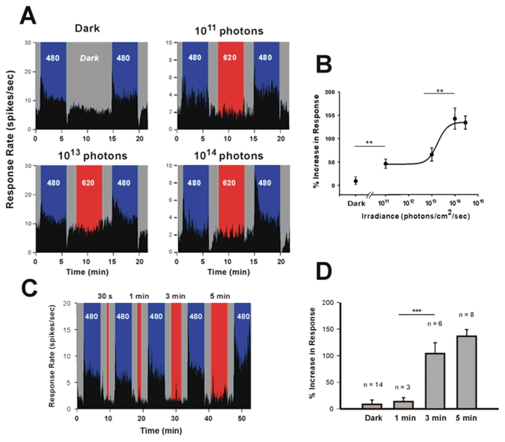
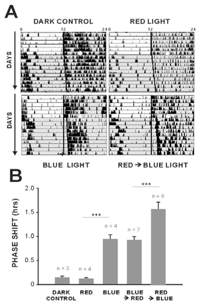
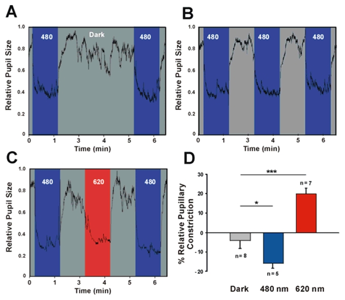
In mammals, nonvisual responses to light have been shown to involve intrinsically photosensitive retinal ganglion cells (ipRGC) that express melanopsin and that are modulated by input from both rods and cones. Recent in vitro evidence suggests that melanopsin possesses dual photosensory and photoisomerase functions, previously thought to be a unique feature of invertebrate rhabdomeric photopigments. In cultured cells that normally do not respond to light, heterologous expression of mammalian melanopsin confers light sensitivity that can be restored by prior stimulation with appropriate wavelengths. Using three different physiological and behavioral assays, we show that this in vitro property translates to in vivo, melanopsin-dependent nonvisual responses. We find that prestimulation with long-wavelength light not only restores but enhances single-unit responses of SCN neurons to 480-nm light, whereas the long-wavelength stimulus alone fails to elicit any response. Recordings in Opn4-/- mice confirm that melanopsin provides the main photosensory input to the SCN, and furthermore, demonstrate that melanopsin is required for response enhancement, because this capacity is abolished in the knockout mouse. The efficiency of the light-enhancement effect depends on wavelength, irradiance, and duration. Prior long-wavelength light exposure also enhances short-wavelength-induced phase shifts of locomotor activity and pupillary constriction, consistent with the expression of a photoisomerase-like function in nonvisual responses to light.


Figures

Figure 1
Figure 1
Enhancement of the short-wavelength light response of representative single SCN neurons (A, B) following long-wavelength light exposure. In this sequence of stimulations the first 480 nm stimulation serves as a reference for the second 480 nm exposure, and subsequently the second as a reference for the third stimulation. (A) shows that the first 480 nm light exposure (5 min) dramatically increases the firing rate of a single neuron (6.84+/−0.1 spikes/sec). During subsequent darkness (dark) the firing rate of the neuron returns to the mean baseline level (3.3 ±0.2 spikes/sec) but again increases during the second 480 nm exposure (5 min) to a rate similar to that observed for the first short-wavelength light exposure (6.21+/− 0.1spikes/sec after dark. Stimulation with 620 nm light (3 min) has no effect on the firing rate of the neuron (3.6+/−0.1) spikes/sec), which is identical to that the baseline rate observed during the periods of darkness. This pre-stimulation with long-wavelength red light results in a 60% increase in the firing rate of the neuron (7.98+/−0.1 spikes/sec), during the following (third) short-wavelength stimulation as compared to the previous 480 nm stimulation of equal irradiance. (B) When an SCN neuron is tested using the same sequence as above with 530 nm light the response rate increases from 18.4 ±0.7 to 28.3 ±0.3 spikes/sec) to a value which is 52.9% greater than the baseline rate but slightly less than the response rate at 480 nm (32.6±0.3 spikes/sec). However, in contrast to a pre-stimulation with red light, the subsequent responsiveness of the neuron to 480 nm light (31.2±0.3 spikes/sec) is slightly decreased by 9.9%. A pre-stimulation exposure with 620 nm light enhances the response of the neuron (43.4 ±0.4 spikes/sec) to the following short-wavelength stimulation by 96.0% similar to the increase seen in the previous neuron shown in A.. Both neurons shown in A and B also exhibit a clear phasic ON response to light onset with 480 and 530 nm, which is absent at 620 nm stimulation. Irradiance values are 1014 photons/cm2/s for 480 nm and 530 nm and 3.0 × 10¹⁴ photons/cm2/s for 620 nm.
Figure 2
Figure 2
The wavelength dependence of the enhancing effect of light is shown in the sequences of responses of a single SCN neuron to blue light exposure (A) and summarized for SCN neurons stimulated with different wavelengths in random order (B). The relative response rate of the neuron to a 480 nm stimulus (3 min duration, 1014 photons/cm2/s) is compared before and after a pre-stimulation phase (3 min, 1014 photons/cm2/s) using a different wavelength flanked by 2 min dark periods necessary to establish a stable baseline level. A sequence of stimulations with wavelengths from 530 nm to 620 nm shows two wavelength dependent trends. During the pre-stimulation exposure, the firing rate response of the neuron decreases with increased wavelength associated with a progressive decrease of the phasic excitatory ON and inhibitory OFF responses that are absent at wavelengths > 560 nm. In contrast, the short-wavelength response of the neuron progressively increases with longer wavelengths. The dashed lines show regression lines fitted through the mean response level illustrating the two trends of decreased response to the progressively longer wavelength reference exposures (y = −4.08× + 42.18, r2 = 0.93) and the increased responsiveness during the consecutive 480 nm light test exposures (y = 5.83x + 35.66, r2 = 0.98). The relative spectral efficiency (B) is shown for the long wavelength enhancing effect by using different wavelengths to assess percent variation in the response for 480nm (^) as reference and test pulses and in the opposite direction by using wavelengths from 400−500 nm (○) as the reference and test pulses before and after 620 nm exposure. Several examples of this latter effect for individual neurons are illustrated in C. In A and C, the dark gray shading corresponds to complete darkness.
Figure 3
Figure 3
Mice lacking melanopsin also lack the capacity for response enhancement by light. Neurons of the SCN in Opn4−/− mouse conserve the capacity to respond to white light (tungsten light source), although response amplitude is significantly reduced (A). This is shown in (B) which compares typical normalized responses to a pulse of white light in the wild-type and in the Opn4−/− mouse. The reduction in responsiveness is mainly observed for the sustained response, since the initial phasic-on response is present in both strains (A, B). When exposed to the sequence of prior light stimulation using 620 nm light, neurons in the SCN of the Opn4−/− mouse fail to show any potentiation of the response to the 480 nm test stimulus (C). Note that the phasic-ON response is reduced by about 50% during the second 480 nm stimulation (same neurone shown in A and C). D shows that the mean percent change in response to short wavelength light for this stimulation sequence (shown in C) leads to a 132.7±12.1% (n=5) increase in wild-type mice, but 27.3±20.1% (n=11) decrease in the Opn4−/− mouse. (*** =p<0.001, t-test)
Figure 4
Figure 4
The enhancing effect of prior long-wavelength light on short-wavelength light responsiveness is irradiance and duration dependent. Increasing irradiance levels of the 620 nm long-wavelength pre-stimulation (5 min) results in an increase in the short-wavelength responsiveness of single SCN neurons (A). After 9 minutes dark exposure the firing rate at 480 nm (1014 photons/cm2/sec, 5 min) is slightly increased from baseline (10.1%). Irradiances of 1011, 1013 and 1014 photons/cm2/sec lead to an increase of the firing rate during the second 480 nm stimulation of 37.6 %, 79.5%, and 188.9% respectively for the neurons illustrated in A. During each long wavelength stimulation the firing rate of the neuron is similar to the baseline firing rate during darkness. Note also the distinct phasic-ON response to 480 nm light in all four neurons and the phasic-OFF response to light in 3 of the 4 neurons which are absent during red light stimulation. The summary dose-response curve (B) shows the pooled data from several neurons (randomly stimulated with different irradiances). The firing rate increases significantly by 45.8±8.1% (n=9, t test p<0.003**) at the lowest irradiance used (1011 photons/cm2/sec) compared to the dark control (7.2±6.6%, n=14). This enhancing effect rises sharply after 1013 photons/cm2/sec (66.7±11.1%, n=11) and saturates at roughly a 1 log unit higher irradiance (142.8±22.5% at 1014 photons/cm2/sec, n=5, ANOVA, post hoc Newman-Keuls test p<0.003**) level resulting in a 150% increase in the firing rate compared to a previous short-wavelength stimulation (curve fit using a 4-parameter Naka-Rushton function). The enhancing effect of long wavelength light to short wavelength light (480 nm, 1014 photons/cm2/s) responsiveness is also stimulus-duration dependent as illustrated by the responses of a single neuron (C) to successively increased durations of pre-stimulation using a 620 nm light (3×10¹⁴ photons/cm2/s, light gray shading). The histogram (D) summarizes for pooled data from several neurons stimulated with different durations. Although an effect of long wavelength light is seen after 1 minute (11.7 ±6.0%, n=3), a significant enhancing effect requires at least 3–5 minutes duration (102.5 ±24.6%, n=6 and 129.0 ±13.8%, n=8, ANOVA followed by Newman-Keuls post hoc-test, p<0.001***). For variance homogeneity in statistical analyses data were first log-transformed (log (X+1)).
Figure 5
Figure 5
Pre-stimulation with red light enhances the amplitude of a behavioural phase shift to a subsequent light exposure at short-wavelength light. Typical actograms are shown on (A) and mean data are illustrated in the histogram (B). Light pulses (open white circle) were administered at CT16. Similar to the dark control animals, a 15 minute light pulse with 620 nm light (5× 10¹¹ photons/cm2/s) fails to induce a phase shift in the onset of locomotor activity (dark = 0.15 ±0.02 min, red light = 0.13 ±0.02 min, n=3 and 4 respectively, ns). Exposure to a pulse of blue light (480 nm, 15 min, 5 ×10¹¹ photons/cm2/s) induces a significant phase shift of 0.95± 0.11 min (n=4, ANOVA, post hoc Newman-Keuls test p<0.001***). When this same blue light exposure is preceded by the red light pulse which alone fails to induce a behavioural response, a significant increase is observed in the amplitude of the phase shift of locomotor activity (1.57± 0.15 min, n=6, ANOVA, post hoc Newman-Keuls test p<0.001***) to blue light (A, B). In contrast, stimulation with blue light followed by red light produces a phase shift which is equivalent to that observed with the blue light exposure alone (0.93± 0.06 min, n=7).
Figure 6
Figure 6
Consensual pupillary constriction in the mouse in response to 480 nm and 620 nm. Note that pupil surface area is measured continuously at a 30 Hz sampling rate. A first baseline 480 nm exposure with blue light (1 min) is followed by 3 minutes of darkness (A), or 1 min of either 480 nm (B) or 620 nm (C) flanked by 1 min periods of darkness, subsequently followed by the 480 nm test exposure. (D) Dark exposure fails to alter pupillary constriction (−3.9± 4.1%, n=9, ns), whereas exposure to blue light reduces the response by 15.6 ± 2.8%, n=5, ANOVA, post hoc Newman-Keuls test p<0.05*). 620 nm exposure results in an increased amplitude of the response (20.2±2.9%, n=7, ANOVA, post hoc Newman-Keuls test P<0.001***). Irradiance values are 480 nm (1012 photons/cm2/s) and 620 nm (1013photons/cm2/s).

Comment in

References

    1. Aggelopoulos NC, Meissl H. Responses of neurones of the rat suprachiasmatic nucleus to retinal illumination under photopic and scotopic conditions. J Physiol. 2000;523:211–222. - PMC - PubMed
    1. Arendt D. Evolution of eyes and photoreceptor cell types. Int J Dev Biol. 2003;47:563–571. - PubMed
    1. Arendt D, Tessmar-Raible K, Snyman H, Dorresteijn AW, Wittbrodt J. Ciliary photoreceptors with a vertebrate-type opsin in an invertebrate brain. Science. 2004;306:869–871. - PubMed
    1. Arshavsky V. Like night and day: rods and cones have different pigment regeneration pathways. Neuron. 2002;36:1–3. - PubMed
    1. Belenky MA, Smeraski CA, Provencio I, Sollars PJ, Pickard GE. Melanopsin retinal ganglion cells receive bipolar and amacrine cell synapses. J Comp Neurol. 2003;460:380–393. - PubMed

Publication types

LinkOut - more resources