

Idiopathic spinal epidural lipomatosis: urgent decompression in an atypical case
- PMID: 17876611
- PMCID: PMC2525894
- DOI: 10.1007/s00586-007-0465-0
Idiopathic spinal epidural lipomatosis: urgent decompression in an atypical case
Abstract
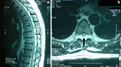
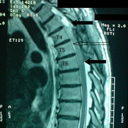
Symptomatic spinal epidural lipomatosis (SEL) is very rare and frequently associated to chronic exogenous steroid use, obesity and Cushing syndrome. The idiopathic cases where no identifiable association with SEL are found constitute only 17% of all cases. The usual clinical manifestations of this entity consist of dorsal or lumbar pain with paresthesias and weakness in lower limbs, but acute symptoms of myelopathy are exceptional. We report a case of acute paraparesis and urinary retention caused by thoracic SEL in a 55-year-old male who did not have any recognized predisposing factor for this condition. Urgent surgical decompression was performed in order to relieve the symptoms. Slow but progressive improvement was assessed after surgery. We consider this case to be exceptional due to the needing to perform an urgent decompressive laminectomy to treat a rapidly progressive myelopathy caused by idiopathic SEL.
Figures
Similar articles
-
Paget disease of the spine manifested by thoracic and lumbar epidural lipomatosis: magnetic resonance imaging findings.Spine (Phila Pa 1976). 2007 Dec 1;32(25):E789-92. doi: 10.1097/BRS.0b013e31815b7eb8. Spine (Phila Pa 1976). 2007. PMID: 18245996
-
Spinal epidural lipomatosis--a brief review.J Clin Neurosci. 2008 Dec;15(12):1323-6. doi: 10.1016/j.jocn.2008.03.001. Epub 2008 Oct 26. J Clin Neurosci. 2008. PMID: 18954986 Review.
-
Minimally invasive excision of lumbar epidural lipomatosis using a spinal endoscope.Minim Invasive Neurosurg. 2008 Feb;51(1):43-6. doi: 10.1055/s-2007-1004569. Minim Invasive Neurosurg. 2008. PMID: 18306131
-
Spinal epidural lipomatosis: two new idiopathic cases and a review of the literature.J Spinal Disord. 1994 Aug;7(4):343-9. J Spinal Disord. 1994. PMID: 7949703 Review.
-
Idiopathic spinal epidural lipomatosis.Br J Neurosurg. 2005 Jun;19(3):265-7. doi: 10.1080/02688690500210086. Br J Neurosurg. 2005. PMID: 16455531
Cited by
-
Spinal cord compression secondary to idiopathic thoracic epidural lipomatosis in an adolescent: A case report and review of literature.Int J Surg Case Rep. 2017;37:225-229. doi: 10.1016/j.ijscr.2017.06.041. Epub 2017 Jul 6. Int J Surg Case Rep. 2017. PMID: 28710985 Free PMC article.
-
Solitary epidural lipoma with ipsilateral facet arthritis causing lumbar radiculopathy.Asian Spine J. 2012 Sep;6(3):203-6. doi: 10.4184/asj.2012.6.3.203. Epub 2012 Aug 21. Asian Spine J. 2012. PMID: 22977701 Free PMC article.
-
The Clinical Characteristics of Spinal Epidural Lipomatosis in the Lumbar Spine.Anesth Pain Med. 2018 Oct 20;8(5):e83069. doi: 10.5812/aapm.83069. eCollection 2018 Oct. Anesth Pain Med. 2018. PMID: 30538942 Free PMC article.
-
Unusual presentation of spinal lipomatosis.Int Med Case Rep J. 2014 Sep 23;7:139-41. doi: 10.2147/IMCRJ.S54456. eCollection 2014. Int Med Case Rep J. 2014. PMID: 25285024 Free PMC article.
-
Clinical and radiological characteristics of spinal epidural lipomatosis: A retrospective review of 90 consecutive patients.J Clin Orthop Trauma. 2022 Aug 13;32:101988. doi: 10.1016/j.jcot.2022.101988. eCollection 2022 Sep. J Clin Orthop Trauma. 2022. PMID: 36035782 Free PMC article.
References
-
- Berman M, Feldman S, Alter M, Zilber N, Kahana E. Acute transverse myelitis: incidence and etiologic considerations. Neurology. 1981;31:966–971. - PubMed
Publication types
MeSH terms
LinkOut - more resources
Full Text Sources
