Endoscopic ultrasound-guided fine-needle aspiration cytology diagnosis of solid pseudopapillary tumor of the pancreas: a case report and literature review
- PMID: 17876886
- PMCID: PMC4434650
- DOI: 10.3748/wjg.v13.i38.5158
Endoscopic ultrasound-guided fine-needle aspiration cytology diagnosis of solid pseudopapillary tumor of the pancreas: a case report and literature review
Abstract
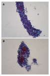
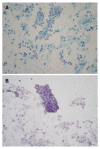
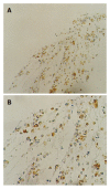
We describe the clinical, imaging and cytopathological features of solid pseudopapillary tumor of the pancreas (SPTP) diagnosed by endoscopic ultrasound-guided (EUS-guided) fine-needle aspiration (FNA). A 17-year-old woman was admitted to our hospital with complaints of an unexplained episodic abdominal pain for 2 mo and a short history of hypertension in the endocrinology clinic. Clinical laboratory examinations revealed polycystic ovary syndrome, splenomegaly and low serum amylase and carcinoembryonic antigen (CEA) levels. Computed tomography (CT) analysis revealed a mass of the pancreatic tail with solid and cystic consistency. EUS confirmed the mass, both in body and tail of the pancreas, with distinct borders, which caused dilation of the peripheral part of the pancreatic duct (major diameter 3.7 mm). The patient underwent EUS-FNA. EUS-FNA cytology specimens consisted of single cells and aggregates of uniform malignant cells, forming microadenoid structures, branching, papillary clusters with delicate fibrovascular cores and nuclear overlapping. Naked capillaries were also seen. The nuclei of malignant cells were round or oval, eccentric with fine granular chromatin, small nucleoli and nuclear grooves in some of them. The malignant cells were periodic acid Schiff (PAS)-Alcian blue (+) and immunocytochemically they were vimentin (+), CA 19.9 (+), synaptophysin (+), chromogranin (-), neuro-specific enolase (-), a1-antitrypsin and a1-antichymotrypsin focal positive. Cytologic findings were strongly suggestive of SPTP. Biopsy confirmed the above cytologic diagnosis. EUS-guided FNA diagnosis of SPTP is accurate. EUS findings, cytomorphologic features and immunostains of cell block help distinguish SPTP from pancreatic endocrine tumors, acinar cell carcinoma and papillary mucinous carcinoma.
Figures


Similar articles
-
Endoscopic ultrasound-guided fine needle aspiration cytology diagnosis of solid pseudopapillary tumor of the pancreas: a report of 3 cases.Acta Cytol. 2010 Sep-Oct;54(5):701-6. doi: 10.1159/000325236. Acta Cytol. 2010. PMID: 20968159
-
Endoscopic ultrasound-guided fine-needle aspiration cytology diagnosis of solid-pseudopapillary tumor of the pancreas: a rare neoplasm of elusive origin but characteristic cytomorphologic features.Am J Clin Pathol. 2004 May;121(5):654-62. doi: 10.1309/DKK2-B9V4-N0W2-6A8Q. Am J Clin Pathol. 2004. PMID: 15151205
-
Endoscopic ultrasound-guided fine-needle aspiration cytology in the diagnosis of intraductal papillary mucinous neoplasms of the pancreas. A study of 8 cases.JOP. 2007 Nov 9;8(6):715-24. JOP. 2007. PMID: 17993724
-
Needle Tract Seeding: An Overlooked Rare Complication of Endoscopic Ultrasound-Guided Fine-Needle Aspiration.Oncology. 2017;93 Suppl 1:107-112. doi: 10.1159/000481235. Epub 2017 Dec 20. Oncology. 2017. PMID: 29258068 Review.
-
Pitfalls in endoscopic ultrasound-guided fine-needle aspiration and how to avoid them.Adv Anat Pathol. 2005 Mar;12(2):62-73. doi: 10.1097/01.pap.0000155053.68496.ad. Adv Anat Pathol. 2005. PMID: 15731574 Review.
Cited by
-
Endoscopic ultrasound-guided fine needle aspiration of solid pseudopapillary tumors of the pancreas: a report of three cases.Korean J Intern Med. 2013 Sep;28(5):599-604. doi: 10.3904/kjim.2013.28.5.599. Epub 2013 Aug 14. Korean J Intern Med. 2013. PMID: 24009457 Free PMC article.
-
Solid-pseudopapillary neoplasms of the pancreas: clinical and pathological features of 33 cases.Surg Today. 2013 Feb;43(2):148-54. doi: 10.1007/s00595-012-0260-3. Epub 2012 Jul 24. Surg Today. 2013. PMID: 22825652
-
Hyaline globules in neuroendocrine and solid-pseudopapillary neoplasms of the pancreas: a clue to the diagnosis.Am J Surg Pathol. 2011 Jul;35(7):981-8. doi: 10.1097/PAS.0b013e31821a9a14. Am J Surg Pathol. 2011. PMID: 21677537 Free PMC article.
-
A Solid Pseudopapillary Tumour of the Head of Pancreas: A Rare Case Report Diagnosed by Fine Needle Aspiration Cytology.J Clin Diagn Res. 2016 Jun;10(6):ED06-8. doi: 10.7860/JCDR/2016/19456.7929. Epub 2016 Jun 1. J Clin Diagn Res. 2016. PMID: 27504299 Free PMC article.
-
Accuracy of diagnosis of solid pseudopapillary tumor of the pancreas on fine needle aspiration: A multi-institution experience of ten cases.Cytojournal. 2015 Dec 4;12:29. doi: 10.4103/1742-6413.171140. eCollection 2015. Cytojournal. 2015. PMID: 26884802 Free PMC article.
References
-
- Pettinato G, Manivel JC, Ravetto C, Terracciano LM, Gould EW, di Tuoro A, Jaszcz W, Albores-Saavedra J. Papillary cystic tumor of the pancreas. A clinicopathologic study of 20 cases with cytologic, immunohistochemical, ultrastructural, and flow cytometric observations, and a review of the literature. Am J Clin Pathol. 1992;98:478–488. - PubMed
-
- Brázdil J, Hermanová M, Kren L, Kala Z, Neumann C, Růzicka M, Nenutil R. [Solid pseudopapillary tumor of the pancreas: 5 case reports] Rozhl Chir. 2004;83:73–78. - PubMed
Publication types
MeSH terms
LinkOut - more resources
Full Text Sources
Medical

