. 2007 Oct 9;46(40):11369-76.
doi: 10.1021/bi7005477. Epub 2007 Sep 18.

Biochemical characterization of a SET and transposase fusion protein, Metnase: its DNA binding and DNA cleavage activity

Affiliations

Biochemical characterization of a SET and transposase fusion protein, Metnase: its DNA binding and DNA cleavage activity

Yaritzabel Roman et al. Biochemistry. .

Abstract

Metnase (SETMAR) is a SET and transposase fusion protein that promotes in vivo end joining activity and mediates genomic integration of foreign DNA. Recent studies showed that Metnase retained most of the transposase activities, including 5'-terminal inverted repeat (TIR)-specific binding and assembly of a paired end complex, and cleavage of the 5'-end of the TIR element. Here we show that R432 within the helix-turn-helix motif is critical for sequence-specific recognition, as the R432A mutation abolishes its TIR-specific DNA binding activity. Metnase possesses a unique DNA nicking and/or endonuclease activity that mediates cleavage of duplex DNA in the absence of the TIR sequence. While the HTH motif is essential for the Metnase-TIR interaction, it is not required for its DNA cleavage activity. The DDE-like motif is crucial for its DNA cleavage action as a point mutation at this motif (D483A) abolished its DNA cleavage activity. Together, our results suggest that Metnase's DNA cleavage activity, unlike those of other eukaryotic transposases, is not coupled to its sequence-specific DNA binding.


Figures

Figure 1
Figure 1
A. Schematic diagram of human Metnase (SETMAR). The PreSET domain contains a cysteine- and histidine-rich putative Zn++ binding motif. The SET domain has the histone lysine methyltransferase motif. The Transposase domain contains the conserved HTH and DDE-like motifs. B. DNA sequence of the 5’-terminal inverted repeats (TIR). The two Metnase binding sites are indicated as gray color. C. A gel mobility shift analysis of the Metnase-TIR complex. Invitro DNA binding activity of Metnase with 5’-32P-TIR DNA was examined in the presence of increasing amounts of either unlabeled poly(dI-dC) or TIR. See the Materials and Methods for details. D. A gel mobility shift analysis of the Metnase-TIR interaction. Indicated amount of Metnase was added to 5’-32P-labeled TIR (200 fmol) for the Metnase-TIR interaction on a gel mobility shift assay.
Figure 2
Figure 2. Sequence specificity of the Metnase-TIR interaction
A. DNA sequences of TIR and four mutants. Mutation sites were indicated as underlined bold letters. B. TIR-specific interaction of Metnase with DNA. Reaction mixtures contained 200 fmol of 32P-labeled TIR (lanes 1–3), MARx11 (lanes 4–6), MAR1M (lanes 7–9), MAR2M (lanes 10–12), and MAR3M (lanes 13–15) in the presence of increasing amounts of wt-Metnase (1.0 pmol in lanes 2, 5, 8, 11, and 14; 2.0 pmol in lanes 3, 6, 9, 12, and 15).
Figure 3
Figure 3. R432 within the HTH motif is essential for the Metnase-TIR interaction
A. A schematic diagram of wt-Metnase and two deletion mutants, dS-Metnase(433–671) & dS-Metnase(329–671). B. Alignment of the HTH motif within the transposase domain of Metnase and a Drosophila transposase, Mos1. Identical and similar amino acids are marked with * and closed circle, respectively. C.In vitro DNA binding activity of wt-Metnase and the mutants. TIR-specific interaction of wt-Metnase, two deletion mutants [dS-Metnase(329–671) and dS-Metnase(433–671)], and the R432A. Reaction mixtures containing wt-Metnase (lanes 2–3), dS-Metnase(329–671) (lanes 4–5), dS-Metnase(433–671) (lanes 6–-7), and R432A (lanes 8–9) were incubated with 200 fmol of 32P-labeled TIR for their interaction with TIR DNA. Where indicated, either 1.0 pmol (lanes 2, 4, 6, & 8) or 2.0 pmol (lanes 3, 5, 7, & 9) of purified protein was included.
Figure 4
Figure 4. Metnase (SETMAR) possesses DNA nicking/endonuclease activity that targets no defined consensus sequence
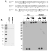
A. SDS-PAGE (10 %) and silver staining of Metnase-containing fractions from a heparin Sepharose column chromatography. B. Western blot analysis of Metnase-containing fractions shown in panel B using an anti-FLAG monoclonal antibody. C. Western blot analysis of Metnase-containing fractions using an anti-Metnase polyclonal antibody. D. DNA nicking and endonuclease activity of Metnase-containing fractions visualized on an agarose gel (1%). Replicative Form (RF) I, -II, and -III represent closed circular (supercoiled), open-circular (nicked), and linearized pBS DNA, respectively. Lanes M1 (RFI + RFII), M2 (RFIII), and M3 (RFI) are the marker DNAs. E. Analysis of Metnase-mediated DNA cleavage products. Following incubation of Metnase with RFI DNA (100 ng of pBS), the linearized DNA products (lane 1) were isolated from 1% agarose gel and labeled with [γ-32P]-ATP and T4 PNK. This labeled RFIII DNA was incubated with either EcoR I (lane 2) or Sca I (lane 3) at 37°C for 60 min. Following incubation, DNA samples were analyzed by 2.0 % agarose gel electrophoresis and autoradiography. The cleavage products were marked with an asterisk. F. DNA sequence analysis of Metnase-mediated DNA cleavage products. To determine the sequence of the Metnase cleavage sites,the linearized DNA products (lane 1 in panel A) were treated with EcoR I. DNA fragments were cloned into thepBluescript II SK+ (Stratagene), and the plasmid DNA containing inserts smaller than 200 nts were sequenced for cleavage site analysis.
Figure 5
Figure 5. The HTH motif is not essential for Metnase-mediated DNA nicking & endonuclease activity
A. Wt-Metnase and three deletion mutants [dS-Metnase(433–671), dS-Metnase(329–671), & dT-Metnase(1–414)]. B. 10% SDS-PAGE (silver-staining) of purified wt-Metnase and three deletion mutants used in this study. C. Reaction mixtures containing 100 ng of RFI DNA was incubated with increasing amount of either wt-Metnase or the mutants (0.7 pmol in lanes 2, 4, 6, & 8; 1.4 pmol in lanes 3, 5, 7, & 9), and analyzed for DNA cleavage by 1% agarose gel electrophoresis.
Figure 6
Figure 6. Metnase-mediated DNA cleavage occurs independent of TIR sequence
A. Reaction mixtures (20 μl) containing 20 fmol of 5’-32P-labeled TIR (lanes 1–3), MARx11 (lanes 4–6), or MAR3M (lanes 7–9) were incubated with 0 (lanes 1, 4, & 7), 0.1 μg (lanes 2, 5, & 8), and 0.2 μg (lanes 3, 6, & 9) of wt-Metnase in the presence of 1 mM MgCl2. After incubation at 37°C for 60 min, reaction mixtures were analyzed by 16% denaturing PAGE for DNA cleavage. Lane M contained 5’-labeled size markers generated by the Maxam & Gilbert chemical cleavage (G and G+A reactions) (30). B. Metnase-mediated DNA cleavage of the TIR DNA at a TA- (lanes 1–3), AT- (lanes 4–6), GC- (lanes 7–9), or CG- (lanes 10–12) dinucleotide at the 5’-end. Where indicated, 0 (lanes 1, 4, 7, & 10), 0.1 μg (lanes 2, 5, 8, & 11), and 0.2 μg (lanes 3, 6, 9, & 12) of wt-Metnase.
Figure 7
Figure 7
An equilibrium binding of Metnase to TIR and non-TIR (MAR3M) DNA. Using the fluorescent spectrophotometer (Varian), a change in fluorescence polarization (anisotropy) was measured in a 500μl assay format containing various amounts of Metnase and a fluorescein- labeled 32-mer of either TIR or MAR3M (10 nM).
Figure 8
Figure 8. DDE-like motif within the transposase domain is essential for Metnase’s DNA cleavage activity
A. Alignment of the Metnase’s DDE motif with Drosophila transposase, Mos-1. The DDE motif (#483, 575, & 610) was highlighted with gray color. Conserved sequences are marked with *. B.10% SDS- PAGE (silver-staining) of purified wt-Metnase and the mutant (D483A) used in this study. C. Cleavage of TIR and MAR3M with wt-Metnase and the D483A mutant. Reaction mixtures (20 μl) containing 20 fmol of 5’-32P-labeled DNA substrate were incubated with wt-Metnase (0.4 μg, lanes 2 & 7; 0.8 μg, lanes 3 & 8) or the D483A mutant (0.4 μg, lanes 4 & 9; 0.8 μg, lanes 5 & 10). Cleavage products were analyzed by 16% denaturing PAGE for DNA cleavage. Lane M contained 5’-labeled size markers.

Similar articles

Cited by

References

    1. Cordaux R, Udit S, Batzer MA, Feschotte C. Birth of a chimeric primate gene by capture of the transposase gene from a mobile element. Proc Natl Acad Sci U S A. 2006;103:8101–8106. - PMC - PubMed
    1. Lee SH, Oshige M, Durant ST, Rasila KK, Williamson EA, Ramsey H, Kwan L, Nickoloff JA, Hromas R. The SET domain protein Metnase mediates foreign DNA integration and links integration to nonhomologous end-joining repair. Proc Natl Acad Sci U S A. 2005;102:18075–18080. - PMC - PubMed
    1. Robertson HMaZKL. Molecular evolution of an ancient mariner transposon, Hsmar1, in the human genome. Gene. 1997;205:203–217. - PubMed
    1. Jordan IK. Evolutionary tinkering with transposable elements. Proc Natl Acad Sci U S A. 2006;103:7941–7942. - PMC - PubMed
    1. Craigie R. HIV integrase, a brief overview from chemistry to therapeutics. J Biol Chem. 2001;276:23213–23216. - PubMed

Publication types

MeSH terms

LinkOut - more resources