Review
. 2007 Oct;88(5):311-24.
doi: 10.1111/j.1365-2613.2007.00535.x.

Central nervous system capillary haemangioblastoma: the pathologist's viewpoint

Affiliations
Review

Central nervous system capillary haemangioblastoma: the pathologist's viewpoint

Mahmoud R Hussein. Int J Exp Pathol. 2007 Oct.

Abstract

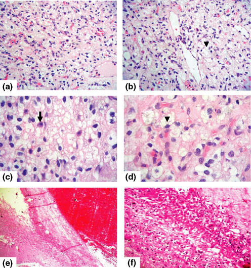
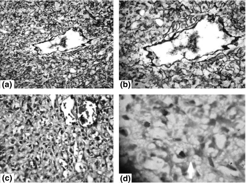
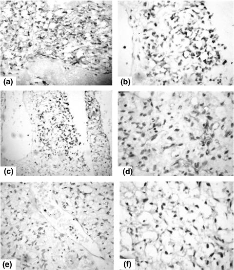
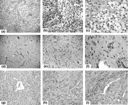
Haemangioblastomas are rare neoplasms of uncertain histogenesis. They represent 1.5-2.5% of intracranial tumours. While the cerebellum is by far the most frequent site, these lesions also tend to occur in the medulla and spinal cord. Most haemangioblastomas are sporadic but up to one quarter are associated with Von Hippel-Lindau disease (VHL). Although a fairly good number of haemangioblastomas were reported, a lack of side-by-side analysis of these reports has resulted in tentative conclusions that merely offer a first glimpse at their clinicopathologic diversity and histogenesis. To remedy this issue, this study presents a literature review concerning these lesions. Medline literature including both relevant monographs and clinicopathological case series. Haemangioblastomas occur either as a part of VHL disease (25-30%, inherited mutation of VHL gene on 3p25-26 chromosome) or as sporadic tumours (often with somatic mutation of VHL gene). They have diverse clinicopathologic presentations with cerebellar lesions having a better prognosis than their brainstem counterparts. Immunostaining is important for separation of haemangioblastomas from other tumours with similar histology. The rich vascularity of haemangioblastomas is due to overexpression of vascular endothelial growth factors. Moreover, 'stromal' cells represent the neoplastic cells of haemangioblastomas and are capable of forming blood islands with extramedullary haematopoiesis.


Figures

Figure 1
Figure 1
Histological features of cerebellar capillary haemangioblastoma. The tumour is composed of a juxtaposition of variable sized vascular spaces (capillaries with variable sized, closely packed, thin walled vessels), large neoplastic stromal cells (arrowhead) with pink to clear foamy cytoplasm with fine vacuoles, hyperchromatic nuclei and numerous mast cells (arrow) (a–d: H&E stain, ×100, ×100, ×400 and ×1000 respectively). No atypia; no fibrillar cells, no necrosis, and no mitotic figures are seen. A distinct margin with the cerebellar tissue is evident (c–d, H&E stain, ×200 and ×400 respectively).
Figure 2
Figure 2
Histochemical features of cerebellar capillary haemangioblastoma. The vascular pattern of the haemangioblastoma is accentuated by staining both with Reticulin (a–b, ×200 and ×400 respectively) and Masson trichrome (c, ×200). Some mast cells are seen amid the stromal cells (d, ×1000, Giemsa stain).
Figure 3
Figure 3
Immunohistochemical features of cerebellar capillary haemangioblastoma. The stromal and vacuolated cells show positive staining for S-100 (2-a, ×200), neuron specific enolase (2-b, ×400), Inhibin (2-c, ×200) and GFAP (d, ×400). The positive staining for Glial fibrillary acidic protein may be reasoned to diffusion of GFAP antigen or entrapment of reactive astrocytes into the tumorous tissue. The tumour cells lack reactivity for epithelial markers including cytokeratin (2-e, ×400) and epithelial membrane antigen (2-f, ×400).The negative staining for epithelial markers together with positive reactivity for Inhibin A help separate haemangioblastomas from metastatic clear cell carcinoma such as renal cell carcinomas.
Figure 4
Figure 4
Histological features of renal cell carcinoma, fibrillary astrocytoma and haemangiopericytoma. In the metastatic renal cell carcinoma, the neoplastic cells are polygonal with sharp ‘hard’ cell borders (‘vegetable cells’, due to cytoplasmic retraction), voluminous clear to eosinophilic cytoplasm, irregular, and lower grade nuclei containing vesicular chromatin and small nucleoli; surrounded by thin walled vessels and embedded in a delicate fibrovascular stroma. The cells are arranged in compact architecture of nests and broad alveoli/trabeculae. Moderate cytological atypia; no fibrillar cells, no necrosis, and no mitotic figures are seen (a–c: H&E stain, ×100, ×200 and ×400 respectively). In fibrillary astrocytoma, the neoplastic glial tissue is hypercellular and composed of fibrillary neoplastic cells with nuclei having greater angularity and density than normal CNS cells, and frequently indent each other (d, e, f, H&E stain, ×40, ×100 and ×400 respectively). The haemangiopericytoma is composed of sheets of spindled or round/oval tumour cells that arrange themselves around prominent, small, thin-walled blood vessels (staghorn vascular pattern lined by flat endothelial cells). The neoplastic cells have a relatively uniform hypercellularity, cells are homogeneous with abundant cytoplasm, indistinct cell borders, oval nuclei, inconspicuous nucleoli, mild pleomorphism and minimal nuclear atypia (g, h, i, H&E stain, ×40, ×400 and ×400 respectively).

Similar articles

Cited by

References

    1. Acikalin MF, Oner U, Tel N, Pasaoglu O, Altinel F. [Round table: infratentorial hemangioblastoma] Neurochirurgie. 1985;31:91–149. - PubMed
    1. Acikalin MF, Oner U, Tel N, Pasaoglu O, Altinel F. Supratentorial hemangioblastoma: a case report and review of the literature. Arch. Pathol. Lab. Med. 2003;127:e382–e384. - PubMed
    1. Altinoz MA, Santaguida C, Guiot MC, Del Maestro RF. Spinal hemangioblastoma containing metastatic renal cell carcinoma in von Hippel-Lindau disease. Case report and review of the literature. J. Neurosurg. Spine. 2005;3:495–500. - PubMed
    1. Arao T, Okada Y, Tanikawa T, et al. A case of von Hippel-Lindau disease with bilateral pheochromocytoma, renal cell carcinoma, pelvic tumor, spinal hemangioblastoma and primary hyperparathyroidism. Endocr. J. 2002;49:181–188. - PubMed
    1. Baccari P, Colombo R, Spotti D, Giovannelli M, Corlatti M, Staudacher C. Multidisciplinar surgical treatment in a case of von Hippel-Lindau with CNS hemangioblastoma, bilateral adrenal pheochromocytoma and multiple bilateral renal cell carcinoma – a case report. Contrib. Nephrol. 2001;136:281–283. - PubMed

MeSH terms

Substances

LinkOut - more resources