. 2007 Oct;88(5):351-60.
doi: 10.1111/j.1365-2613.2007.00539.x.

The mouse mammary carcinoma 4T1: characterization of the cellular landscape of primary tumours and metastatic tumour foci

Affiliations

The mouse mammary carcinoma 4T1: characterization of the cellular landscape of primary tumours and metastatic tumour foci

Sally A DuPré et al. Int J Exp Pathol. 2007 Oct.

Abstract

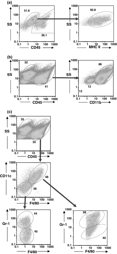
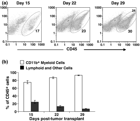
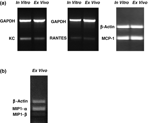
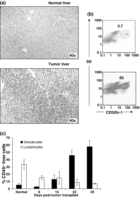
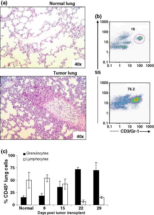
The murine mammary carcinoma 4T1 causes a leukemoid reaction with profound granulocytosis coincident with the production of tumour-derived growth factors. Here, we study the evolving cellular landscape of primary tumours and metastatic tumour foci and correlate haematopoietic cell infiltration with the production of tumour-derived chemokines. Flow cytometric analysis of enzyme digested primary tumours at different times after transplantation revealed a progressively increasing CD45(+) haematopoietic cell infiltrate consisting predominantly of CD11b(+) myeloid cells. Most of these cells had an F4/80(+)/CD11c(+) phenotype, many of which also stained Gr-1(+). Smaller numbers of Gr-1(+)CD11b(+) granulocytes and lymphoid cells were also identified. Progressive increases in Gr-1(+) granulocytes were observed in enzymatic digests of livers and lungs with metastatic tumour foci. Cultured 4T1 tumour cells expressed mRNA transcripts for the myeloid cell chemokines RANTES, MCP-1 and KC, and enzymatically digested cells from primary 4T1 tumours partially depleted of CD45(+) cells expressed transcripts for these chemokines and also MIP-1alpha and MIP-1beta. These data demonstrate that 4T1 tumour-bearing mice have mixed myeloid cell infiltrates of primary tumours and granulocytic infiltrates of metastatic organs. This pathologic presentation correlated with the expression of tumour-derived chemokines.


Figures

Figure 1
Figure 1
CD11b+ myeloid cells are the predominate infiltrating cells in primary 4T1 tumours. Primary tumours were digested with an enzyme cocktail, dissociated into single cell suspensions, stained with FITC-anti-CD45 and PE-anti-I-Ad, PE-Cy5-anti-CD3e, PE-Cy5-anti-Gr-1, PE-Cy7-anti-CD8a (a), FITC-anti-Gr-1, PE-anti-CD11b, PE-Cy5-anti-CD45 (b), and PE-Cy5-anti-CD45, PE-anti-CD11c, FITC-anti-Gr-1, PE-Cy7-streptavidin, and biotin-anti-F4/80 (c), and examined by flow cytometry. (a) Tumour cells were identified as CD45/MHC II+ cells (92.8% of CD45 population). (b) CD45+ infiltrating cells are predominantly CD11b+ myeloid cells. (c) CD45+ cells stained with both F4/80 and CD11c, and many stained with Gr-1. A population of Gr-1+ cells did not stain with either F4/80 or CD11c, together with a Gr-1 negative population that represented lymphoid cells.
Figure 2
Figure 2
The cellular landscape of the 4T1 tumour changes over time. (a) Flow cytometric dot plots of CD45+ tumour-infiltrating cells on days 15, 22, and 29 post-tumour transplant. (b) Single cell suspensions of primary tumour cells and infiltrates were labelled with FITC-anti-CD45, PE-anti-CD11b, PE-Cy5-anti-Gr-1, PE-Cy5-anti-CD3e and PE-Cy7-anti-CD8a. CD11b+ myeloid cells and lymphoid and other cells are shown as percentages of CD45+ tumour-infiltrating haematopoietic cells. Bars represent mean ± SD for groups of three mice in one of two similar experiments.
Figure 3
Figure 3
Chemokine mRNA expression in cultured 4T1 tumour cells and 4T1 tumour cells from primary tumours partially depleted of CD45+ cells. (a) Ethidium bromide-stained 1.5% agarose gels show expression of mRNA transcripts for the myeloid cell CC chemokines KC, RANTES and MCP-1 in 1 × 10⁶ cultured 4T1 cells and 1 × 10⁶ cells from enzymatically digested primary tumours partially depleted of CD45+ cells as described in the Materials and methods. (b) Expression of mRNA transcripts for the CC chemokines MIP-1α and MIP-1β in 4T1 tumour cells from enzymatically digested primary tumours partially depleted of CD45+ cells as described in the Materials and methods.
Figure 4
Figure 4
Granulocytic infiltrates in livers of 4T1 tumour-bearing mice. (a) H&E stained sections from livers of normal and 29-day tumour-bearing mice. (b) Bivariate flow cytometric dot plots showing the increase in infiltrating Gr-1+ granulocytes in livers of tumour-bearing mice. Livers were digested with an enzyme cocktail, made into a single cell suspension, stained with FITC-anti-CD45, PE-anti-CD4, PE-anti-CD19, PE-Cy5-anti-CD3e, PE-Cy5-anti-Gr-1 and PE-Cy7-anti-CD8a, and analysed by flow cytometry. Total liver cells were first gated on CD45, and then examined for Gr-1 staining. Numbers represent Gr-1+ cells as a percent of CD45+ cells in the liver. (c) Granulocytes (Gr-1+) and lymphocytes (CD3+ T lymphocytes and CD19+ B lymphocytes) as a percentage of CD45+ cells in livers of normal mice and mice at different times after tumour transplantation. Bars represent mean ± SD (four mice per group) from a representative experiment.
Figure 5
Figure 5
Granulocytic infiltrates in lungs of 4T1 tumour-bearing mice. (a) H&E stained sections from lungs of normal and 29-day tumour-bearing mice. (b) Bivariate flow cytometric dot plots showing the increase in infiltrating Gr-1+ granulocytes in lungs of tumour-bearing mice. Lungs were digested with an enzyme cocktail, made into a single cell suspension, stained with FITC-anti-CD45, PE-anti-CD4, PE-anti-CD19, PE-Cy5-anti-CD3e, PE-Cy5-anti-Gr-1 and PE-Cy7-anti-CD8a, and analysed by flow cytometry. Total lung cells were first gated on CD45, then Gr-1. Numbers represent Gr-1+ cells as a percent of CD45+ cells. (C) Granulocytes (Gr-1+) and lymphocytes (CD3+ T lymphocytes and CD19+ B lymphocytes) as a percentage of CD45+ cells in lungs of normal mice and mice at different times after tumour transplantation. Bars represent mean ± SD (four mice per group) from a representative experiment.

Similar articles

Cited by

References

    1. Adler EP, Lemken CA, Katchen NS, Kurt RA. A dual role for tumor-derived chemokine RANTES (CCL5) Immunol. Lett. 2003;90:187–194. - PubMed
    1. Aeed PA, Nakajima M, Welch DR. The role of polymorphonuclear leukocytes (PMN) on the growth and metastatic potential of 13762NF mammary adenocarcinoma cells. Int. J. Cancer. 1988;42:748–759. - PubMed
    1. Almand B, Clark JI, Nikitina E, et al. Increased production of immature myeloid cells in cancer patients: a mechanism of immunosuppression in cancer. J. Immunol. 2001;166:678–689. - PubMed
    1. Angulo I, de las Heras FG, Garcia-Bustos JF, Gargallo D, Munoz-Fernandez MA, Fresno M. Nitric oxide-producing CD11b(+)Ly-6G(Gr-1)(+)CD31(ER-MP12)(+) cells in the spleen of cyclophosphamide-treated mice: implications for T-cell responses in immunosuppressed mice. Blood. 2000;95:212–220. - PubMed
    1. Aslakson CJ, Miller FR. Selective events in the metastatic process defined by analysis of the sequential dissemination of subpopulations of a mouse mammary tumor. Cancer Res. 1992;52:1399–1405. - PubMed

Publication types

MeSH terms