. 2007 Oct;40(5):656-70.
doi: 10.1111/j.1365-2184.2007.00458.x.

Cell cycle-dependent expression of potassium channels and cell proliferation in rat mesenchymal stem cells from bone marrow

Affiliations

Cell cycle-dependent expression of potassium channels and cell proliferation in rat mesenchymal stem cells from bone marrow

X L Deng et al. Cell Prolif. 2007 Oct.

Abstract

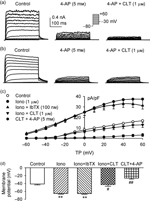
Objective: Recently, our team has demonstrated that voltage-gated delayed rectifier K(+) current (IK(DR)) and Ca(2+)-activated K(+) current (I(KCa)) are present in rat bone marrow-derived mesenchymal stem cells; however, little is known of their physiological roles. The present study was designed to investigate whether functional expression of IK(DR) and I(KCa) would change with cell cycle progression, and whether they could regulate proliferation in undifferentiated rat mesenchymal stem cells (MSCs).

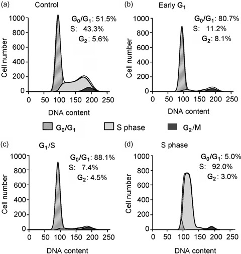
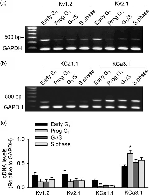
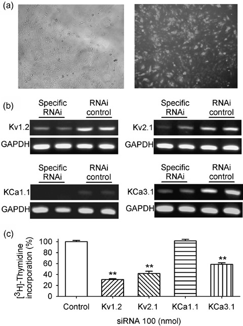
Materials and methods: Membrane potentials and ionic currents were recorded using whole-cell patch clamp technique, cell cycling was analysed by flow cytometry, cell proliferation was assayed with DNA incorporation method and the related genes were down-regulated by RNA interference (RNAi) and examined using RT-PCR.

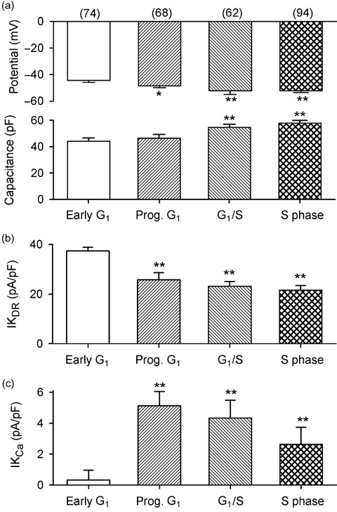
Results: It was found that membrane potential hyperpolarized, and cell size increased during the cell cycle. In addition, IK(DR) decreased, while I(KCa) increased during progress from G(1) to S phase. RT-PCR revealed that the mRNA levels of Kv1.2 and Kv2.1 (likely responsible for IK(DR)) reduced, whereas the mRNA level of KCa3.1 (responsible for intermediate-conductance I(KCa)) increased with the cell cycle progression. Down-regulation of Kv1.2, Kv2.1 or KCa3.1 with the specific RNAi, targeted to corresponding gene inhibited proliferation of rat MSCs.

Conclusion: These results demonstrate that membrane potential, IK(DR) and I(KCa) channels change with cell cycle progression and corresponding alteration of gene expression. IK(DR) and intermediate-conductance I(KCa) play an important role in maintaining membrane potential and they participate in modulation of proliferation in rat MSCs.


Figures

Figure 1
Figure 1
Pharmacological separation of K+ channel currents in rat MSCs. (a) Membrane currents recorded in a cell with 300‐ms voltage steps from –80 to between –50 and +60, and then back to –30 mV as shown in the inset (0.2 Hz). Two components of outwards currents were observed in this cell. One gradually activating current was delayed rectifier K+ current (IKDR), sensitive to inhibition by 5 mm 4‐AP, and another component with noisy oscillation was Ca2+‐activated K+ current (IKCa) sensitive to inhibition by 1 µm clotrimazole (CLT). (b) Membrane current recorded in another cell with the same voltage protocol. Current was inhibited by 5 mm 4‐AP, the remaining current not sensitive to clotrimazole, suggesting that only IKDR is present in this cell. (c) I‐V relationships of membrane currents in rat MSCs (n = 8) expressing both IKDR and IKCa. Application of 1 µm ionomycin (Iono) increased membrane current, iberiotoxin (IbTX, 100 nm) slightly decreased current at +30 to +60 mV, and clotrimazole (1 µm) reversed ionomycin‐induced current and produced a further reduction of membrane current. Remaining current was suppressed by co‐application of clotrimazole and 5 mm 4‐AP. (d) Mean values of membrane potentials determined in current clamp mode in the same rat MSCs as in (c), control, after application of 1 µm ionomycin, co‐application of ionomycin and 100 nm iberiotoxin, combination of ionomycin with 1 µm clotrimazole, and clotrimazole plus 5 mm 4‐AP.
**P < 0.01 vs control; #P < 0.05, ##P < 0.01 vs ionomycin plus iberiotoxin.
Figure 2
Figure 2
Cell cycle distribution in rat MSCs determined by flow cytometry. (a) Untreated control rat MSCs. (b) Cells from early G1, treated with starvation medium (0.5% FBS) for 24 h. (c) Cells from the end of G1, treated with regular culture medium (10% FBS) containing 2 mm thymidine for 24 h. (d) Cells from S phase, cultured with normal culture medium (10% FBS) for 8–10 h after 24 h of thymidine treatment.
Figure 3
Figure 3
Cell cycle‐dependent changes in membrane potential, cell size, IKDR and IKCa in rat MSCs. (a) Membrane potential (upper panel) hyperpolarized in rat MSCs from progressing (Prog) G1 to S phase, and cell size (lower panel, defined by membrane capacitance, Cm) increased in cells from progressing G1 to S phase. (b) IKDR density reduced in cells from progressing G1 to S phase. (c) IKCa density increased in cells progressing from G1 to S phase.
Figure 4
Figure 4
Cell cycle‐dependent changes of mRNA levels of K+ channel α‐subunits in rat MSCs. (a,b) Original gels showing expression of Kv1.2, Kv2.1, KCa1.1 and KCa3.1 mRNA from different cell cycle phases (Prog: progressing). (c) Mean values of cDNA levels (relative to GAPDH) of Kv1.2, Kv2.1, KCa1.1 and KCa3.1 from different cycling phases. *P < 0.05 versus early G1 (n = 4 different treatments).
Figure 5
Figure 5
Effects of down‐regulation of K+ channels with specific RNAi on cell proliferation in rat MSCs. (a) Images showing an example of transfecting efficiency with fluorescent RNA duplex, phase contrast (left panel) and fluorescence (right panel). (b) Original gels showing reduced messenger RNA levels of Kv1.2, Kv2.1, KCa1.1 and KCa3.1 with the corresponding specific RNAi, compared to RNAi control. (c) Cell proliferation was reduced by the down‐regulation of the specific RNAi of Kv1.2, Kv2.1 or KCa3.1, but not KCa1.1.
** P < 0.01 vs control

Similar articles

Cited by

References

    1. Bruder SP, Jaiswal N, Haynesworth SE (1997) Growth kinetics, self‐renewal, and the osteogenic potential of purified human mesenchymal stem cells during extensive subcultivation and following cryopreservation. J. Cell. Biochem. 64, 278–294. - PubMed
    1. Bruggemann A, Stuhmer W, Pardo LA (1997) Mitosis‐promoting factor‐mediated suppression of a cloned delayed rectifier potassium channel expressed in Xenopus oocytes. Proc. Natl Acad. Sci. USA 94, 537–542. - PMC - PubMed
    1. Cahalan MD, Wulff H, Chandy KG (2001) Molecular properties and physiological roles of ion channels in the immune system. J. Clin. Immunol. 21, 235–252. - PubMed
    1. Cahill KS, Toma C, Pittenger MF, Kessler PD, Byrne BJ (2003) Cell therapy in the heart: cell production, transplantation, and applications. Methods Mol. Biol. 219, 73–81. - PubMed
    1. Caplan AI, Bruder SP (2001) Mesenchymal stem cells: building blocks for molecular medicine in the 21st century. Trends Mol. Med. 7, 259–264. - PubMed

Publication types

MeSH terms