. 2007 Oct;40(5):706-20.
doi: 10.1111/j.1365-2184.2007.00467.x.

The in vitro behaviour and patterns of colony formation of murine epithelial stem cells

Affiliations

The in vitro behaviour and patterns of colony formation of murine epithelial stem cells

D Tudor et al. Cell Prolif. 2007 Oct.

Abstract

Objective: The mechanisms of renewal of skin and mucosal epithelia in vivo are associated with hierarchies of stem and amplifying cells organized in distinct spatial patterns. Stem and amplifying characteristics persist after isolation and growth of human keratinocytes in vitro but the pattern for murine keratinocytes has been less clear.

Materials and methods: Murine keratinocytes were grown in low calcium media and examined for their patterns of colony morphologies.

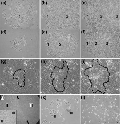
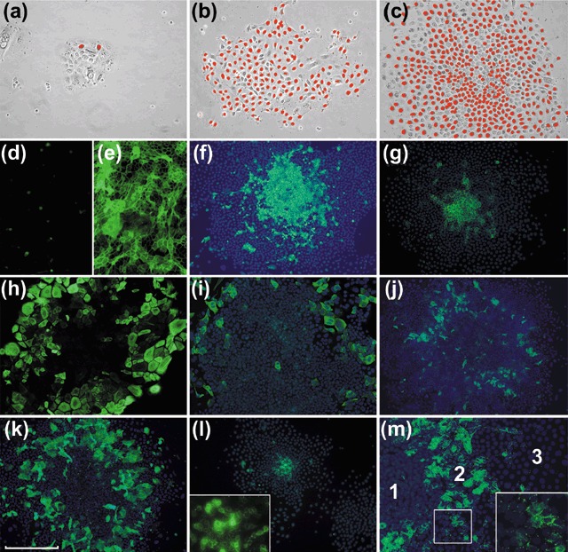
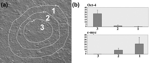
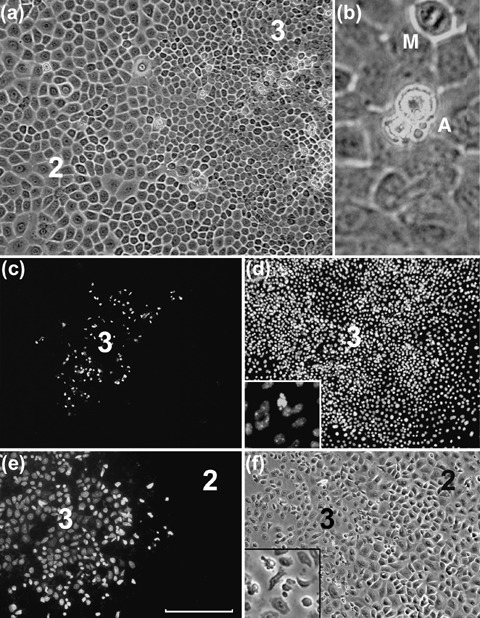
Results: We consistently identified three types of colonies, one of which contains concentric zones of amplifying and differentiated cells surrounding a central zone of cells that have patterns of expression and behavioural characteristic of stem cells. This zonal organization facilitated analysis of stem cell formation and loss. Cells in the central stem cell zone undergo rapid symmetric divisions but expansion of this population is partially limited by their peripheral transition into amplifying cells. A striking feature of central zone cells is their enhanced apoptotic susceptibility and stem cell expansion limited by consistently high background rates of apoptosis. This occasionally reaches catastrophic levels with elimination of the entire central zone.

Conclusion: In vitro amplification of stem cells for the generation of engineered tissue has tended to focus on control of asymmetric division but these findings suggest that development of mechanisms protecting stem cells from apoptotic changes are also likely to be of particular value.


Figures

Figure 1
Figure 1
Colony formation in cultures of mouse epithelial cells. (a–c) Type I, II and III colonies in a culture of ear epidermal cells 14 days after plating. The type 1 colony consists only of large ovoid or flattened cells that are loosely scattered. The type 2 colony consists of a peripheral rim of type 1 cells enclosing a zone of type 2 cells that are smaller, rounded and closely packed together. The type 3 colony has a similar distribution of type 1 and 2 cells but has an additional central zone of smaller and more refractile type 3 cells. (d–e) Similar series of colonies in a culture of mucosal epithelial cells from tongue. (g–h) Images from a 48‐h time lapse video of a Type III colony in a culture of footpad epidermal cells. The zone of type 3 cells, which has been outlined, shows an approximate doubling of cells during both the first (g to h) and second (h to i) 24‐h periods. (j) Low power image of a large Type III colony (palatal epithelium) showing large zones of type 2 and 3 cells demarcated by a clear boundary (arrows). At higher magnification (inset) the abrupt change in cell size at this boundary is seen. (k) A primary culture of palatal epithelial cells 5 days after plating showing a mixture of early Type I, II and III colony forms. (l) A culture of ear epidermal cells grown in E medium lacks distinct colony formation but shows a spectrum of cells from small and rounded to large, flattened and elongated (scale bar; for a–f,k,l = 150 µm, g–i = 120 µm, for j = 600 µm).
Figure 2
Figure 2
Proliferation and differentiation in murine colonies. Staining for BrdU cell labelling in Type I, II and III colonies (a–c, respectively) shows little labelling of flattened type 1 cells and high levels of labelling of type 2 and 3 cells. The Type III colony shown in (c) has a central area of lesser labelling, an appearance occasionally seen in large colonies. Panels d–m show IF staining of colonies with mAbs against various markers, with or without Hoechst nuclear counterstain. Cultures grown in low (< 0.06 mm) calcium media (d) show little staining for Dsg but there is marked expression after growth for 72 h (e) at a calcium concentration of 1.2 mm. Type III colonies in cultures of palatal keratinocytes stained for K15 (f) and footpad keratinocytes stained for E‐cadherin (g) with strong staining mainly restricted to the central zones of type 3 cells. A Type III colony of buccal keratinocytes (h) shows staining of large flattened peripheral cells for K18. A colony of palatal keratinocytes (i) shows a similar pattern for K19. Colonies of buccal keratinocytes stained for K4 (j) and K16 (k) show the presence of differentiated flattened suprabasal cells. A Type II colony stained for connexin 26 (l) shows staining of central type 2 cells which at higher magnification can be seen to be largely punctate and at the cell peripheries. A Type III colony shows similar staining of the zone of type 2 cells but not of type 1 and 3 cells (scale bar; for a–c = 150 µm, d–m = 250 µm).
Figure 3
Figure 3
Expression of Oct‐4 and c‐myc in relation to cell zones. (a) Laser capture image showing the boundaries of the zones of cells collected from a Type III colony. (b) Q‐PCR data for Oct‐4 and c‐myc for type 1, 2 and 3 cells. Each bar represents the mean of five independent samples ± SD expressed as calculated copy number per cell.
Figure 4
Figure 4
Apoptotic patterns in murine colonies. (a) The central region of a large colony of palatal epithelium showing the region of junction between zones of type 3 and type 2 cells. Some highly refractile cells are seen, almost entirely restricted to the zone of type 3 cells. At higher magnification (b) these cells are seen to have the characteristic fragmentation and blebbing of apoptotic cells (A). A mitotic figure (M) is also apparent. The central zone of a palatal epithelial colony stained for caspase‐3 (c) and with Hoechst stain (d) shows restriction of caspase‐3 staining to the zone of type 3 cells. At higher magnification (inset, d) Hoechst staining shows a blebbed appearance of caspase‐stained cells. (e,f) A colony of footpad keratinocytes shows caspase‐3 staining and altered morphology of almost the entire zone of type 3 cells (scale bar; for a = 100 µm, b = 25 µm, c,d = 300 µm, e,f = 150 µm).
Figure 5
Figure 5
Diagrammatic representation of events in murine colonies. (a) Homeostatic maintenance of epidermis in vivo is thought to be associated with an asymmetric stem cell division pattern in which each division produce one stem cell and one differentiating cell that undergoes a series of amplification divisions before terminally differentiating. (b) Murine colonies in vitro differ in that SCs undergo a high proportion of symmetric divisions (A) that expand the SC population. Loss of SCs occurs by transition into amplifying cells. This transition may be associated with asymmetric divisions, as illustrated at (B), but is not necessarily so. The population of amplifying cells also expands by symmetrical divisions (C) before transition into appropriate (d1) or inappropriate (d2) differentiation pathways. (c) Each cell zone of Type III colonies is characterized by different activities and the occurrence of apoptosis in Zone III is an additional factor reducing of the rate of expansion of the SC population and may increase to such a level that all SCs are eliminated, generating a Type II colony that is no longer self‐renewing.

Similar articles

Cited by

References

    1. Arai F, Hirao A, Ohmura M, Sato H, Matsuoka S, Takubo K, Ito K, Koh GY, Suda T (2004) Tie2/angiopoietin‐1 signaling regulates hematopoietic stem cell quiescence in the bone marrow niche. Cell 118, 149–161. - PubMed
    1. Barrandon Y, Green H (1985) Cell size as a determinant of the clone‐forming ability of human keratinocytes. Proc. Natl Acad. Sci. USA 82, 5390–5394. - PMC - PubMed
    1. Barrandon Y, Green H (1987) Three clonal types of keratinocyte with different capacities for multiplication. Proc. Natl Acad. Sci. USA 84, 2302–2306. - PMC - PubMed
    1. Bickenbach JR (1981) Identification and behavior of label‐retaining cells in oral mucosa and skin. J. Dent. Res. 60, 1611–1620. - PubMed
    1. Bickenbach JR, Chism E (1998) Selection and extended growth of murine epidermal stem cells in culture. Exp. Cell Res. 244, 184–195. - PubMed

Publication types