. 2007 Oct;40(5):780-94.
doi: 10.1111/j.1365-2184.2007.00462.x.

Human ovarian surface epithelial cells immortalized with hTERT maintain functional pRb and p53 expression

Affiliations

Human ovarian surface epithelial cells immortalized with hTERT maintain functional pRb and p53 expression

N F Li et al. Cell Prolif. 2007 Oct.

Abstract

Objective: Cell immortalization is considered to be a prerequisite status for carcinogenesis. Normal human ovarian surface epithelial (OSE) cells, which are thought to be the origin of most of human ovarian carcinomas, have a very limited lifespan in culture. Establishment of immortalized OSE cell lines has, in the past, required inactivation of pRb and p53 functions. However, this often leads to increased chromosome instability during prolonged culture.

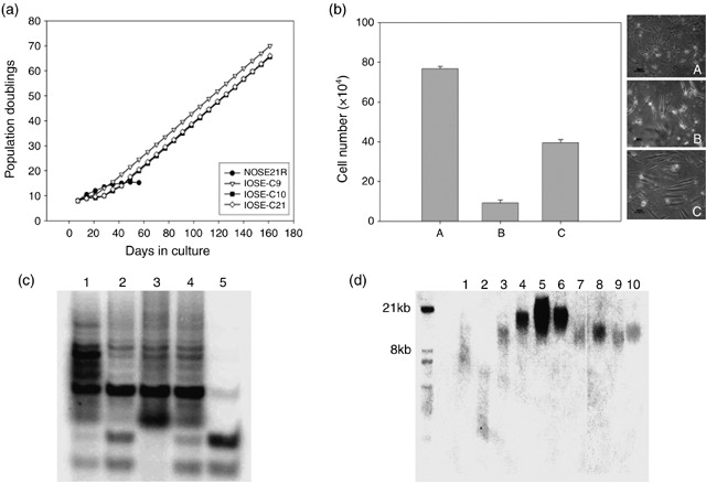
Materials and methods: In this study, we have used a retroviral infection method to overexpress human telomerase reverse transcriptase (hTERT) gene, in primary normal OSE cells, under optimized culture conditions.

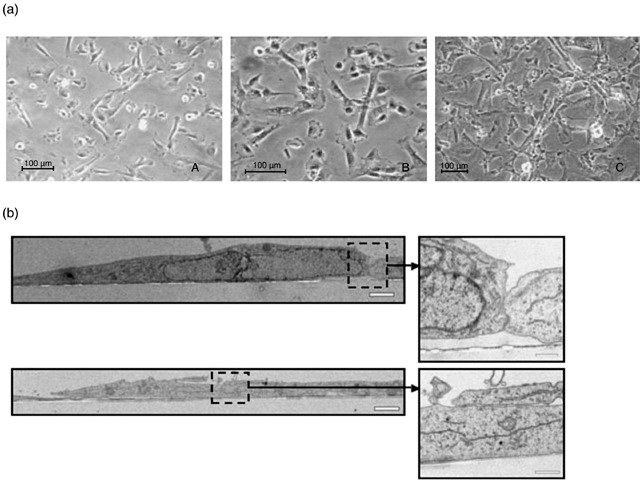
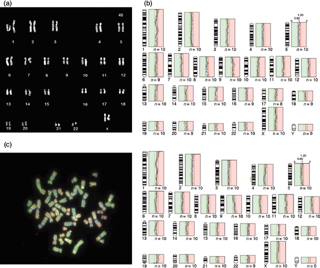
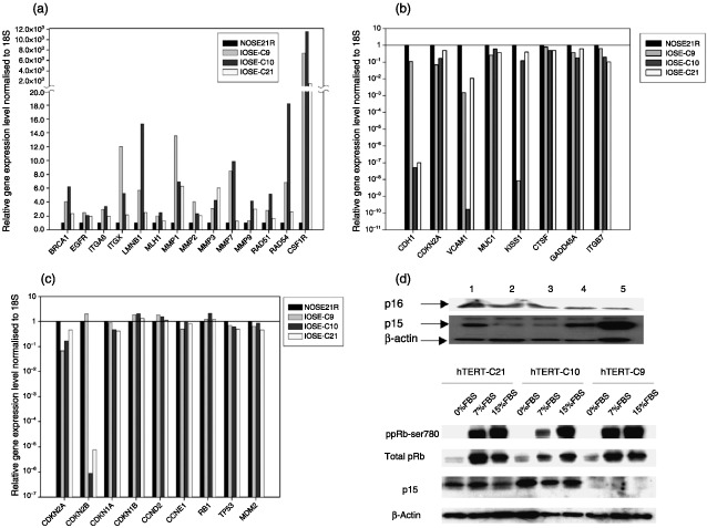
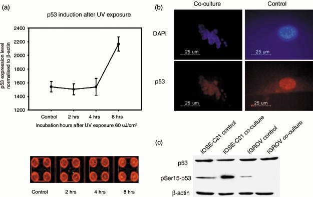
Results: In vitro and in vivo analysis of hTERT-immortalized cell lines confirmed their normal epithelial characteristics. Gene expression profiles and functional analysis of p16(INK4A), p15(INK4B), pRb and p53 confirmed the presence of their intact functions. Our study suggests that inactivation of pRb and p53 is not necessary for OSE immortalization. Furthermore, down-regulation of p15(INK4B) in the immortalized cells may indicate a functional role for this protein in them.

Conclusion: These immortal OSE cell lines are likely to be an important tool for studying human OSE biology and carcinogenesis.


Figures

Figure 1
Figure 1
hTERT‐immortalized cell lines show prolonged lifespan with activated telomerase function. (a) Growth curve of hTERT‐immortalized OSE lines (IOSE‐C9, C10, C21) and parental normal OSE cells (NOSE21R) in 180 days of culture. IOSE‐C9, C10 and C21 cells show stable population growth rates 30 days after subcloning. (b) Serum dependence and growth factor dependence analysis. After culturing the hTERT immortal line IOSE‐C21 in complete medium (A), serum‐free medium (B) and growth factor‐free medium (C) for 14 days, cells underwent population growth arrest in serum‐free medium, and slowed their expansion in growth factor‐free medium, but with a change to elongated morphology. Compared to cultures in complete medium, there were significant decreases of live cell populations in both serum and growth factor withdrawn cultures (P < 0.001). (c) TRAP assay show positive telomerase activity in all three immortal lines. 1: Positive control; 2: IOSE‐C21; 3: IOSE‐C9; the missing control band indicates the existence of PCR inhibitor in reaction mix. 4: IOSE‐C10; 5: NOSE21R (parental cells). The lowest two bands of each lane present the PCR endogenous control. Lanes 1–4 show the telomere ladder with 6 base pair (T2AG3) difference between each band, which is a positive indication of telomerase activity. (d) Telomere length assays showed relatively longer telomere length in the three immortal cell lines compared to the parental cells. 1: Telomere ‘high‐level’ control (supplied by the commercial kit); 2: Telomere ‘low‐level’ control (supplied by the commercial kit); 3: NOSE21R (parental cells); 4: IOSE‐C21 cells at passage 9; 5: IOSE‐C21 cells at passage 14; 6: IOSE‐C21 cells at passage 18; 7: IOSE‐C10 cells at passage 4; 8: IOSE‐C10 cells at passage 5; 9: IOSE‐C9 cells at passage 4; 10: IOSE‐C9 cells at passage 5.
Figure 2
Figure 2
hTERT immortal lines maintain typical epithelial morphology. (a) Light microscopy illuminates that cells of all three immortal lines, IOSE‐C21 (A), IOSE‐C9 (B) and IOSE‐C10 (C), maintain typical epithelial morphology. (b) Transmission electron microscopy (TEM) revealed that the epithelial cells formed a monolayer with desmosome structures visible at sites of cell–cell contact. Inserts are the enlarged images of the areas highlighted by the dashed lines. Scale Bar = 2 µm and 0.5 µm (inserts).
Figure 3
Figure 3
M‐FISH karyotyping and CGH analysis of IOSE cell lines. (a) Representative karyotype of the IOSE‐C21 cell line. (b) Average CGH profile of IOSE‐C10 (passage 18) showed no significant chromosomal alteration at threshold 0.8–1.2 (green: parental NOSE21R cells; red: IOSE cell lines). (c) Representative metaphase CGH hybridization image of IOSE‐C9 passage 18 (left) and average CGH profile of this sample showing an interstitial deletion of chromosome 4. The gain of material at chromosome 1p is a consistent artefact of CGH analysis, and it is not considered as significant alteration.
Figure 4
Figure 4
Semiquantitative real‐time RT‐PCR analysis of 94 cancer related genes. (a) Genes with relative expression levels increased in all three IOSE lines compared to parental NOSE21R cells. (b) Genes with relative expression levels decreased in all three IOSE lines compared to parental NOSE21R cells. The expression levels of genes in parental NOSE21R cells were calibrated to 1 (1.00E+00). (c) Relative expression levels of Rb, TP53 and cell cycle control pathway‐associated genes. Expression levels of genes in parental NOSE21R cells were calibrated to 1 (1.00E+00). (d) Western blot analysis confirmed the low protein expression levels of p15 in IOSE‐C21 line when cultured at low density but significant increases in expression in confluent culture; however, p16 protein levels remained undetectable. 1: NOSE21R parental cells; 2: IOSE‐C21 passage 9 at 70% confluence; 3: IOSE‐C21 passage 14 at 80% confluence; 4: IOSE‐C21 passage 18 at 95% confluence; 5: IOSE‐C21 cells in 5% serum medium. (e) When serum was deprived, all three IOSE lines showed a decreased ratio of phospho‐Rb to total Rb protein and two lines (IOSE‐C21, IOSE‐C10) showed increased p15 expression.
Figure 5
Figure 5
hTERT immortal lines maintain intact p53 function. (a) In‐cell Western assay indicates that after exposure of IOSE‐C21 (passage 18) cells to ultraviolet (UV) damage, p53 protein levels increased significantly 8 h after UV exposure (P < 0.01) as indicated by the mean intensity of p53 signal normalized to β‐actin signal (p53: red fluorescence staining; actin: green fluorescent staining) in four replicates. (b) IOSE‐C21 cells underwent apoptosis with highly expressed p53 in the nucleus when co‐cultured with human macrophages. (c) Phospho‐p53 is up‐regulated in IOSE‐C21 cells but not in the cancer line IGROV‐3 cells when co‐cultured with human macrophages.

Similar articles

Cited by

References

    1. Alvero AB, Fishman DA, Qumsiyeh MB, Garg M (2004) Telomerase prolongs the lifespan of normal human ovarian surface epithelial cells without inducing neoplastic phenotype. J. Soc. Gynecol. Investig. 11, 553–561. - PubMed
    1. Auersperg N, Maines‐Bandiera SL, Dyck HG, Kruk PA (1994) Characterization of cultured human ovarian surface epithelial cells: phenotypic plasticity and premalignant changes. Lab. Invest. 71, 510–518. - PubMed
    1. Bodnar AG, Ouellette M, Frolkis M, Holt SE, Chiu CP, Morin GB (1998) Extension of life‐span by introduction of telomerase into normal human cells. Science 279, 349–352. - PubMed
    1. Bond JA, Haughton MF, Rowson JM, Smith PJ, Gire V, Wynford‐Thomas D (1999) Control of replicative life span in human cells: barriers to clonal expansion intermediate between M1 senescence and M2 crisis. Mol. Cell. Biol. 19, 3103–3114. - PMC - PubMed
    1. Davies BR, Steele IA, Edmondson RJ, Zwolinski SA, Saretzki G, Von Zglinicki T (2003) Immortalisation of human ovarian surface epithelium with telomerase and temperature‐sensitive SV40 large T antigen. Exp. Cell Res. 288, 390–402. - PubMed

Publication types

MeSH terms