. 2007;8(9):R196.
doi: 10.1186/gb-2007-8-9-r196.

Drawing the tree of eukaryotic life based on the analysis of 2,269 manually annotated myosins from 328 species

Affiliations

Drawing the tree of eukaryotic life based on the analysis of 2,269 manually annotated myosins from 328 species

Florian Odronitz et al. Genome Biol. 2007.

Abstract

Background: The evolutionary history of organisms is expressed in phylogenetic trees. The most widely used phylogenetic trees describing the evolution of all organisms have been constructed based on single-gene phylogenies that, however, often produce conflicting results. Incongruence between phylogenetic trees can result from the violation of the orthology assumption and stochastic and systematic errors.

Results: Here, we have reconstructed the tree of eukaryotic life based on the analysis of 2,269 myosin motor domains from 328 organisms. All sequences were manually annotated and verified, and were grouped into 35 myosin classes, of which 16 have not been proposed previously. The resultant phylogenetic tree confirms some accepted relationships of major taxa and resolves disputed and preliminary classifications. We place the Viridiplantae after the separation of Euglenozoa, Alveolata, and Stramenopiles, we suggest a monophyletic origin of Entamoebidae, Acanthamoebidae, and Dictyosteliida, and provide evidence for the asynchronous evolution of the Mammalia and Fungi.

Conclusion: Our analysis of the myosins allowed combining phylogenetic information derived from class-specific trees with the information of myosin class evolution and distribution. This approach is expected to result in superior accuracy compared to single-gene or phylogenomic analyses because the orthology problem is resolved and a strong determinant not depending on any technical uncertainties is incorporated, the class distribution. Combining our analysis of the myosins with high quality analyses of other protein families, for example, that of the kinesins, could help in resolving still questionable dependencies at the origin of eukaryotic life.


Figures

Figure 1
Figure 1
Taxon and class related statistics of the myosin dataset. (a) The pie-chart shows the number of myosins for each class. (b) The charts show the number of species and the number of myosins for a set of selected taxa. Exact numbers are given in brackets.
Figure 2
Figure 2
Phylogenetic tree of the myosin motor domains. The phylogenetic tree was built from the multiple sequence alignment of 1,984 myosin motor domains. The complete tree with bootstrap values and sequence descriptors is available as Additional data file 2. The expanded view shows the myosin sequences of class-VI and their distribution in taxa. Every other myosin class has been analyzed in a similar way. Labels at branches are bootstrap values (1,000 total boostraps). The scale bar corresponds to estimated amino acid substitutions per site. The tree was drawn using FigTree v1.0 [40].
Figure 3
Figure 3
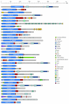
Schematic diagram of the domain structures of representative members of the 35 myosin classes. The sequence name of the representative member is given in the motor domain of the respective myosin. A color key to the domain names and symbols is given on the right except for the myosin domain, which is colored in blue. The abbreviations for the domains are: C1, protein kinase C conserved region 1; CBS, cystathionine-beta-synthase; Cyt-b5, cytochrome b5-like Heme/Steroid binding domain; DIL, dilute; FERM, band 4.1, ezrin, radixin, and moesin; FYVE, zinc finger in Fab1, YOTB/ZK632.12, Vac1, and EEA1; IQ motif, isoleucine-glutamine motif; MyTH1, myosin tail homology 1; MyTH4, myosin tail homology 4; PB1, Phox and Bem1p domain; PDZ, PDZ domain; PH, pleckstrin homology; Pkinase, protein kinase domain; PX, phox domain; RA, Ras association (RalGDS/AF-6) domain; RCC1, regulator of chromosome condensation; RhoGAP, Rho GTPase-activating protein; SH2, src homology 2; SH3, src homology 3; UBA, ubiquitin associated domain; WD40, WD (tryptophan-aspartate) or beta-transducin repeats.
Figure 4
Figure 4
Schematic diagram of the domain structures of the orphan myosins of the Fungi/Metazoa lineage. The sequence names of the ophan myosins are given in the motor domain of the respective myosins. Color keys to the domain names and symbols are given on the right except for the myosin domain, which is colored in blue. Myosin names next to domain representations list orthologs from closely related species or orthologs from the same species. These sequences have a similar domain organization. Sequences that are not orthologs and have not resulted from recent gene duplications are shown separately, although their domain organizations might be very similar. The myosin domains without names on the bottom symbolize that only head fragments are available for the sequences listed on the right. The exclamation mark on the left side of some sequences signifies that the corresponding sequences (especially the tail regions) have not completely been validated because of missing comparative genome sequences. Those sequences and corresponding tail domain predictions might change with upcoming genome sequences of related species. Abbreviations for the domains are: SAM, sterile alpha motif; Vicilin-N, Vicilin amino-terminal region; WW, tryptophan-tryptophan motif domain; Y phosphatase, protein tyrosine phosphatase, catalytic domain.
Figure 5
Figure 5
Schematic diagram of the domain structures of the orphan myosins from the Alveolata lineage. The sequence names of the ophan myosins are given in the motor domain of the respective myosins. Color keys to the domain names and symbols are given on the right except for the myosin domain, which is colored in blue. Myosin names next to domain representations list orthologs from closely related species or orthologs from the same species. These sequences have a similar domain organization. Sequences that are not orthologs and have not resulted from recent gene duplications are shown separately, although their domain organizations might be very similar. The myosin domains without names on the bottom symbolize that only head fragments are available for the sequences listed on the right. The exclamation mark on the left side of some sequences signifies that the corresponding sequences (especially the tail regions) have not completely been validated because of missing comparative genome sequences. Those sequences and corresponding tail domain predictions might change with upcoming genome sequences of related species. Abbreviations for the domains are: HDAC interact, histone deacetylase (HDAC) interacting.
Figure 6
Figure 6
Schematic diagram of the domain structures of the orphan myosins from Stramenopiles. The sequence names of the ophan myosins are given in the motor domain of the respective myosins. Color keys to the domain names and symbols are given on the right except for the myosin domain, which is colored in blue. Myosin names next to domain representations list orthologs from closely related species or orthologs from the same species. These sequences have a similar domain organization. Sequences that are not orthologs and have not resulted from recent gene duplications are shown separately, although their domain organizations might be very similar. Abbreviations for the domains are: CH, Calponin homology domain; GAF, domain present in phytochromes and cGMP-specific phosphodiesterases; HEAT repeat, named after the proteins huntingtin, elongation factor 3 (EF3), the 65 kDa alpha regulatory subunit of protein phosphatase 2A (PP2A) and the yeast PI3-kinase TOR1; Mis14, kinetochore protein Mis14 like.
Figure 7
Figure 7
Schematic diagram of the domain structures of the orphan myosins of species not belonging to one of the other taxa. Both alleles of Trypanosoma cruzi have been assembled independently, providing two slightly different copies of each myosin gene. None of the Myo-F versions is complete and the presented domain organization of Myo-F is the result of a merged version of both myosins. The sequence names of the ophan myosins are given in the motor domain of the respective myosins. Color keys to the domain names and symbols are given on the right except for the myosin domain, which is colored in blue. Myosin names next to domain representations list orthologs from closely related species or orthologs from the same species. These sequences have a similar domain organization. Sequences that are not orthologs and have not resulted from recent gene duplications are shown separately, although their domain organizations might be very similar. Abbreviations for the domains are: AAA, ATPase family associated with various cellular activities; DnaJ domain, named after the prokaryotic heat shock protein DnaJ; RhoGEF, Rho GDP/GTP exchange factor.
Figure 8
Figure 8
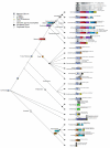
Schematic drawing of the evolution of myosin diversity. The tree has been constructed based on the combination of the phylogenetic information obtained from the analysis of single myosin classes as well as the analysis of the class distribution of major taxa (see Materials and methods). Thus, branch lengths do not correspond to any scale. Nodes that have already been suggested are symbolized by filled circles. Nodes that we propose base on the analysis of the myosins are represented by open circles. The exact myosin contents of several representative organisms are given. The myosin inventory of all 328 organisms is available from Additional data file 3.
Figure 9
Figure 9
Schematic drawing of the evolution of myosin diversity in the Fungi/Metazoa lineage based on the 'accepted' taxonomy. The inventions and losses of the myosin classes have been plotted onto the 'accepted' phylogeny of the Eukaryotes available at NCBI. Branch lengths do not correspond to any scale.
Figure 10
Figure 10
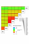
Asynchronous evolution of mammalian myosin proteins. The matrix illustrates the normalized distances between corresponding sequences. Asynchronous evolution is observed if the pattern of the deviation from the mean is different. For example, the pattern from rat to the other mammalian species is very similar, illustrating their synchronous evolution in general. However, there are differences in the patterns of some class-I myosins between rat and mouse and opossum, indicating their asynchronous evolution. In contrast, the sequence comparison patterns of cow and the other mammals are very different, indicating the asynchronous evolution of all cow myosin genes. The abbreviations for the organisms are: Rn, Rattus norvegicus; Mm, Mus musculus; Pat, Pan troglodytes; Hs, Homo sapiens; Mam, Macaca mulatto; Caf, Canis familiaris; Bt, Bos Taurus; Md, Monodelphis domestica.
Figure 11
Figure 11
Asynchronous evolution of fungi myosin proteins. The matrix is shown in a similar way as in Figure 10. The consensus tree from the analysis of the single myosin class trees is shown. The obtained polytomic tree is the result of the asynchronous evolution of the different species. The abbreviations for the organisms are: Bad, Batrachochytrium dendrobatidis JEL423; Sc_c, Saccharomyces cerevisiae S288c; Sc_a, Saccharomyces cerevisiae YJM789; Sc_b, Saccharomyces cerevisiae RM11-1a; Sap, Saccharomyces paradoxus NRRL Y-17217; Smi, Saccharomyces mikatae IFO 1815; Sak, Saccharomyces kudriavzevii IFO 1802; Sab_b, Saccharomyces bayanus MCYC 623; Sab_a, Saccharomyces bayanus 623-6C; Sk_a, Saccharomyces kluyveri NRRL Y-12651; Kw, Kluyveromyces waltii NCYC 2644; Kl, Kluyveromyces lactis NRRL Y-1140; Erg, Eremothecium gossypii ATCC 10895; Nac, Naumovia castellii NRRL Y-12630; Cgl, Candida glabrata CBS138; Loe, Lodderomyces elongisporus NRLL YB-4239; Deh, Debaryomyces hansenii CBS767; Cad, Candida dubliniensis CD36; Ca_a, Candida albicans SC5314; Ca_b, Candida albicans WO-1; Ct_a, Candida tropicalis MYA-3404; Cap, Candida parapsilosis; Cll, Clavispora lusitaniae ATCC 42720; Pig, Pichia guilliermondii ATCC 6260; Pcs, Pichia stipitis CBS 6054; Rha, Rhizopus arrhizus RA 99-880; Phb, Phycomyces blakesleeanus; Fnd_c, Filobasidiella neoformans var. neoformans JEC21; Fnd_b, Filobasidiella neoformans var. neoformans B-3501A; Fna_b, Filobasidiella neoformans var. neoformans H99; Fnb_b, Filobasidiella neoformans var. bacillispora R265; Lab, Laccaria bicolor S238N; Cpc, Coprinopsis cinerea okayama7#130; Phc, Phanerochaete chrysosporium RP-78; Um_a, Ustilago maydis 521; Um_b, Ustilago maydis FB1; Spr, Sporobolomyces roseus IAM 13481; Alb, Alternaria brassicicola ATCC 96836; Pn, Phaeosphaeria nodorum SN15; Mg, Mycosphaerella graminicola; Chg, Chaetomium globosum CBS 148.51; Poa, Podospora anserina; Nc, Neurospora crassa OR74A; Mag, Magnaporthe grisea 70-15; Gz, Gibberella zeae PH-1; Gim, Gibberella moniliformis 7600; Nh, Nectria haematococca MPVI; Scs, Sclerotinia sclerotiorum 1980; Bof, Botryotinia fuckeliana B05.10; Hj, Hypocrea jecorina QM9414; Cop, Coccidioides posadasii C735; Coi_a, Coccidioides immitis RS; Coi_c, Coccidioides immitis RMSCC 2394; Ur, Uncinocarpus reesii 1704; Ajc_b, Ajellomyces capsulatus NAmII G217B; Ajc_a, Ajellomyces capsulatus NAmII G186AR; Ajc_c, Ajellomyces capsulatus NAmI WU24; Nef, Neosartorya fischeri NRRL 181; Asf, Aspergillus fumigatus Af293; Asc, Aspergillus clavatus NRRL 1; An, Aspergillus niger ATCC 1015; Ast, Aspergillus terreus NIH2624; Ao, Aspergillus oryzae RIB40; Af, Aspergillus flavus NRRL3357; En, Emericella nidulans FGSC A4; Asa, Ascosphaera apis USDA-ARSEF 7405; Yl, Yarrowia lipolytica CLIB99; Sp, Schizosaccharomyces pombe 972h-; Sj, Schizosaccharomyces japonicus yFS275.
Figure 12
Figure 12
Evolution of the first myosins. The first myosin, called ur-myosin, is expected to consist only of the myosin motor domain. By domain fusion it generated the IQ motif either directly carboxy-terminal to the motor domain (2), or after a gene duplication event (1). After a further gene duplication event, this myosin developed to the class-I myosins as well as the ancestor of most of the other myosin classes after fusion with an SH3 domain (which developed into the amino-terminal SH3-like domain).

References

    1. Embley TM, Martin W. Eukaryotic evolution, changes and challenges. Nature. 2006;440:623–630. doi: 10.1038/nature04546. - DOI - PubMed
    1. Yang S, Doolittle RF, Bourne PE. Phylogeny determined by protein domain content. Proc Natl Acad Sci USA. 2005;102:373–378. doi: 10.1073/pnas.0408810102. - DOI - PMC - PubMed
    1. Doolittle RF. Evolutionary aspects of whole-genome biology. Curr Opin Struct Biol. 2005;15:248–253. doi: 10.1016/j.sbi.2005.04.001. - DOI - PubMed
    1. Jeffroy O, Brinkmann H, Delsuc F, Philippe H. Phylogenomics: the beginning of incongruence? Trends Genet. 2006;22:225–231. doi: 10.1016/j.tig.2006.02.003. - DOI - PubMed
    1. Ciccarelli FD, Doerks T, von Mering C, Creevey CJ, Snel B, Bork P. Toward automatic reconstruction of a highly resolved tree of life. Science. 2006;311:1283–1287. doi: 10.1126/science.1123061. - DOI - PubMed

Publication types

LinkOut - more resources