Review
. 2008 Apr;86(14 Suppl):E207-16.
doi: 10.2527/jas.2007-0473. Epub 2007 Sep 18.

Defining the transcriptional signature of skeletal muscle stem cells

Affiliations
Review

Defining the transcriptional signature of skeletal muscle stem cells

Z Yablonka-Reuveni et al. J Anim Sci. 2008 Apr.

Abstract

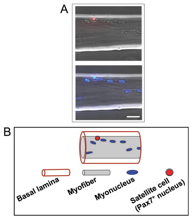
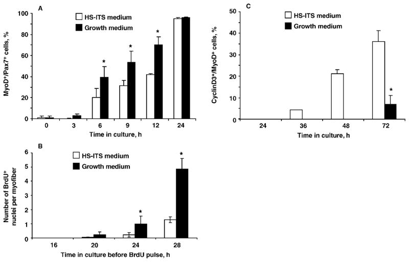
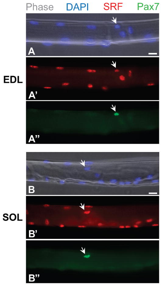
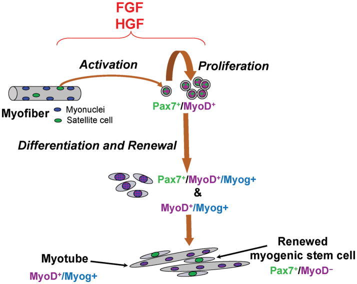
Satellite cells, the main source of myoblasts in postnatal muscle, are located beneath the myofiber basal lamina. The myogenic potential of satellite cells was initially documented based on their capacity to produce progeny that fused into myotubes. More recently, molecular markers of resident satellite cells were identified, further contributing to defining these cells as myogenic stem cells that produce differentiating progeny and self-renew. Herein, we discuss aspects of the satellite cell transcriptional milieu that have been intensively investigated in our research. We elaborate on the expression patterns of the paired box (Pax) transcription factors Pax3 and Pax7, and on the myogenic regulatory factors myogenic factor 5 (Myf5), myogenic determination factor 1 (MyoD), and myogenin. We also introduce original data on MyoD upregulation in newly activated satellite cells, which precedes the first round of cell proliferation. Such MyoD upregulation occurred even when parent myofibers with their associated satellite cells were exposed to pharmacological inhibitors of hepatocyte growth factor and fibroblast growth factor receptors, which are typically involved in promoting satellite cell proliferation. These observations support the hypothesis that most satellite cells in adult muscle are committed to rapidly entering myogenesis. We also detected expression of serum response factor in resident satellite cells prior to MyoD expression, which may facilitate the rapid upregulation of MyoD. Aspects of satellite cell self-renewal based on the reemergence of cells expressing Pax7, but not MyoD, in myogenic cultures are discussed further herein. We conclude by describing our recent studies using transgenic mice in which satellite cells are traced and isolated based on their expression of green fluorescence protein driven by regulatory elements of the nestin promoter (nestin-green fluorescence protein). This feature provides us with a novel means of studying satellite cell transcriptional signatures, heterogeneity among muscle groups, and the role of the myogenic niche in directing satellite cell self-renewal.


Figures

Figure 1
Figure 1
Resident satellite cells can be traced by their expression of the paired box (Pax) transcription factor, Pax7. A) Immunolabeling of a myofiber isolated from the extensor digitorum longus muscle of the adult mouse and reacted with an antibody against Pax7 (obtained from Developmental Studies Hybridoma Bank, University of Iowa, Iowa City, IA). The top panel depicts merged phase and immunostained images; the bottom panel depicts parallel merged phase and 4′,6-diamidino-2-phenylin-dole-stained images, which show myonuclei. Bar = 15 μm. B) Illustration of the myofiber shown in panel A, which schematically depicts the Pax7-positive (Pax7+) satellite cell positioned beneath the basal lamina, corresponding with the immunostained panel above. The continuous basal lamina is formed around myofibers in late stages of embryogenesis, correlating with the appearance of satellite cells (Yablonka-Reuveni, 1995). It is conceivable that unique interactions between the satellite cell and the myofiber basal lamina may establish the satellite cell niche, regulate satellite cell lineage commitment, and control the extent of proliferation, differentiation, and renewal.
Figure 2
Figure 2
Kinetics of satellite cell activation [myogenic determination factor 1 (MyoD) expression; panel A], proliferation [bromodeoxyurine (BrdU) uptake; panel B], and differentiation [cyclin D3 (cyclinD3) expression; panel C] in myofiber cultures isolated from the extensor digitorum longus muscle of the adult mouse and maintained in growth medium or in serum-poor medium containing 2% horse serum (HS) supplemented with insulin-transferrin-sodium selenite (ITS; i.e., HS-ITS medium). Significant differences (P < 0.05) between treatment groups within panels A, B, and C are marked with asterisks. A) Single myofiber cultures were analyzed by double immunofluorescence for Pax7 and MyoD (using mouse and rabbit primary antibodies, respectively), and cells were monitored for the number of cells expressing both MyoD and Pax7 [MyoD-positive (MyoD+)/Pax7-positive (Pax7+)] compared with the total number of satellite cells (i.e., all Pax7+ cells). Data for the various time points were evaluated for statistical differences using the nonparametric Friedman test for repeated measures. Results are from 3 independent experiments and depict the average number (±SD) of MyoD+/Pax7+ cells out of the total number of Pax7+ cells; a minimum of 42 myofibers were analyzed per time point. B) Individual myofiber cultures received a 4-h pulse of 2.5 μM BrdU at the time points indicated on the x-axis and were then fixed and processed for immunofluorescent detection of nuclei that incorporated BrdU (BrdU-positive, BrdU+), thus representing cells in the S phase of the cell cycle during the exposure to BrdU. Data for the various time points were evaluated for statistical differences by using Student’s t-test. Results are from 4 independent experiments and depict the mean (±SD) for a minimum of 47 myofibers per time point. C) Emergence of differentiating myoblasts [cells positive for both cyclinD3 and MyoD (cyclinD3+/MyoD+) out of all MyoD+ cells] in cultures of isolated myofibers. Myofibers were analyzed by double immunofluorescence for MyoD and cyclinD3 (using mouse and rabbit primary antibodies, respectively). Data for the various time points were evaluated for differences by using Student’s t-test. Results are from 3 independent experiments and depict the mean (±SD) for a minimum of 48 myofibers per time point.
Figure 3
Figure 3
Expression of serum response factor (SRF) by satellite cells in freshly isolated myofibers from extensor digitorum longus (EDL, panels A, A′, and A″) and soleus (SOL, panels B, B′, and B″) muscles harvested from the adult mouse. Myofibers were analyzed by double immu-noflurescence for the paired box transcription factor 7 (Pax7) and SRF by using mouse and rabbit primary antibodies, respectively. Myonuclei and satellite cell nuclei are positive for SRF and 4′,6-diamidino-2-phenylindole (DAPI), whereas satellite cells (arrows) are detected by their specific expression of Pax7. Bar = 15 μm.
Figure 4
Figure 4
Our working model of satellite cell activation, proliferation, differentiation, and self-renewal based on the expression patterns of the paired box transcription factor 7 (Pax7), myogenic determination factor 1 (MyoD), and myogenin (Myog). The present model is derived from our reports on the myogenesis of chicken, rat, and mouse satellite cells (Yablonka-Reuveni et al., 1999b; Kastner et al., 2000; Halevy et al., 2004; Shefer et al., 2006; Day et al., 2007). The proliferating myoblast population is represented by the Pax7-positive (Pax7+)/MyoD-positive (MyoD+) mononuclear cells. Nuclei that are MyoD+/Myog-positive (Myog+) (and no longer express Pax7) are found within differentiated mononuclear cells and myotubes, whereas a minor population of Pax7+/MyoD+/Myog+ cells represents a transitional stage within recently differentiated myoblasts; newly formed myotubes occasionally display Pax7+/ MyoD+/Myog+ nuclei as well. Renewed cells [Pax7+/MyoD-negative (MyoD)] also express green fluorescence protein (GFP) when satellite cell cultures are established from transgenic mice expressing GFP driven by regulatory elements of the nestin gene (nestin-GFP). Nestin-GFP transgene upregulation in mononuclear cells indicates reentry into the satellite cell niche. FGF = fibroblast growth factor; HGF = hepatocyte growth factor.

References

    1. Anderson JE. The satellite cell as a companion in skeletal muscle plasticity: Currency, conveyance, clue, connector and colander. J Exp Biol. 2006;209:2276–2292. - PubMed
    1. Andres V, Walsh K. Myogenin expression, cell cycle withdrawal, and phenotypic differentiation are temporally separable events that precede cell fusion upon myogenesis. J Cell Biol. 1996;132:657–666. - PMC - PubMed
    1. Barber TD, Barber MC, Cloutier TE, Friedman TB. Pax3 gene structure, alternative splicing and evolution. Gene. 1999;237:311–319. - PubMed
    1. Beauchamp JR, Heslop L, Yu DSW, Tajbakhsh S, Kelly RG, Wernig A, Buckingham ME, Partridge TA, Zammit PS. Expression of CD34 and Myf5 defines the majority of quiescent adult skeletal muscle satellite cells. J Cell Biol. 2000;151:1221–1234. - PMC - PubMed
    1. Bischoff R. Interaction between satellite cells and skeletal muscle fibers. Development. 1990;109:943–952. - PubMed

Publication types

MeSH terms