. 2007 Sep 25;104(39):15382-7.
doi: 10.1073/pnas.0707456104. Epub 2007 Sep 18.

The E-protein Tcf4 interacts with Math1 to regulate differentiation of a specific subset of neuronal progenitors

Affiliations

The E-protein Tcf4 interacts with Math1 to regulate differentiation of a specific subset of neuronal progenitors

Adriano Flora et al. Proc Natl Acad Sci U S A. .

Abstract

Proneural factors represent <10 transcriptional regulators required for specifying all of the different neurons of the mammalian nervous system. The mechanisms by which such a small number of factors creates this diversity are still unknown. We propose that proteins interacting with proneural factors confer such specificity. To test this hypothesis we isolated proteins that interact with Math1, a proneural transcription factor essential for the establishment of a neural progenitor population (rhombic lip) that gives rise to multiple hindbrain structures and identified the E-protein Tcf4. Interestingly, haploinsufficiency of TCF4 causes the Pitt-Hopkins mental retardation syndrome, underscoring the important role for this protein in neural development. To investigate the functional relevance of the Math1/Tcf4 interaction in vivo, we studied Tcf4(-/-) mice and found that they have disrupted pontine nucleus development. Surprisingly, this selective deficit occurs without affecting other rhombic lip-derived nuclei, despite expression of Math1 and Tcf4 throughout the rhombic lip. Importantly, deletion of any of the other E-protein-encoding genes does not have detectable effects on Math1-dependent neurons, suggesting a specialized role for Tcf4 in distinct neural progenitors. Our findings provide the first in vivo evidence for an exclusive function of dimers formed between a proneural basic helix-loop-helix factor and a specific E-protein, offering insight about the mechanisms underlying transcriptional programs that regulate development of the mammalian nervous system.


Conflict of interest statement

The authors declare no conflict of interest.

Figures

Fig. 1.
Fig. 1.
Identification of Tcf4 as a functional partner of Math1. (A) A β-galactosidase assay confirms the interaction between atonal and Tcf4 in yeast. Positive (p53+ T antigen) and negative (p53 or the atonal bait, Ato) controls are shown. (B) Coimmunoprecipitation of Math1 and Tcf4 in NIH 3T3 cells. Samples subjected to immunoprecipitation are shown on top of the gel; antibodies are indicated at the bottom. Anti-Flag and anti-V5 antibody detected tagged Math1 and Tcf4 proteins, respectively (arrows). Molecular markers are shown. (C) Autoradiography of a nondenaturing acrylamide gel used for EMSA; transfected constructs are indicated at the bottom. The top indicates which samples were incubated with antibodies against tagged Math1 and Tcf4 proteins or with unlabeled oligonucleotide. The arrows indicate the complexes formed between nuclear protein extracts and labeled oligonucleotide. Lanes are numbered at the bottom. (D) Bar diagram shows luciferase data. The expression constructs transfected in NIH 3T3 cells are indicated at the bottom. Bars show the fold induction over luciferase activity from cells transfected with empty vector pCDNA3. The standard deviation is shown.
Fig. 2.
Fig. 2.
In situ hybridization of sequential sagittal sections of a mouse embryo at embryonic day 14.5. The antisense probes are shown on the right. The section shown in A has been hybridized with the Math1 antisense probe, and the section shown in B has been hybridized with the Tcf4 antisense probe. uRL, upper RL; lRL, lower RL; Cb, cerebellum.
Fig. 3.
Fig. 3.
β-Galactosidase staining of structures derived from the RL of Math1+/lacZ mice. The pictures show dorsal (A–A″), lateral (B–B″), and ventral (C–C″) views of P0 hindbrains. The arrows point to the AES, and the arrowhead points to the pontine nucleus (PN). ECN, external cuneate nucleus; Cb, cerebellum; LRt, lateral reticulate nucleus. The genotype of the animals is shown at the top.
Fig. 4.
Fig. 4.
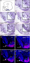
Comparison of the phenotypes of Math1−/− and Tcf4−/− mice. The outline of the sections is shown on the left of each row of images, and the framed portions represent the regions shown in the pictures. Arrowheads in A and B indicate the lateral reticulate (LRt) and external cuneate (ECN) nuclei, respectively. The arrowheads in C indicate the external granule layer cells (EGL), and the arrow points to the lower RL (lRL). In D, the arrowheads indicate the pontine nucleus (PN), and the arrows show the location of the AES. Pax6 staining is revealed as a red signal over the blue color of the nuclear stain TOTO3. The structures missing in Math1−/− animals are indicated between parentheses. The genotypes of the animals are shown at the top.
Fig. 5.
Fig. 5.
Genetic interaction between Math1 and Tcf4. A schematic of the coronal sections is shown in A. The framed portion represents the region analyzed by Nissl and Pax6 staining. (B–F) Nissl staining of coronal sections at the level of the caudal pontine nucleus of hindbrains collected from P0 mice. The brackets show the region corresponding to the location of the AES. The arrows indicate the cells of the AES. Magnifications of the region between brackets are shown in the Insets. PN, pontine nucleus. At least six animals per genotype were analyzed; genotypes are shown in each panel. (G–J) Pax6 staining of the same region analyzed by Nissl staining. Pax6 expression is revealed by the red color superimposed on the blue nuclear staining. Arrowheads indicate the position of the AES.
Fig. 6.
Fig. 6.
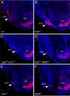
Analysis of pontine nuclear neuron differentiation in mice lacking different E-proteins. The cells of the anterior migratory stream (AES) are visualized by immunofluorescence staining using the anti-Pax6 antibody. Pax6 expression is represented by the red staining in the pictures. Cell nuclei are stained in blue. The arrowheads are pointing to the AES. The genotype of the animals is shown in the left bottom part of each panel. PN, pontine nucleus.

Similar articles

Cited by

References

    1. Guillemot F. Curr Opin Cell Biol. 2005;17:639–647. - PubMed
    1. Ross SE, Greenberg ME, Stiles CD. Neuron. 2003;39:13–25. - PubMed
    1. Bertrand N, Castro DS, Guillemot F. Nat Rev Neurosci. 2002;3:517–530. - PubMed
    1. Parras CM, Schuurmans C, Scardigli R, Kim J, Anderson DJ, Guillemot F. Genes Dev. 2002;16:324–338. - PMC - PubMed
    1. Guillemot F, Lo LC, Johnson JE, Auerbach A, Anderson DJ, Joyner AL. Cell. 1993;75:463–476. - PubMed

Publication types

MeSH terms

Substances