. 2007 Sep 25;104(39):15560-5.
doi: 10.1073/pnas.0705891104. Epub 2007 Sep 18.

Hydrogen sulfide attenuates myocardial ischemia-reperfusion injury by preservation of mitochondrial function

Affiliations

Hydrogen sulfide attenuates myocardial ischemia-reperfusion injury by preservation of mitochondrial function

John W Elrod et al. Proc Natl Acad Sci U S A. .

Abstract

The recent discovery that hydrogen sulfide (H(2)S) is an endogenously produced gaseous second messenger capable of modulating many physiological processes, much like nitric oxide, prompted us to investigate the potential of H(2)S as a cardioprotective agent. In the current study, we demonstrate that the delivery of H(2)S at the time of reperfusion limits infarct size and preserves left ventricular (LV) function in an in vivo model of myocardial ischemia-reperfusion (MI-R). This observed cytoprotection is associated with an inhibition of myocardial inflammation and a preservation of both mitochondrial structure and function after I-R injury. Additionally, we show that modulation of endogenously produced H(2)S by cardiac-specific overexpression of cystathionine gamma-lyase (alpha-MHC-CGL-Tg mouse) significantly limits the extent of injury. These findings demonstrate that H(2)S may be of value in cytoprotection during the evolution of myocardial infarction and that either administration of H(2)S or the modulation of endogenous production may be of clinical benefit in ischemic disorders.


Conflict of interest statement

Conflict of interest statement: Funding for this study was provided by Ikaria, Inc. (Seattle, WA), and the hydrogen sulfide donor was also provided by Ikaria. D.J.L. is currently serving as a paid consultant to Ikaria, and C.S. is currently employed by Ikaria.

Figures
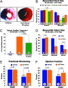
Fig. 1.
Fig. 1.
H2S donor therapy in MI-R. (A) Representative mid-myocardial cross sections of TTC-stained hearts for vehicle and 50 μg/kg H2S donor. Dark blue area (i.e., Evan's blue-stained), nonischemic zone; remaining area, AAR; white area, infracted tissue; red (i.e., TTC-positive), viable myocardium. (B) Doses of (10–500 μg/kg) or vehicle were injected i.v. at the time of reperfusion. INF/AAR revealed a U-shaped dose–response curve. (C) Serum cardiac troponin-I (cTnI) in sham-, vehicle-, and H2S (50 μg/kg)-treated mice. (D) Myocardial infarct size after 45-min LCA ischemia and 72-h reperfusion. H2S-treated mice displayed a significant reduction in INF/AAR as well as INF/LV. (E) Percent fractional shortening (%FS) after myocardial infarction. Postinfarction H2S-treated mice displayed a significant improvement in %FS as compared with vehicle-treated mice. (F) Percent ejection fraction (%EF) after myocardial infarction. Postinfarction H2S-treated mice displayed a significant improvement in %EF as compared with vehicle-treated mice (*, P < 0.05, **, P < 0.01, ***, P < 0.001 vs. BASE). (Circles inside bars denote n per group.)
Fig. 2.
Fig. 2.
H2S therapy limits the extent of myocardial inflammation. Representative H&E-stained histological images after 45 min LCA ischemia and 24 h reperfusion. (A) Vehicle-treated mice displayed a high degree of hemorrhage and infiltrating leukocytes within the ischemic zone. (B) Histopathology was attenuated in the myocardial sections of mice treated with H2S donor (50 μg/kg). (C) MPO activity was significantly decreased in mice treated with H2S donor 4 h after reperfusion. (D) Leukocyte rolling was reduced down to control levels in mice receiving thrombin plus H2S donor at all time points (**, P < 0.01 vs. all other groups, n = 7/group; circles inside bars denote n per group).
Fig. 3.
Fig. 3.
H2S donor inhibits cardiac mitochondrial respiration and preserves mitochondrial function in vitro. (A) Cardiac mitochondrial respiratory rate. Mitochondria isolated from WT mouse hearts were analyzed for oxygen consumption in the presence of H2S. H2S (1–50 μM) dose-dependently reduced mitochondrial respiration. (***, P < 0.001, n = 4 per group). (B) Representative tracings of O2 consumption in vehicle- and H2S-treated (10 μM) mitochondria. Whereas both groups had similar rates of O2 consumption before hypoxia, H2S-treated mitochondria displayed a significantly greater rate of O2 consumption 30 min after hypoxia as evident by a steeper slope of the red line. (C) Mitochondrial recovery 30 min after hypoxia. Percentage recovery of respiration rate was significantly greater in mitochondria treated with H2S donor. Circles inside bars denote n per group.
Fig. 4.
Fig. 4.
H2S preserves mitochondrial function and structure after in vivo MI-R. Mitochondria were isolated from hearts after 45 min LCA ischemia and 24 h reperfusion. (A) Mice treated with H2S at the time of reperfusion displayed a significant improvement in mitochondrial complex I efficiency as determined by using pyruvate and malate as substrate. (B) The efficiency of complex II was assessed by using succinate and G3P as substrate and inhibiting complex I with rotenone. The same H2S donor-treated mice displayed significantly greater rate of O2 consumption after MI-R (*, P < 0.05; **, P < 0.01; ***, P < 0.001 vs. sham). Circles inside bars denote n per group. (C) After 45 min of ischemia and 24 h reperfusion, myocardial samples were qualitatively assessed by transmission electron microscopy for structural mitochondrial changes. Longitudinal sections of vehicle-treated hearts displayed uniform mitochondrial swelling with disorganized cristae and decreased matrix density. The presence of amorphous matrix densities or granular dense bodies, a distinctive feature of irreversible myocardial cell injury after reperfusion, can also be seen in a number of mitochondria in vehicle-treated samples. H2S donor-treated hearts displayed little change in mitochondrial structure. Generally, mitochondria seemed to be highly dense with well organized cristae with little distinguishable differences from sham samples.
Fig. 5.
Fig. 5.
H2S reduces cardiomyocyte apoptosis in vitro and in vivo after MI/R. (A) Isolated adult cardiomyocytes were subjected to 6 h hypoxia and 12 h reoxygenation. Groups receiving the H2S donor (100 μM) at the time of reoxygenation displayed a significant reduction in caspase-3 activity (circles inside bars denote n per group). (B) After 45 min LCA ischemia and 4 h reperfusion, mouse heart samples from sham, vehicle, and H2S donor-treated mice were evaluated for TUNEL-positive nuclei. H2S donor-treated mice were found to have a 59% reduction in the number of TUNEL-positive nuclei as compared with vehicle-treated animals (P < 0.001, n = 6 per group).
Fig. 6.
Fig. 6.
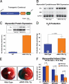
αMHC-CGL-Tg mice are protected against MI-R injury. (A) Transgenic construct. (B) Myocardial cystathionase RNA expression. αMHC-CGL-Tg mice and their WT littermates were analyzed for increased myocardial CGL message. Tg mice were found to have a significant increase in CGL mRNA as corrected by the housekeeping gene GAPDH with no alteration in the expression of cystathionine β-synthase (CBS). (C) Myocardial protein expression. Increase in message translated to a significant increase in protein expression as can be seen in the immunoblot and calculated optical density. (D) H2S production. The increase in CGL enzyme translated into ≈2-fold increase in H2S production by myocardial homogenates of αMHC-CGL-Tg mice as assessed by an H2S specific electrode (circles inside bars denote n per group). (E) Representative mid-myocardial cross sections of TTC-stained hearts for WT and αMHC-CGL-Tg mice after 45 min LCA ischemia and 72 h reperfusion. (F) Myocardial infarct size (45 min ischemia and 72 h reperfusion). Transgenic mice displayed a reduction in infarct size per area-at-risk (INF/AAR) as compared with WT littermates. Infarct per LV (INF/LV) was also significantly reduced.

Similar articles

Cited by

References

    1. Abe K, Kimura H. J Neurosci. 1996;16:1066–1071. - PMC - PubMed
    1. Beauchamp RO, Jr, Bus JS, Popp JA, Boreiko CJ, Andjelkovich DA. Crit Rev Toxicol. 1984;13:25–97. - PubMed
    1. Pryor WA, Houk KN, Foote CS, Fukuto JM, Ignarro LJ, Squadrito GL, Davies KJ. Am J Physiol. 2006;291:R491–R511. - PubMed
    1. Kimura H. Biochem Biophys Res Commun. 2000;267:129–133. - PubMed
    1. Boehning D, Snyder SH. Annu Rev Neurosci. 2003;26:105–131. - PubMed

Publication types

MeSH terms