. 2007 Oct 1;179(7):4849-56.
doi: 10.4049/jimmunol.179.7.4849.

A novel inflammatory pathway involved in leukocyte recruitment: role for the kinin B1 receptor and the chemokine CXCL5

Affiliations

A novel inflammatory pathway involved in leukocyte recruitment: role for the kinin B1 receptor and the chemokine CXCL5

Johan Duchene et al. J Immunol. .

Abstract

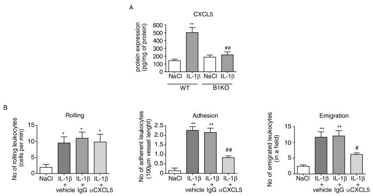
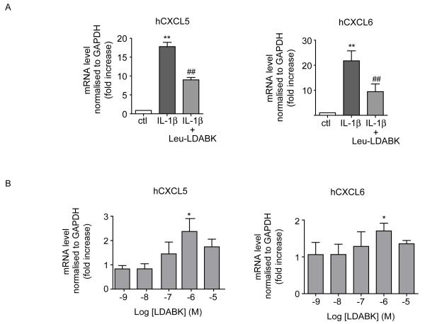
The kinin B1 receptor is an inducible receptor not normally expressed but induced by inflammatory stimuli and plays a major role in neutrophil recruitment, particularly in response to the cytokine IL-1beta. However, the exact mechanism involved in this response is unclear. The aim of this study was to dissect the molecular mechanism involved, in particular to determine whether specific ELR-CXCL chemokines (specific neutrophil chemoattractants) played a role. Using intravital microscopy, we demonstrated that IL-1beta-induced leukocyte rolling, adherence, and emigration in mesenteric venules of wild-type (WT) mice, associated with an increase in B1 receptor mRNA expression, were substantially attenuated (>80%) in B1 receptor knockout mice (B1KO). This effect in B1KO mice was correlated with a selective down-regulation of IL-1beta-induced CXCL5 mRNA and protein expression compared with WT mice. Furthermore a selective neutralizing CXCL5 Ab caused profound suppression of leukocyte emigration in IL-1beta-treated WT mice. Finally, treatment of human endothelial cells with IL-1beta enhanced mRNA expression of the B1 receptor and the human (h) CXCL5 homologues (hCXCL5 and hCXCL6). This response was suppressed by approximately 50% when cells were pretreated with the B1 receptor antagonist des-Arg9-[Leu8]-bradykinin while treatment with des-Arg9-bradykinin, the B1 receptor agonist, caused a concentration-dependent increase in hCXCL5 and hCXCL6 mRNA expression. This study unveils a proinflammatory pathway centered on kinin B1 receptor activation of CXCL5 leading to leukocyte trafficking and highlights the B1 receptor as a potential target in the therapeutics of inflammatory disease.


Figures

Fig 1
Fig 1. IL-1β-induced PMN recruitment is abolished in B1KO mice
(A) Leukocyte-endothelial cell interactions in mouse mesenteric postcapillary venules in vivo in response to IL-1β (5ng, i.p.) in WT and B1KO mice were measured by intravital microscopy. The different parameters of rolling, adhesion and emigration of leukocytes were measured in WT and B1KO mice 4h after treatment with saline (i.p.) or IL-1β. (B) The arrows show rolling (1), adherent (2) and emigrated (3) cells. (C) PMN accumulation was determined by measurement of MPO activity. Mesenteric tissue were removed from WT and B1KO mice 4h following treatment with saline or IL-1β (5ng, i.p.) and 25ng of protein extract used from each animal used to measure MPO activity. The data are expressed as the unit of MPO per g of total protein. (D) The inducibility of B1 receptor mRNA expression in mesenteric tissue in response to IL-1β (5ng, i.p.) was assessed in WT and B1KO mice by quantitative real-time RT-PCR. The data are expressed as fold increase compared to control (WT NaCl) normalised to β–actin. Data are mean ± SEM for n=6 animals per group. ** P<0.01 saline versus treated values. ## P<0.01 WT versus B1KO values.
Fig 2
Fig 2. IL-1β-induced ELR-CXCL chemokine mRNA expression is attenuated in B1KO mice
mRNA expression of CXCL1, CXCL7, CXCL2 and CXCL5 assessed using quantitative real-time RT-PCR of mesenteric tissue from WT and B1KO mice treated with saline or IL-1β (5ng, i.p., 2h).The data are expressed as fold increase above control (WT NaCl) normalised to β–actin. Data shown are mean ± SEM for n=6 animals per group. ** P<0.01 saline versus treated values. ## P<0.01 WT versus B1KO values. * P<0.05 saline versus treated values. # P<0.05 WT versus B1KO values.
Fig 3
Fig 3. CXCL5 plays a major role in IL-1β-induced leukocyte recruitment
(A) CXCL5 protein was measured by ELISA in mesenteric tissue removed from WT and B1KO mice 4h after treatment with saline or with IL-1β (5ng, i.p.). The data are expressed as CXCL5 per mg of total protein (pg/mg of protein). Data shown are mean ± SEM for n=6 animals per group ** P<0.01 saline versus treated values. ## P<0.01 WT versus B1KO values. (B) Leukocyte-endothelial cell interactions in WT mouse mesenteric postcapillary venules were measured by intra-vital microscopy in vivo 4h following treatment with IL-1β (5ng, i.p.) in mice pre-injected (30min prior to IL-1β) with saline, control IgG or anti-CXCL5 antibody (20μg/mice, i.p.). Rolling, adhesion and emigration of leukocytes were measured. Data shown are mean ± SEM for n=6 animals per group ** P<0.01 saline versus treated values. ## P<0.01 WT versus treated values. * P<0.05 saline versus treated values. # P<0.05 WT versus treated values.
Fig 4
Fig 4. Temporal relationship between endothelial kinin B1 receptor, hCXCL5, hCXCL6 and kallikrein/kininogen expression in IL-β-treated endothelial cells
(A) Time course (0, 4h, 8h and 24h) of B1 receptor, hCXCL5 and hCXCL6 mRNA expression in response to IL-1β (1ng/mL) in HUVEC. Expression of B1 receptor, hCXCL5 and hCXCL6 were measured by quantitative real-time RT-PCR. (B) Protein expression of B1 receptor in HUVEC after 8h of IL-1β treatment by western blotting. (C) mRNA expression of kininogen and kallikrein in HUVEC after 4h of IL-1β treatment was measured by quantitative real-time RT-PCR. The data are expressed as the fold increase above control (non-treated cells) normalised to GAPDH for mRNA and to tubulin for protein. Data shown are mean ± SEM for n = 4. * P<0.05 control versus treated values. ** P<0.01 control versus treated values.
Fig 5
Fig 5. Kinin B1 receptor-induced hCXCL5 and hCXCL6 expression in HUVEC
(A) Effect of B1 antagonist (Lys-[Leu8]des-Arg9-BK at 10μM, 15min pre-treatment at t=0) on IL-1β (1ng/mL, 8h)-induced hCXCL5 and hCXCL6 mRNA expression in HUVEC. (B) Effect of B1 agonist (Lys-des-Arg9-BK, 4h, 1-10,000 nM) on hCXCL5 and hCXCL6 mRNA expression. The data are expressed as the fold increase above control (non-treated cells) normalised to GAPDH for mRNA. Data are mean ± SEM for n = 4. ** P<0.01 saline versus treated values. * P<0.05 saline versus treated values. ## P<0.01 IL-1β versus IL-1β + Leu-LDBK values.

Similar articles

Cited by

References

    1. Luster AD, Alon R, von Andrian UH. Immune cell migration in inflammation: present and future therapeutic targets. Nature immunology. 2005;6:1182–1190. - PubMed
    1. Brown KA, Brain SD, Pearson JD, Edgeworth JD, Lewis SM, Treacher DF. Neutrophils in development of multiple organ failure in sepsis. Lancet. 2006;368:157–169. - PubMed
    1. Tedgui A, Mallat Z. Cytokines in atherosclerosis: pathogenic and regulatory pathways. Physiol Rev. 2006;86:515–581. - PubMed
    1. Ahluwalia A, Perretti M. Involvement of bradykinin B1 receptors in the polymorphonuclear leukocyte accumulation induced by IL-1 beta in vivo in the mouse. J Immunol. 1996;156:269–274. - PubMed
    1. McLean PG, Ahluwalia A, Perretti M. Association between kinin B(1) receptor expression and leukocyte trafficking across mouse mesenteric postcapillary venules. J Exp Med. 2000;192:367–380. - PMC - PubMed

Publication types

MeSH terms