. 2007 Dec 15;110(13):4261-7.
doi: 10.1182/blood-2007-04-086611. Epub 2007 Sep 18.

Accelerated atherogenesis and neointima formation in heparin cofactor II deficient mice

Affiliations

Accelerated atherogenesis and neointima formation in heparin cofactor II deficient mice

Cristina P Vicente et al. Blood. .

Abstract

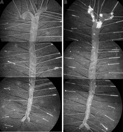
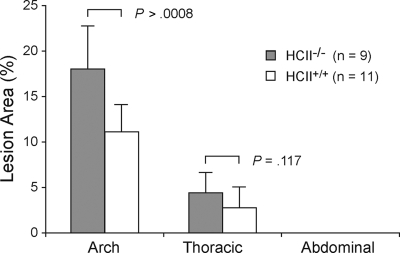
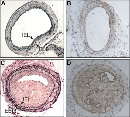
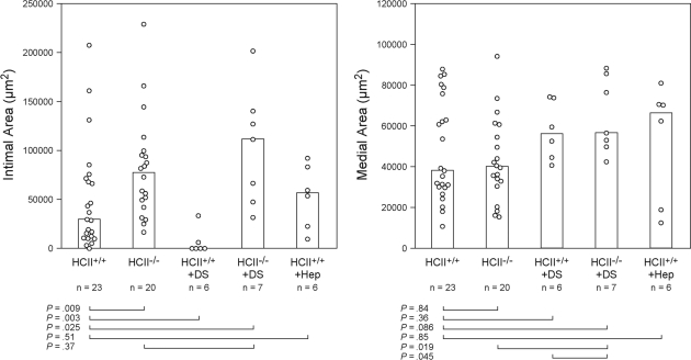
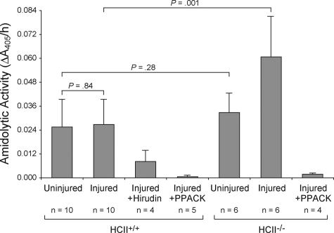
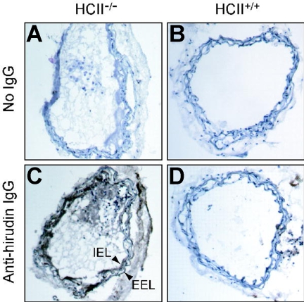
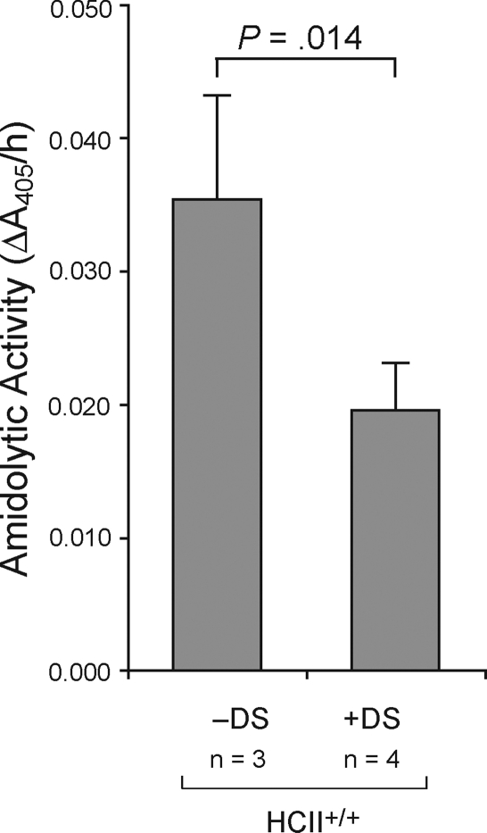
Heparin cofactor II (HCII) is a plasma protein that inhibits thrombin when bound to dermatan sulfate or heparin. HCII-deficient mice are viable and fertile but rapidly develop thrombosis of the carotid artery after endothelial injury. We now report the effects of HCII deficiency on atherogenesis and neointima formation. HCII-null or wild-type mice, both on an apolipoprotein E-null background, were fed an atherogenic diet for 12 weeks. HCII-null mice developed plaque areas in the aortic arch approximately 64% larger than wild-type mice despite having similar plasma lipid and glucose levels. Neointima formation was induced by mechanical dilation of the common carotid artery. Thrombin activity, determined by hirudin binding or chromogenic substrate hydrolysis within 1 hour after injury, was higher in the arterial walls of HCII-null mice than in wild-type mice. After 3 weeks, the median neointimal area was 2- to 3-fold greater in HCII-null than in wild-type mice. Dermatan sulfate administered intravenously within 48 hours after injury inhibited neointima formation in wild-type mice but had no effect in HCII-null mice. Heparin did not inhibit neointima formation. We conclude that HCII deficiency promotes atherogenesis and neointima formation and that treatment with dermatan sulfate reduces neointima formation in an HCII-dependent manner.


Figures

Figure 1
Figure 1
Atherosclerotic lesions. Typical appearance of aortas from apoE+/+HCII−/− (A) and apoE−/−HCII−/− (B) mice. Mice were fed a Western diet beginning at 8 weeks of age and were killed at 20 weeks of age. To show the entire aorta in a single image, each panel is a composite of 3 adjacent microscopic fields. Atherosclerotic plaques are present in the aortic arch and to a lesser extent in the thoracic aorta of the apoE−/−HCII−/− mouse (B).
Figure 2
Figure 2
Atherosclerotic plaque formation in aortas of HCII+/+ and HCII−/− mice in the apoE-null background. Lesion areas are expressed as percentages of the total intimal surface area of the aortic arch, the thoracic aorta, and the abdominal aorta. No lesions were observed in HCII+/+ or HCII−/− mice in the wild-type apoE background. Mean lesion areas are indicated. Error bars equal 1 SD.
Figure 3
Figure 3
Common carotid arteries of HCII−/− mice 3 weeks after mechanical dilation with a beaded wire probe. (A,C) Cross sections stained with Verhoeff van Gieson stain for elastin. (B,D) Cross sections stained with a monoclonal anti–α-smooth muscle actin IgG. (C,D) Injured (left) arteries. (A,B) Contralateral uninjured (right) arteries. IEL indicates internal elastic lamina; EEL, external elastic lamina.
Figure 4
Figure 4
Neointimal and medial areas of carotid arteries from HCII+/+ and HCII−/− mice 3 weeks after injury. Some mice were given 4 equal doses of dermatan sulfate (+ DS, 20 μg/g body weight) or heparin (+ Hep, 0.125 μg/g body weight) 10 minutes, 12 hours, 24 hours, and 48 hours after injury. The columns indicate the median values for each group.
Figure 5
Figure 5
Amidolytic activity associated with carotid arteries. The left (injured) and right (uninjured) carotid arteries were harvested 30 minutes after injury, opened longitudinally, rinsed, and incubated with the chromogenic substrate tosyl-Gly-Pro-Arg-p-nitroanilide. The rate of substrate hydrolysis was determined by the change in absorbance at 405 nm. Changes in A405 were linear over the time course of the experiment. Phe-Pro-Arg-chloromethylketone (PPACK, 10 nM) or recombinant hirudin (10 units/mL) was added along with the substrate as indicated. Mean values are indicated. Error bars equal 1 SD.
Figure 6
Figure 6
Binding of hirudin to arterial sections. Frozen sections were prepared from carotid arteries of HCII−/− (A,C) or HCII+/+ (B,D) mice harvested 1 hour after injury. The sections were then incubated with hirudin in vitro, and bound hirudin was detected with sheep anti–hirudin IgG and a secondary antibody linked to peroxidase (C,D). Controls in which the primary antibody was omitted are shown (A,B). A thrombus is visible in the arterial lumen of the HCII−/− mouse. IEL indicates internal elastic lamina; EEL, external elastic lamina.
Figure 7
Figure 7
Decreased amidolytic activity in arteries of mice treated with dermatan sulfate. Dermatan sulfate (20 μg/g) or an equal volume of normal saline was administered intravenously to wild-type mice 5 minutes after carotid injury. The injured arterial segments were harvested 30 minutes later and assayed for thrombin activity with tosyl-Gly-Pro-Arg-p-nitroanilide. Mean values are indicated. Error bars equal 1 SD.

Similar articles

Cited by

References

    1. Libby P. Inflammation in atherosclerosis. Nature. 2002;420:868–874. - PubMed
    1. Tracy RP. Thrombin, inflammation, and cardiovascular disease: an epidemiologic perspective. Chest. 2003;124:49S–57S. - PubMed
    1. Coughlin SR. Protease-activated receptors in the cardiovascular system. Cold Spring Harb Symp Quant Biol. 2002;67:197–208. - PubMed
    1. Farb A, Sangiorgi G, Carter AJ, et al. Pathology of acute and chronic coronary stenting in humans. Circulation. 1999;99:44–52. - PubMed
    1. Marmur JD, Thiruvikraman SV, Fyfe BS, et al. Identification of active tissue factor in human coronary atheroma. Circulation. 1996;94:1226–1232. - PubMed

Publication types