Review
. 2007 Oct;8(10):786-97.
doi: 10.1038/nrm2258.

Melanosomes--dark organelles enlighten endosomal membrane transport

Affiliations
Review

Melanosomes--dark organelles enlighten endosomal membrane transport

Graça Raposo et al. Nat Rev Mol Cell Biol. 2007 Oct.

Abstract

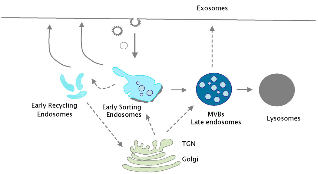
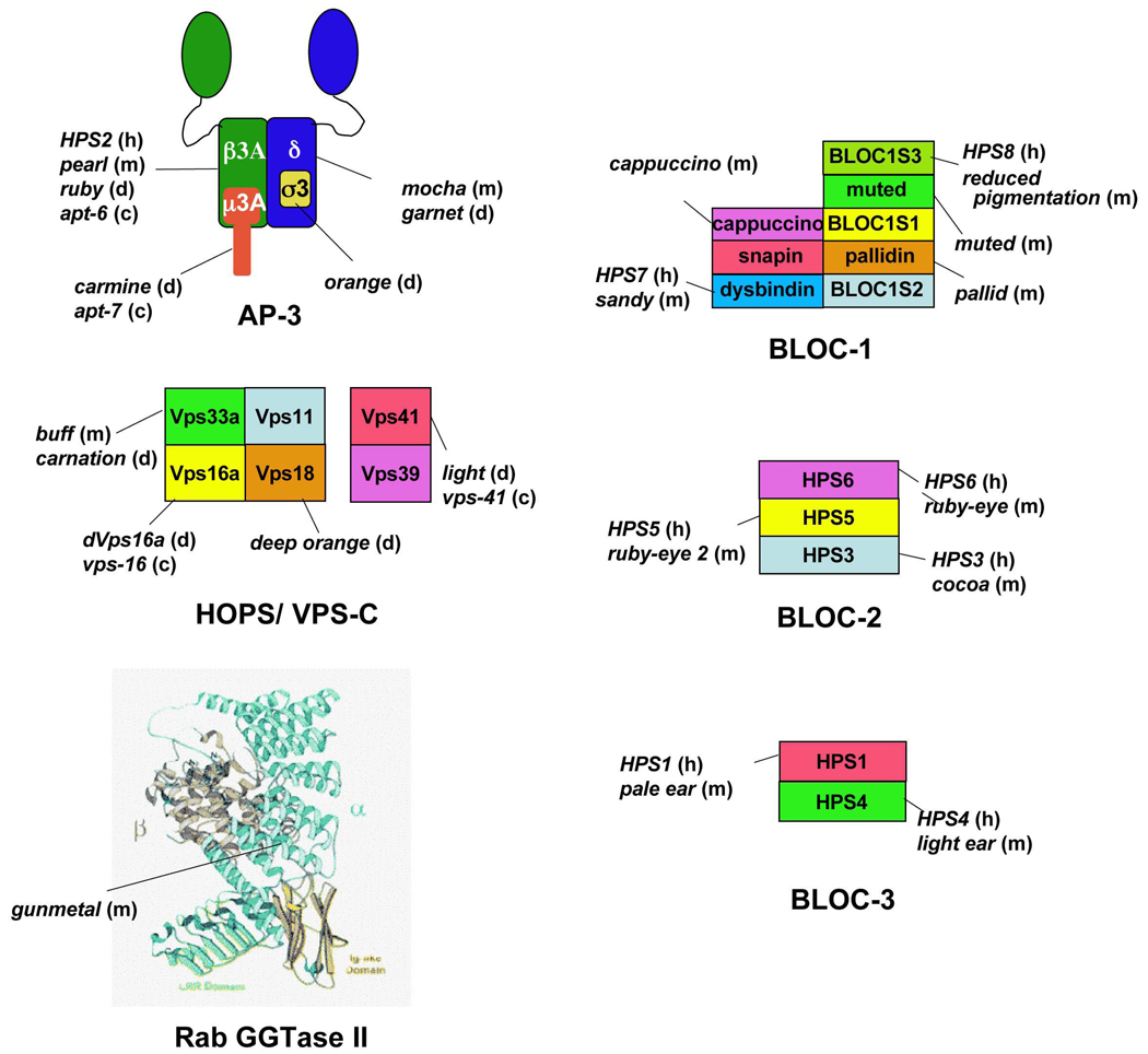
Melanosomes are tissue-specific lysosome-related organelles of pigment cells in which melanins are synthesized and stored. Analyses of the trafficking and fate of melanosomal components are beginning to reveal how melanosomes are formed through novel pathways from early endosomal intermediates. These studies unveil generalized structural and functional modifications of the endosomal system in specialized cells, and provide unexpected insights into the biogenesis of multivesicular bodies and how compartmentalization regulates protein refolding. Moreover, genetic disorders that affect the biogenesis of melanosomes and other lysosome-related organelles have shed light onto the molecular machinery that controls specialized endosomal sorting events.


Figures

Figure 1
Figure 1. Ultrastructural characterization of melanosomes
Electron microscopy analyses of MNT-1 human melanoma cells fixed by high pressure freezing before cryosubstitution and epon embedding. a. The four stages of melanosome development. Note the dense bilayered coat (arrowhead) and intralumenal vesicles (arrows) of stage I melanosomes, the proteinaceous fibrils (arrows) of stage II, and the melanin deposition in stages III and IV. b. Typical field of MNT-1 cytoplasm near the nucleus, which contains all four stages of melanosomes. M, mitochondria; N, nucleus. Bar, 0,5 µm.
Figure 2
Figure 2. Model for formation of premelanosome fibrils
The melanocyte-specific protein, Pmel (the primary structure of which is shown at the bottom) is present on the limiting membrane and the internal vesicles of stage I melanosomes/vacuolar early endosomes. Within these structures, Pmel is cleaved to Mα and Mβ fragments by a proprotein convertase (PC; bottom left). Mα fragments that dissociate from membranes begin to form small, irregular fibrils that become fully organized within stage II melanosomes. Only Mα and derived fragments (bottom right) are reproducibly detected in stage II melanosomes, whereas late endosomes are essentially devoid of detectable Pmel fragments. The figure depicts one of several hypothetical models to explain how cargo segregates from stage I melanosomes to both stage II melanosomes and late endosomes, in which each “daughter” organelle bears distinct cohorts of internal membranes. Pmel lumenal, transmembrane (TM) and cytoplasmic (cyt) domains are indicated at bottom. The RPT domain, consisting of partial direct repeats required for fibril formation, is highlighted. Squares and circles represent N- and O-linked glycosylation, respectively.
Figure 3
Figure 3. Endosomal protein complexes that regulate cargo sorting to melanosomes
a–c. Adaptor proteins and BLOCs on endosomes. Whole mount preparations (a), and ultrathin cryosections (b and c) of MNT-1 human melanoma cells were double immunogold labeled using protein A conjugated to 10 or 15 nm gold particles as indicated. Prior to fixation, cells internalized transferrin conjugated either to horseradish peroxidase and treated with substrate to highlight the early endosomal network (a), or to biotin (Tf-biot) for immunogold labelling of early endosomes (b). Arrows, labelling for AP-3, AP-1 and BLOC-1. Note the predominant labelling for AP-3 and AP-1 near melanosomes and for BLOC-1 in Tf-biot filled tubules. D–E, TYRP1 is trapped in endosomes in BLOC-1 mutants. Early endosomes of immortalized melanocytes from BLOC-1-deficient muted mice that were untransfected (BLOC-1 mutant, E) or “rescued” with the missing Muted subunit (Wild-type, D) were loaded with Transferrin-FITC (Tf-FITC). Ultrathin cryosections of fixed cells were immunogold labeled with anti-TYRP1 and anti-FITC with 10 or 15 nm gold particles. In mutant cells, both TYRP1 and Tf-FITC accumulate in expanded vacuolar endosomes (End.). Rescued cells are normally pigmented and bear numerous mature melanosomes, many close to Tf/Tryp1-containing endosomes. Bars: 200 nm. Panels A, B, D and E are reproduced and modified with permission from refs. (A), 69 (B) and 68 (D, E).
Figure 4
Figure 4. Model of melanosome maturation
Schematic diagram of endosomal and melanosomal organelles of all melanocytic stages, and the biosynthetic transport pathways among them for three representative melanosome cargoes – PMEL, tyrosinase, and TYRP1. Divergence of endocytic cargo internalized by fluid phase and destined for lysosomes is also shown. Transport pathways for each cargo are indicated by solid or dashed lines as indicated in the legend. All melanosome cargoes derive from the Golgi and traverse vacuolar and/or tubular elements of early endosomes either directly or through the cell surface. From vacuolar elements, PMEL fibrils (green) segregate to stage II melanosomes and endocytic cargo segregates to late endosomes and eventually lysosomes. From tubuar elements of early endosomes, tyrosinase and TYRP1 are targeted to maturing melanosomes where they promote melanin deposition (orange). AP-3 coats initiate one endosome-to-melanosome pathway for a cohort of tyrosinase; BLOC-1, BLOC-2, and perhaps AP-1 regulate a second pathway traversed by a separate cohort of tyrosinase and by TYRP1. RAB32 and RAB38 might function at multiple sites along either or both of these pathways. The pathways taken by other melanosome components and the function of additional complexes defective in Hermansky-Pudlak syndrome within these pathways are not yet known.
Box 2
Box 2
Box 3: Figure
Box 3: Figure

References

    1. Gruenberg J. The endocytic pathway: a mosaic of domains. Nature Reviews, Mol. Cell Biol. 2001;2:721–730. - PubMed
    1. Maxfield FR, McGraw TE. Endocytic recycling. Nature Rev. Mol. Cell Biol. 2004;5:121–132. - PubMed
    1. Spritz RA, Chiang PW, Oiso N, Alkhateeb A. Human and mouse disorders of pigmentation. Curr. Opin. Genet. Dev. 2003;13:284–289. - PubMed
    1. Jeffery G. Architecture of the optic chiasm and the mechanisms that sculpt its development. Physiol. Rev. 2001;81:1393–1414. - PubMed
    1. Wakamatsu K, Ito S. Advanced chemical methods in melanin determination. Pigment Cell Res. 2002;15:162–173. - PubMed

Publication types