

8-plex quantitation of changes in cerebrospinal fluid protein expression in subjects undergoing intravenous immunoglobulin treatment for Alzheimer's disease
- PMID: 17880003
- PMCID: PMC3594777
- DOI: 10.1002/pmic.200700316
8-plex quantitation of changes in cerebrospinal fluid protein expression in subjects undergoing intravenous immunoglobulin treatment for Alzheimer's disease
Abstract
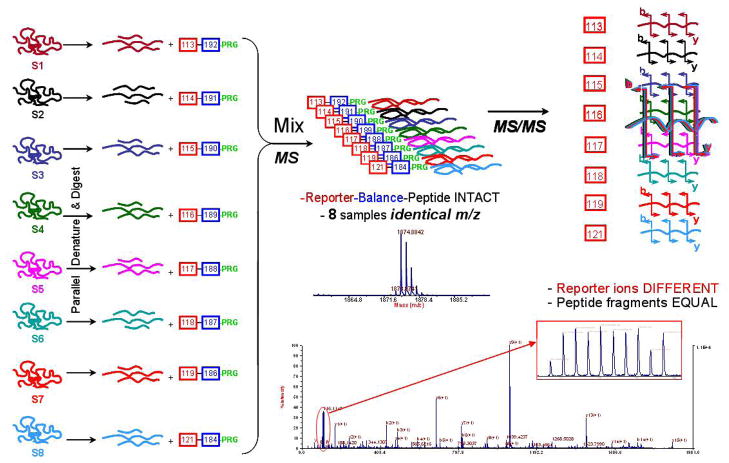
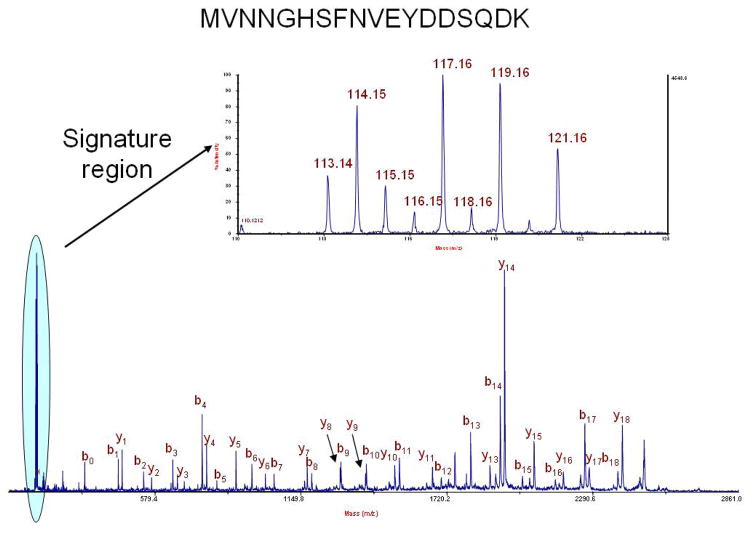
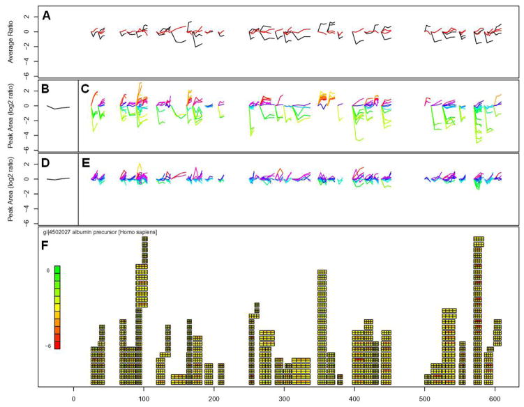
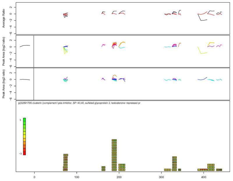
An 8-plex version of an isobaric reagent for the quantitation of proteins using shotgun methods is presented. The 8-plex version of the reagent relies on amine-labeling chemistry of peptides similar to 4-plex reagents. MS/MS reporter ions at 113, 114, 115, 116, 117, 118, 119, and 121 m/z are used to quantify protein expression. This technology which was first applied to a test mixture consisting of eight proteins and resulted in accurate quantitation, has the potential to increase throughput of analysis for quantitative shotgun proteomics experiments when compared to 2- and 4-plex methods. The technology was subsequently applied to a longitudinal study of cerebrospinal fluid (CSF) proteins from subjects undergoing intravenous Ig treatment for Alzheimer's disease. Results from this study identify a number of protein expression changes that occur in CSF after 3 and 6 months of treatment compared to a baseline and compared to a drug washout period. A visualization tool was developed for this dataset and is presented. The tool can aid in the identification of key peptides and measurements. One conclusion aided by the visualization tool is that there are differences in considering peptide-based observations versus protein-based observations from quantitative shotgun proteomics studies.
Figures
References
-
- Tonge R, Shaw J, Middleton B, Rowlinson R, et al. Validation and development of fluorescence two-dimensional differential gel electrophoresis proteomics technology. Proteomics. 2001;1:377–396. - PubMed
-
- Choe LH, Lee KH. Quantitative and qualitative measure of intralaboratory two-dimensional protein gel reproducibility and the effects of sample preparation, sample load, and image analysis. Electrophoresis. 2003;24:3500–3507. - PubMed
-
- Lee KH. Proteomics: a technology-driven and technology-limited discovery science. Trends Biotechnol. 2001;19:217–222. - PubMed
-
- McDonald WH, Yates JR. Shotgun proteomics: integrating technologies to answer biological questions. Curr Opin Mol Ther. 2003;5:302–309. - PubMed
-
- Ross PL, Huang YN, Marchese JN, Williamson B, et al. Multiplexed protein quantitation in Saccharomyces cerevisiae using amine-reactive isobaric tagging reagents. Mol Cell Proteomics. 2004;3:1154–1169. - PubMed
Publication types
MeSH terms
Substances
Grants and funding
LinkOut - more resources
Full Text Sources
Other Literature Sources
Medical
