. 2007 Oct;5(10):e251.
doi: 10.1371/journal.pbio.0050251.

A ubiquitin ligase complex regulates caspase activation during sperm differentiation in Drosophila

Affiliations

A ubiquitin ligase complex regulates caspase activation during sperm differentiation in Drosophila

Eli Arama et al. PLoS Biol. 2007 Oct.

Erratum in

  • PLoS Biol. 2007 Nov;5(11):e291

Abstract

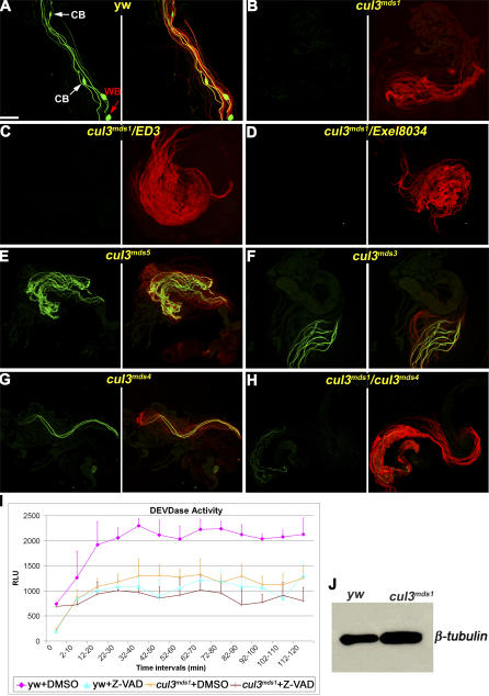
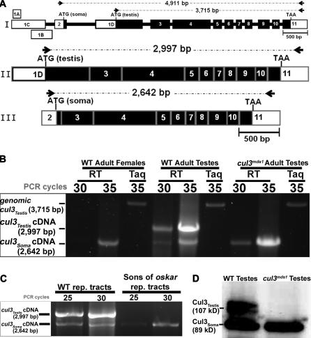
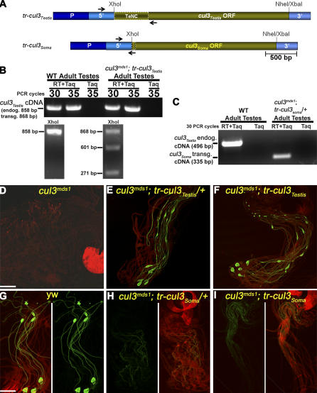
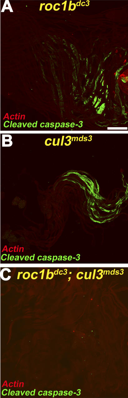
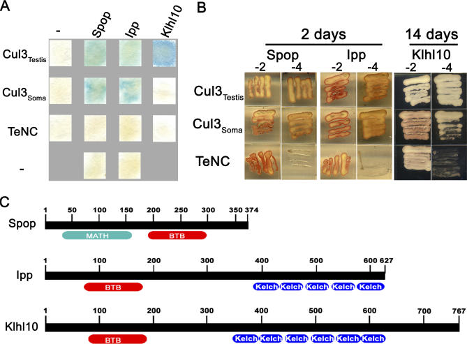
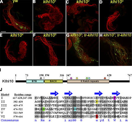
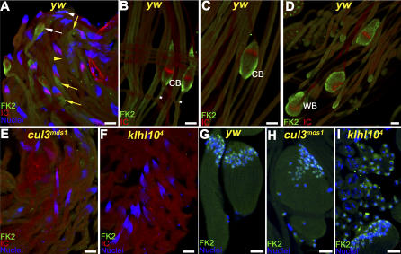
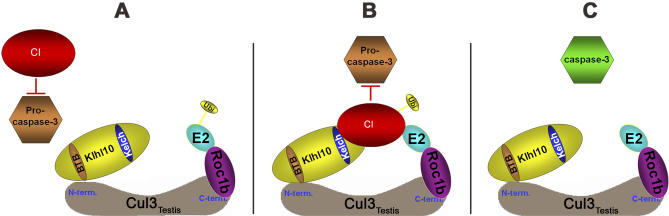
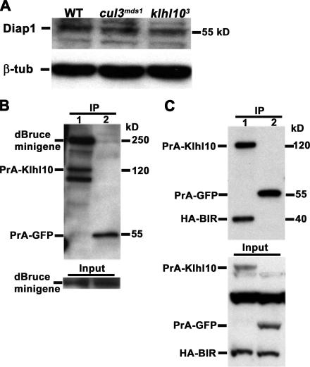
In both insects and mammals, spermatids eliminate their bulk cytoplasm as they undergo terminal differentiation. In Drosophila, this process of dramatic cellular remodeling requires apoptotic proteins, including caspases. To gain further insight into the regulation of caspases, we screened a large collection of sterile male flies for mutants that block effector caspase activation at the onset of spermatid individualization. Here, we describe the identification and characterization of a testis-specific, Cullin-3-dependent ubiquitin ligase complex that is required for caspase activation in spermatids. Mutations in either a testis-specific isoform of Cullin-3 (Cul3(Testis)), the small RING protein Roc1b, or a Drosophila orthologue of the mammalian BTB-Kelch protein Klhl10 all reduce or eliminate effector caspase activation in spermatids. Importantly, all three genes encode proteins that can physically interact to form a ubiquitin ligase complex. Roc1b binds to the catalytic core of Cullin-3, and Klhl10 binds specifically to a unique testis-specific N-terminal Cullin-3 (TeNC) domain of Cul3(Testis) that is required for activation of effector caspase in spermatids. Finally, the BIR domain region of the giant inhibitor of apoptosis-like protein dBruce is sufficient to bind to Klhl10, which is consistent with the idea that dBruce is a substrate for the Cullin-3-based E3-ligase complex. These findings reveal a novel role of Cullin-based ubiquitin ligases in caspase regulation.


Conflict of interest statement

Competing interests. The authors have declared that no competing interests exist.

Figures

Figure 1
Figure 1. Mutations in cul3Testis Block Caspase Activation and Spermatid Individualization, but Not Axonemal Tubulin Polyglycylation
(A–H) Visualization of active drICE with anti-cleaved caspase-3 antibody (CM1; green) and axonemal tubulin polyglycylation with anti-glycylated tubulin monoclonal antibody (AXO 49; red). These figures are composed of a green layer only in the left panel, and green and red layers combined in the right panel. (A) Wild-type individualizing spermatids stain positively for active effector caspase and polyglycylated axonemal tubulin (white arrows pointing at cystic bulges [CBs] and red arrow pointing at a waste bag [WB]). Elongated spermatids from (B) homozygotes for the null cul3mds1 allele or (C and D) transheterozygotes for cul3mds1 and two different deficiencies that cover the cullin-3 gene, DF(2L)ED3 and DF(2L)Exel8034, respectively, stain for polyglycylation but not for active effector caspase. (E–G) Homozygote mutants for three hypomorphic cul3Testis alleles, cul3mds5, cul3mds3, and cul3mds4, respectively, have spermatid individualization defects but still display some levels of active effector caspase expression. (H) However, the level of active effector caspase expression was dramatically reduced in spermatids from transheterozygote mutants for the null cul3mds1 and either of the hypomorphic alleles, such as cul3mds4. All the figures are in the same magnification; scale bar 200 μm. (I) The diagram depicts a DEVDase activity assay for cul3mds1 −/− testes. Caspase-3–like (DEVDase) activity is detected in wild-type testes and is blocked either after treatment with the caspase-3 inhibitor Z-VAD.fmk or in cul3mds1 −/− testes. DEVDase activity, presented as relative luminescence units (RLUs), was determined on Ac-DEVD-pNA substrate in testis extracts made of 180 wild-type (yw) or cul3mds1 −/− testes treated with Z-VAD or left untreated (DMSO). Readings were obtained every 2 min, and each time interval represents an average (mean ± SEM) of five readings. Note that the level of DEVDase activity in cul3mds1 −/− testes is highly similar to the corresponding level in wild-type testes that were treated with Z-VAD. (J) A Western blot analysis for the assessment of the relative protein amounts used in (I). A portion of the testis extracts in (I) were used as controls to determine the relative amounts of total protein in each extract using the anti-β-Tubulin antibody.
Figure 2
Figure 2. The cul3mds1–5 Alleles Contain Mutations in a New Exon of the cullin-3 Gene
(A) Genomic organization of the cullin-3 locus. Thick bars indicate exons and dotted lines indicate introns. Solid bars indicate coding sequences, whereas open bars indicate UTRs. The Drosophila cullin-3 gene contains 14 exons, nine of which encode the bulk of the protein (exons 3–11) and are shared by both the somatic and testis-specific isoforms. While the three somatic isoforms (cul3Soma) differ in their 5′ UTRs, each beginning with a unique first exon (exons 1A, 1B, and 1C), they share a second, somatic-only exon that contains a start codon (exon 2, green bar). The testis isoform, cul3Testis, begins with a unique first exon (1D, blue bar) that includes both 5' UTR and coding sequences, including a start codon. The relative locations of the molecular lesions in cul3Testis (orange, cul3mds1–5 alleles) and cul3Soma (purple, cul3gft2, 4, GR18 alleles) are shown with stars (see the main text for more details on the precise molecular lesions of the cul3mds1–5 alleles). The molecular alterations of the cul3gft alleles were reported in [71]: cul3gft06430 contains a PZ element insertion 228 nucleotides from exon 2 (the insertion is indicated by a purple triangle). cul3gft[GR18] is missing a single nucleotide causing a premature stop codon at amino acid 167. cul3gft4 bears a C-to-T transversion, which results in an A710-to-V conversion. cul3gft2 contains a five-nucleotide deletion that results in a premature stop codon at amino acid 748 that removes half of the C-terminal Cullin homology domain (CHD). (B) A scheme of the two major mRNA isoforms of cullin-3, cul3Testis, and cul3Soma. (C) Genomic PCR and sequencing analyses of the cullin-3 locus revealed a 181-bp deletion in cul3mds1 (the arrows in A depict the relative locations of the primers used in this gPCR; yw and Canton S strains were used as wild-type controls). (D) For positive control, wild-type spermatids were stained for cleaved caspase-3 expression (green). (E) Consistent with the idea that both the mds and gft alleles affect the same gene, cullin-3, cul3mds1/cul3gft2 transheterozygote mutant spermatids displayed defects in individualization and negatively stained for cleaved caspase-3 (left panel). Spermatids were counter-stained with phalloidin that binds to F-actin in the spermatids' tail (right panel, red; the strong red staining at the bottom corresponds to remnants of the testis sheath). (F–I) Spermatids were stained for cleaved caspase-3 (green in left panels) and for axonemal tubulin polyglycylation (red in right panels). (F) Wild-type control testis positively stained for cleaved caspase-3 and axonemal tubulin polyglycylation. (G–I) Whereas transheterozygous combinations between cul3mds1 and the strong cul3gft[GR18] (G) or cul3gft1 (H) alleles displayed spermatid individualization defects and stained negatively for cleaved caspase-3 and positively for polyglycylation, mutant spermatids from cul3mds1 in trans to the hypomorphic cul3gft4 allele also exhibited individualization defects but displayed reduced level of cleaved caspase-3 expression (I). All the figures were taken at the same magnification; scale bars, 200 μm.
Figure 3
Figure 3. The Expression of cul3Testis Is Restricted to Male Germ Cells
(A) Schematic structures of the Drosophila cullin-3 gene (I) and of cul3Testis (II) and cul3Soma (III) mRNAs. Exons and introns are indicated by thick and thin bars, respectively. Thick black bars indicate coding sequences, whereas open bars indicate UTRs. The locations of the primers used in the comparative RT–PCR experiments in (B and C) are indicated by arrows, and the expected length sizes of the amplified fragments are indicated above each scheme. (B) Analysis of cul3Testis versus cul3Soma expression in the testis and the soma. The above primers (arrows in A) to amplify either a 2,997-bp cul3Testis or a 2,642-bp cul3Soma cDNA fragment were added to one reaction master-mix. The reaction was stopped at different cycle points to identify the linear amplification phase (30 and 35 cycles are indicated). The “RT” columns represent reverse transcriptase followed by PCR reactions, and the “Taq” are the control, PCR-only, reactions. Note that the cul3Testis expression levels were much higher in the wild-type (WT) testes than these of cul3Soma. On the other hand, only cul3Soma transcripts were detected in somatic tissues, which are represented by adult female flies. In addition, no cul3Testis transcripts were detected in cul3mds1 mutant testes, confirming that this is a null cul3Testis allele. (C) The expression of cul3Testis is restricted to the male germ cells. While the expression of cul3Soma was not affected in sons of oskar agametic testes, no cul3Testis expression was detected. (D) Consistent with the RT-PCR analysis, no Cul3Testis protein was detected in cul3mds1 mutant testes on Western blot. Note, however, that expression of the Cul3Soma protein in cul3mds1 mutant testes was unaffected.
Figure 4
Figure 4. cul3Testis but Not cul3Soma Can Restore Caspase Activation and Spermatid Individualization to cul3Testis Null Mutants
(A) Schematic structure of the rescue constructs for cul3Testis −/− male sterile flies. The constructs tr-cul3Testis and tr-cul3Soma are composed of the cul3Testis isoform's promoter region (dark blue, consists of the intronic sequences flanked by exons 2 and 1D) and 5′ UTR (light blue) that were fused upstream of the coding regions (ORFs) of either cul3Testis or cul3Soma followed by the 3′ UTR of cul3Testis. (B and C) Transcriptional expression from the transgenes was confirmed by RT–PCR analyses on RNA from testes of the indicated genotypes. The relative locations of the primers are indicated with black arrows in (A). “RT+Taq” and “Taq” indicate reactions with reverse transcriptase or without it, respectively, to control for possible genomic DNA contamination. (B) To differentiate between the cul3Testis endogenous (endog.) and transgenic (transg.) cDNAs, we cleaved the RT-PCR fragments with XhoI, a unique restriction site in the transgene. Note that the RT-PCR product from cul3mds1; tr-cul3Testis but not from WT testes was cleaved by XhoI, confirming its transgenic source. (C) Transgenic expression of cul3Soma (tr-cul3Soma) in adult testis. Note the absence of the endogenous cul3Testis cDNA band and in contrast, the presence of the transgenic cul3Soma band in cul3mds1; tr-cul3Soma/+ testes. (D–I) Testes stained for cleaved caspase-3 (CM1, green) and spermatid's tail and ICs (phalloidin, red). (D) Mutant spermatids for cul3Testis (cul3mds1 −/−) manifest a block in caspase activation and spermatid individualization. (E) Either one or (F) two copies of transgenic cul3Testis (tr-cul3Testis) restores caspase activation, spermatid individualization, and fertility of cul3mds1 −/− male flies. (G) Wild-type control testes. Note the CBs and WBs (green oval structures). (H–I) In contrast, dramatically reduced CM1-positive cysts are found in cul3mds1 mutants, which ectopically express (H) one or (I) two copies of the cul3Soma transgene (tr-cul3Soma). These spermatids failed to individualize, no CBs and WBs are detected and the males are sterile. Scale bars 200 μm.
Figure 5
Figure 5. Double Mutants for cul3Testis and roc1b Block Caspase Activation During Spermatid Differentiation
Testes stained for cleaved caspase-3 (CM1, green) and spermatid's tail (phalloidin, red). (A) Spermatids in roc1b mutant flies (roc1bdc3 −/−) display severe individualization defects and still display some levels of CM1 staining. (B) Similarly, spermatids in flies homozygous for weak cul3Testis alleles, such as cul3mds3, also display some level of CM1 staining. (C) However, spermatids mutants for both roc1b and cul3Testis manifest a complete block in caspase activation during individualization.
Figure 6
Figure 6. Klhl10, a BTB and Kelch Domains Protein, Preferentially Interacts with Cul3Testis and Not with Cul3Soma
(A) Three BTB-domain proteins, the Drosophila orthologues of Spop, Ipp, and Klhl10, were found to interact with Cul3Testis in a yeast-two-hybrid screen. β-galactosidase filter assay demonstrates that whereas Spop and Ipp can also interact with Cul3Soma, Klhl10 only interacts with Cul3Testis. While the TeNC domain of Cul3Testis is required for this interaction, it is not sufficient to mediate the interaction with Klhl10. (B) Similarly, in a nutrient-omitted medium assay, yeasts with both Cul3Testis and Klhl10 grew rapidly (2 d) on plates that lacked, in addition to leucine and tryptophan (−2), also histidine and adenine (−4 plates). However, yeast with Klhl10 and Cul3Soma grew very poorly on −4 plates. Even after two weeks of incubation, the colony is only partially established. Note that the results for the auxotrophy rescue of Klhl10 are shown not after 2 d but rather after 14 d in order to reflect the weak interaction between Klhl10 and Cul3Soma. (C) Schematic representations of Spop, Ipp, and Klhl10, and the relative locations of their major domains. The BTB-domains of all these proteins are sufficient for binding to Cullin-3.
Figure 7
Figure 7. Mutations in klhl10 Block Caspase Activation and Spermatid Individualization, but Not Axonemal Tubulin Polyglycylation
(A–H) Visualization of active effector caspase with anti-cleaved caspase-3 antibody (CM1; green) and (A–F) axonemal tubulin polyglycylation with anti-glycylated tubulin monoclonal antibody (AXO 49; red) or (G–H) F-actin, which stains the ICs and the spermatids' tails (phalloidin; red). These figures are composed of combined green and red layers. (A) Wild-type individualizing spermatids positively stain for cleaved caspase-3 and polyglycylated axonemal tubulin. (B–F) In a variety of klhl10 −/− alleles, elongated spermatids stain for polyglycylation but not for cleaved caspase-3. (G and H) Transgenic klhl10 construct (tr-klhl10, composed of cul3Testis promoter and 5′ UTR, klhl10 coding region, and cul3Testis 3′ UTR) restores caspase activation, spermatid individualization, and fertility to klhl10 −/− male flies. (I) Schematic representation of the Klhl10 protein. The relative locations of the BTB, BACK, and Kelch domains are depicted by thick bars. Different colored stars depict the locations of the different mutations, and the colors correspond to the colored amino-acids in (J). The molecular nature of the mutations and their color code are as follows: klhl102 (Z2–1331) carries a G1801-to-A transversion that converts glutamic acid (E601, red) to lysine at repeat VI. klhl103 (Z2–0960) carries a G1119-to-A transversion that converts tryptophan (W373, purple) to stop codon, resulting in a deletion of most of the Kelch domain. klhl104 (Z2–2739) carries a C1237-to-T transversion that converts arginine (R413, yellow) to stop codon, which also deletes most of the Kelch repeats. klhl105 (Z2–3284) carries a G1486-to-A transversion that converts glycine (G496, blue) to arginine at repeat IV. klhl106 (Z2–4385) carries a C1439-to-T transversion that converts serine (S480, green) to phenylalanine at repeat IV. On the other hand, klhl107 (Z2–3353) carries a G508-to-A transversion that converts a highly conserved alanine (A170) to threonine in the BTB domain (gray star). (J) Alignment of the six Kelch repeats of Klhl10. The alignment is based on the crystal structure of the Keap1 Kelch domain which folds into a β-propeller structure with 6 blades. The residue range for each blade is indicated at the left. The four conserved β-strands in each blade are indicated above the sequences by arrows. Residues conserved in all six blades are highlighted with dark gray and appear in upper case in the consensus line, whereas residues that are conserved in at least three blades appear in lower case. Any two and above conserved residues are highlighted with light gray. Color highlighted residues are mutated in the various klhl10−/− alleles and correspond to the stars in (I).
Figure 8
Figure 8. The Cul3-Roc1b-Klhl10 Complex Promotes Protein Ubiquitination during Spermatid Individualization
Testes were stained with the anti–multi-ubiquitin monoclonal antibody (FK2) that detects ubiquitinated proteins (green), phalloidin, which marks the individualization complex (IC, red), and DAPI to visualize the nuclei (blue). (A) Before the formation of an IC, very low levels of ubiquitinated proteins are detected (yellow arrowhead). Once a mature IC is assembled in the vicinity of the nuclei, a steep gradient of ubiquitinated protein staining is detected from the very bottom of the nuclei to the tip of the spermatids' tails (yellow arrows). After the caudal translocation of the IC, ubiquitinated proteins are no longer detectable in the post-individualized portion of the spermatids (the region between the nuclei, indicated by a white arrowhead, and an early CB, indicated by a white arrow). (B and C) When the bulk cytoplasm of the spermatids accumulates in a cystic bulge (CB), ubiquitinated proteins are prominent within the CB and the pre-individualized region (white asterisks). (D) Once all the cytoplasm is stripped away, ubiquitinated proteins are detectable only in the waste bag (WB). (E and F) Elongated spermatids from either cul3mds1 or klhl104 mutants, respectively, did not stain for ubiquitinated proteins. (G) Protein ubiquitination is detected in nuclei of early elongating spermatids. (H and I) The pattern of protein ubiquitination at earlier stages of spermatid maturation is not affected in cul3mds1 and klhl104 mutants. Scale bars, 100 μm.
Figure 9
Figure 9. Working Model for a Cullin-3–Based Ubiquitin Ligase Complex in the Testis
The diagram represents the assembly of the testis-specific Cullin-3–containing ubiquitin ligase (E3) and its proposed function in caspase activation during spermatid individualization. (A) The model suggests that once an active Cul3Testis-Roc1b-Klhl10 E3 ubiquitin ligase complex is assembled, it recruits a caspase inhibitor “CI” protein (red) via the Kelch domain of the substrate recruitment protein Klhl10 (yellow). A candidate for this caspase inhibitor is dBruce, a giant BIR-domain-containing ubiquitin-conjugating enzyme [12,35,36,103,104]. dBruce can physically interact via its BIR domain with Klhl10 (see Figure 10), and loss of dbruce function leads to spermatid death [12]. Therefore, dBruce has biochemical and genetic properties expected for the postulated “CI” protein. (B) Next, the ubiquitin-conjugating enzyme (E2, blue), which is recruited by the RING finger protein Roc1b (purple), ubiquitinates the “CI” protein. (C) Subsequent degradation of this inhibitor allows the activation of caspases at the onset of spermatid individualization process.
Figure 10
Figure 10. Diap1 Levels Are Not Affected in the Absence of the Functional Cul3-Roc1b-Klhl10 Complex, but dBruce Can Interact with the Substrate Recruitment Protein Klhl10 in S2 Cells
(A) Diap1 protein levels were not affected in cul3mds1 and klhl103 mutant testes, as assessed by Western blotting of protein extracts from dissected testes. Therefore, Diap1 does not appear to be a major target for the Cul3-based E3-ligase complex. β-tubulin protein levels served as loading control. (B andC) Co-IP experiment in S2 cells indicate that Klhl10 can bind to the BIR domain of dBruce. The immunoprecipitate (IP) is shown at the top, and pre-incubation of whole lysates are shown at the bottom (Input). Cell lysates were incubated with IgG beads which bind to Protein A (PrA). For Western blotting of IPs, (B) anti-dBruce antibody or (C) anti-HA antibody were used. (B) Cells were co-transfected with a dbruce “mini gene” (consisting of the first N-terminal 1,622 amino acids, including the BIR domain, and the last C-terminal 446 amino acids that contain the UBC domain) and (lane 1) PrA-klhl10 or (lane 2) PrA-GFP (see Materials and Methods for details). (C) Cells were co-transfected with HA-tagged dBruce-BIR peptide containing the first N-terminal 387 amino acids of dBruce that includes the BIR domain region (amino acids 251–321). This motif is sufficient to bind to Klhl10 in S2 cells (see Materials and Methods for details).

Comment in

Similar articles

Cited by

References

    1. Abraham MC, Shaham S. Death without caspases, caspases without death. Trends Cell Biol. 2004;14:184–193. - PubMed
    1. Degterev A, Boyce M, Yuan J. A decade of caspases. Oncogene. 2003;22:8543–8567. - PubMed
    1. Lamkanfi M, Festjens N, Declercq W, Berghe TV, Vandenabeele P. Caspases in cell survival, proliferation and differentiation. Cell Death Differ. 2007;14:44–55. - PubMed
    1. Budihardjo I, Oliver H, Lutter M, Luo X, Wang X. Biochemical pathways of caspase activation during apoptosis. Annu Rev Cell Dev Biol. 1999;15:269–290. - PubMed
    1. Salvesen GS, Dixit VM. Caspase activation: The induced-proximity model. Proc Natl Acad Sci U S A. 1999;96:10964–10967. - PMC - PubMed

Publication types

MeSH terms