. 2007 Sep 19:4:23.
doi: 10.1186/1742-2094-4-23.

Sinomenine, a natural dextrorotatory morphinan analog, is anti-inflammatory and neuroprotective through inhibition of microglial NADPH oxidase

Affiliations

Sinomenine, a natural dextrorotatory morphinan analog, is anti-inflammatory and neuroprotective through inhibition of microglial NADPH oxidase

Li Qian et al. J Neuroinflammation. .

Abstract

Background: The mechanisms involved in the induction and regulation of inflammation resulting in dopaminergic (DA) neurotoxicity in Parkinson's disease (PD) are complex and incompletely understood. Microglia-mediated inflammation has recently been implicated as a critical mechanism responsible for progressive neurodegeneration.

Methods: Mesencephalic neuron-glia cultures and reconstituted cultures were used to investigate the molecular mechanisms of sinomenine (SN)-mediated anti-inflammatory and neuroprotective effects in both the lipopolysaccharide (LPS)- and the 1-methyl-4-phenylpyridinium (MPP+)-mediated models of PD.

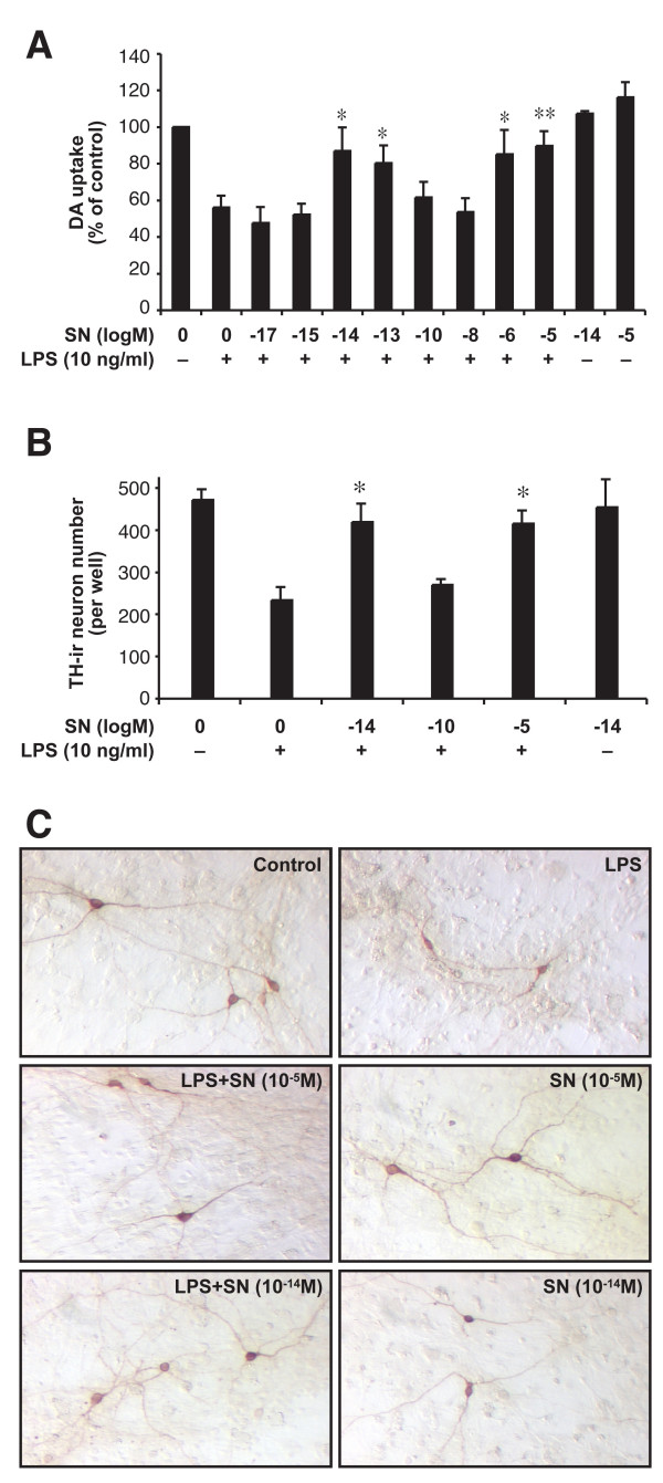
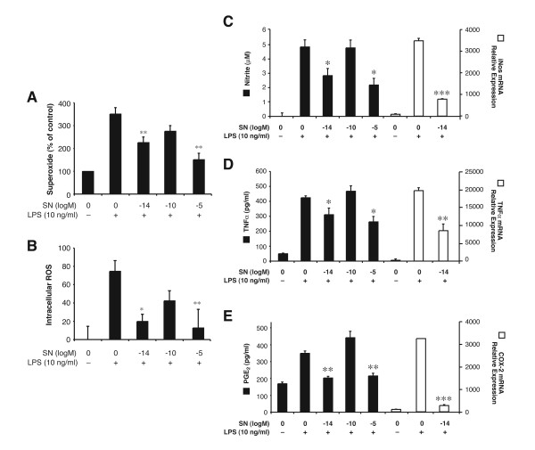
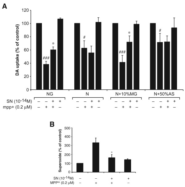
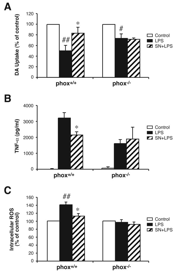
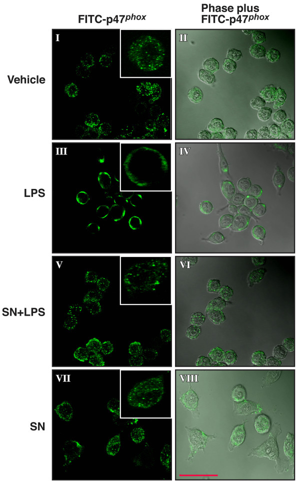
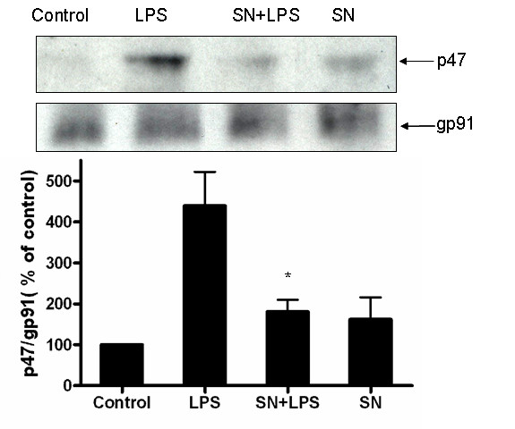
Results: SN showed equivalent efficacy in protecting against DA neuron death in rat midbrain neuron-glial cultures at both micro- and sub-picomolar concentrations, but no protection was seen at nanomolar concentrations. The neuroprotective effect of SN was attributed to inhibition of microglial activation, since SN significantly decreased tumor necrosis factor-alpha (TNF-alpha, prostaglandin E2 (PGE2) and reactive oxygen species (ROS) production by microglia. In addition, from the therapeutic point of view, we focused on sub-picomolar concentration of SN for further mechanistic studies. We found that 10(-14) M of SN failed to protect DA neurons against MPP+-induced toxicity in the absence of microglia. More importantly, SN failed to show a protective effect in neuron-glia cultures from mice lacking functional NADPH oxidase (PHOX), a key enzyme for extracellular superoxide production in immune cells. Furthermore, we demonstrated that SN reduced LPS-induced extracellular ROS production through the inhibition of the PHOX cytosolic subunit p47phoxtranslocation to the cell membrane.

Conclusion: Our findings strongly suggest that the protective effects of SN are most likely mediated through the inhibition of microglial PHOX activity. These findings suggest a novel therapy to treat inflammation-mediated neurodegenerative diseases.


Figures

Figure 1
Figure 1
Both micromolar and sub-picomolar concentrations of SN are neuroprotective against LPS-induced neurotoxicity. Rat primary mesencephalic neuron-glia cultures seeded in a 24-well culture plate with 5 × 10⁵ rat midbrain cells, then pretreated with SN (10-17 to 10-5 M) for 30 min before the addition of 10 ng/ml LPS. Eight days later, the LPS-induced dopaminergic neurotoxicity was quantified by the [3H]-DA uptake assay (A); by immunocytochemical analysis, including TH-IR neuron counts (B); and by representative pictures of immunostained sections (C). Results are expressed as percentage of vehicle-treated control cultures and represent the mean ± SE. for four (A) or three (B, C) independent experiments in triplicate. The mean absolute values of [3H]-DA uptake for vehicle-treated cultures range from 5000 to 7000 cpm. *P < 0.05, **P < 0.01 compared with the LPS-treated cultures.
Figure 2
Figure 2
Effect of SN on LPS-induced production of pro-inflammatory factors and their mRNA expression. Enriched microglia cells were pretreated with vehicle or SN (10-5, 10-10, and 10-14 M) for 30 min before LPS (10 ng/ml) stimulation. Effects of SN are shown on LPS-induced production of superoxide (A, expressed as % of control); and on intracellular ROS (B, expressed as absolute absorbance). Extracellular superoxide was measured as SOD-inhibitable reduction of WST-1, and intracellular ROS was determined by probe DCFH-DA. Supernatant was collected at 24 h for nitrite assay (C), at 3 h for TNF-α assay (D), and at 24 h for PGE2 assay (E). RNA were extracted at 3 h after LPS stimulation; the effect of SN, at sub-picomolar concentrations, on LPS-induced iNOS, TNF-α, and COX-2 mRNA expression (C-E respectively, open bars) are shown. Results are expressed as mean ± SE for three independent experiments performed in triplicate. *P < 0.05, **P < 0.01 compared with the LPS-treated cultures.
Figure 3
Figure 3
SN protects against MPP+-elicited DA neurodegeneration through microglia. SN (10-14 M) or MPP+ (0.2 μM) were added to the following types of cell cultures: (NG): original neuron-glial cultures; (N): neuron-enriched cultures; (N+10%MG): cultures reconstituted by adding 10% of microglia to the neuron-enriched cultures; (N+50%AS): cultures reconstituted by adding 50% of astroglia to the neuron-enriched cultures. Two and 4 days after MPP+ treatment, SN (10-14 M) was added again to the SN-treated cultures. On day 8, the MPP+-induced dopaminergic neurotoxicity was quantified by the [3H]-DA uptake assay (A), and on day 4, the release of superoxide was determined as described in Materials and methods section. Results are expressed as percentage of the vehicle-treated control cultures and represent the mean ± SE. for three independent experiments performed in triplicate. *P < 0.05, compared with MPP+ treated cultures. # P < 0.05, ## P < 0.01 compared with the vehicle-treated control cultures.
Figure 4
Figure 4
Microglial PHOX is critical for sub-picomolar SN neuroprotection. PHOX+/+ and PHOX-/- mouse neuron-glia cultures were pretreated with vehicle or SN (10-14 M) for 30 min, followed by LPS treatment. Neurotoxicity was assessed by measuring DA uptake (A), TNF-α production (B) and intracelluar ROS (C), respectively. Results are expressed as % of the control culture (A and C) and as pg/ml (B), and represent the mean ± SE for 3 individual experiments performed in triplicate in each experiment. *P < 0.05 compared with LPS culture. # P < 0.05, ## P < 0.01 compared with the vehicle-treated control cultures.
Figure 5
Figure 5
Immunofluorescence and confocal microscopical analysis of p47phox localization in LPS-stimulated microlgia cells. HAPI cells were treated with LPS for 10 min in the absence or presence of SN pretreatment for 0.5 h. Cells were immunostained with a rabbit polyclonal antibody against p47phox, then washed and incubated with FITC-conjugated goat anti-rabbit antibody. The signal of p47phox (FITC-p47phox; on left) and the merge view of cell morphology and p47phox (Phase plus FITC-p47phox, on right) are shown. The inset shown in the right corner of each treatment condition shows the location of FITC-p47phox in a single, randomly selected cell. Focal planes spaced at 0.4-μm intervals were imaged with a Zeiss 510 laser scanning confocal microscope (63 × PlanApo 1.4 numerical aperture objective) equipped with LSM510 digital imaging software. Three adjacent focal planes were averaged using Metamorph software. The signal of p47phox (FITC-p47phox; green) and the merge view of cell morphology and p47phox (Phase plus FITC-p47phox) are shown. Scale bar, 50 μm.
Figure 6
Figure 6
Membrane fraction analysis of the effect of SN on cytosolic p47phoxprotein translocation. HAPI cells were pretreated with vehicle or SN (10-14 M) for 30 min, followed by LPS treatment for 10 min. Membrane fractions were isolated to perform western blot analysis, using gp91phox as an internal membrane control. Each experiment was performed three times. *P < 0.05, compared with the LPS-treated cultures.

References

    1. McGeer PL SI, Boyes BE, McGeer EG. Reactive microglia are positive for HLA-DR in the substantia nigra of Parkinson's and Alzheimer's disease brains. Neurology. 1988;38:1285–1291. - PubMed
    1. Liu B, Hong JS. Role of microglia in inflammation-mediated neurodegenerative diseases: mechanisms and strategies for therapeutic intervention. J Pharmacol Exp Ther. 2003;304:1–7. doi: 10.1124/jpet.102.035048. - DOI - PubMed
    1. Rosi S, Ramirez-Amaya V, Vazdarjanova A, Worley PF, Barnes CA, Wenk GL. Neuroinflammation alters the hippocampal pattern of behaviorally induced Arc expression. J Neurosci. 2005;25:723–731. doi: 10.1523/JNEUROSCI.4469-04.2005. - DOI - PMC - PubMed
    1. Chen LC, Smith A, Ben Y, Zukic B, Ignacio S, Moore D, Lee N. Temporal gene expression patterns in G93A/SOD1 mouse. Amyotroph Lateral Scler Other Motor Neuron Disord. 2004;5:164–171. doi: 10.1080/14660820410017091. - DOI - PubMed
    1. Kreutzberg GW. Microglia: a sensor for pathological events in the CNS. Trends Neurosci. 1996;19:312–318. doi: 10.1016/0166-2236(96)10049-7. - DOI - PubMed

Publication types

MeSH terms