. 2007 Sep 20;55(6):970-84.
doi: 10.1016/j.neuron.2007.08.004.

Expectation modulates neural responses to pleasant and aversive stimuli in primate amygdala

Affiliations

Expectation modulates neural responses to pleasant and aversive stimuli in primate amygdala

Marina A Belova et al. Neuron. .

Abstract

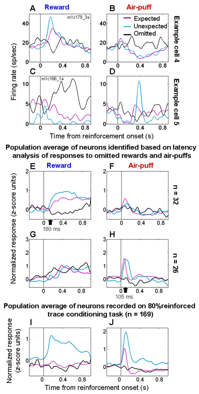
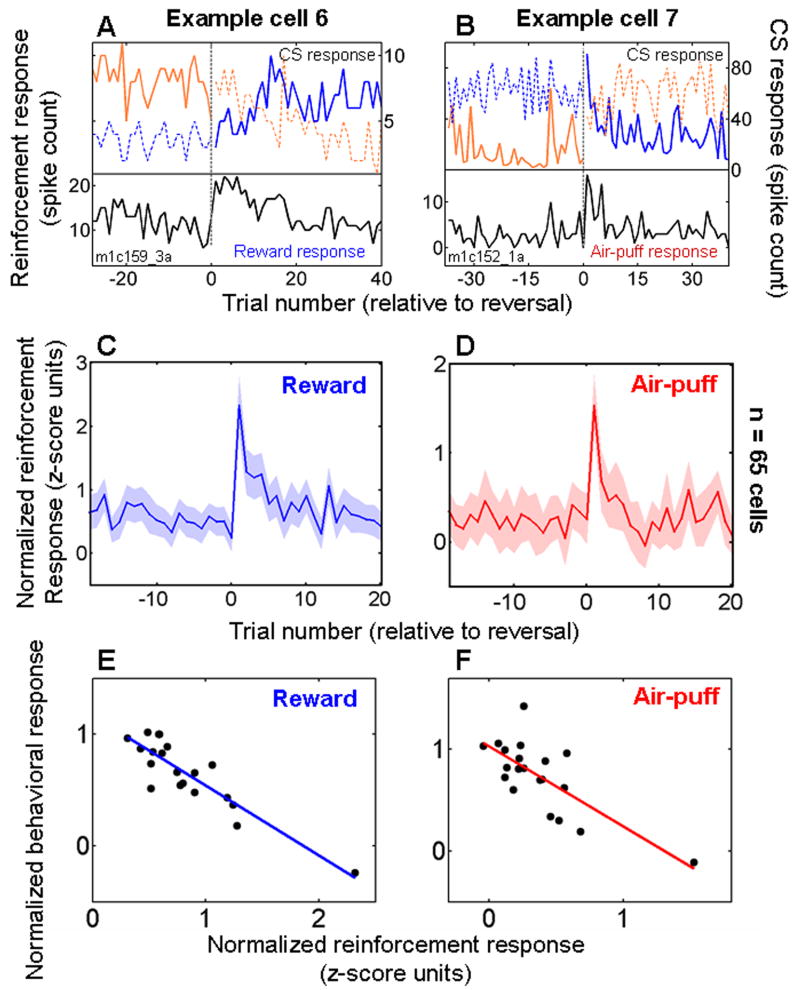
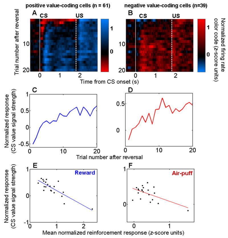
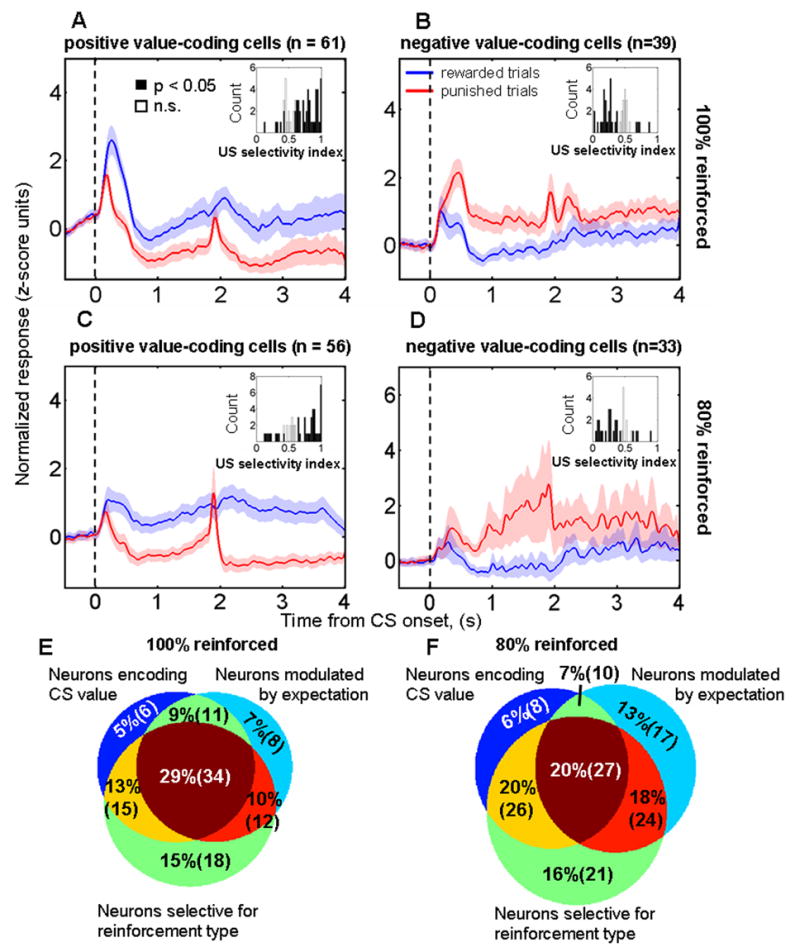
Animals and humans learn to approach and acquire pleasant stimuli and to avoid or defend against aversive ones. However, both pleasant and aversive stimuli can elicit arousal and attention, and their salience or intensity increases when they occur by surprise. Thus, adaptive behavior may require that neural circuits compute both stimulus valence--or value--and intensity. To explore how these computations may be implemented, we examined neural responses in the primate amygdala to unexpected reinforcement during learning. Many amygdala neurons responded differently to reinforcement depending upon whether or not it was expected. In some neurons, this modulation occurred only for rewards or aversive stimuli, but not both. In other neurons, expectation similarly modulated responses to both rewards and punishments. These different neuronal populations may subserve two sorts of processes mediated by the amygdala: those activated by surprising reinforcements of both valences-such as enhanced arousal and attention-and those that are valence-specific, such as fear or reward-seeking behavior.


Figures

Figure 1
Figure 1. Behavioral tasks and recording site reconstruction
A. Trace-conditioning task. Sequence of events for trials. For CSs followed by large rewards or punishments, reinforcement contingencies reverse without warning after initial learning. Not depicted: a third trial type, in which non-reinforcement or a small reward followed a CS. B. Random task. Rewards and air-puffs were presented with equal probability in a random order. C. Anatomical reconstruction of recording sites in monkey L, with amygdala extent and site locations estimated by MRI. Left, coronal slice. Right, sagittal slice. Symbols indicate properties of recorded cells. Green, rEM cells; red, aEM cells; black, nEM cells; blue, no effect of expectation; solid and open circles, 100% and 80% probability trace-conditioning tasks, respectively.
Figure 2
Figure 2. Expectation modulates neural responses to reinforcement in the amygdala
A,B. Peri-Stimulus Time Histograms (PSTHs) (which average neural responses as a function of time across trials) from one amygdala cell exhibiting a stronger response to reward (A) but not air-puff (B) when reinforcement was unexpected (random task, blue) compared to expected (trace-conditioning task, magenta). C,D. PSTHs from a neuron with stronger responses to unexpected air-puff (D) but not to unexpected reward (C). E,F. PSTH from a cell with stronger responses to both valences of unexpected reinforcement. All PSTHs smoothed with a 10 ms moving average.
Figure 3
Figure 3. Valence specific and valence non-specific modulation of reinforcement responses by expectation
A–F. Normalized and averaged population PSTHs showing responses to expected (magenta, trace-conditioning task) and unexpected (blue, random task) rewards (A,C,E) and punishments (B,D,F). A,B, rEM neurons (n = 47). C,D, aEM neurons (n = 35). E,F. nEM neurons (n = 65). Shading, s.e.m. G–I. Difference in normalized activity for unexpected compared to expected rewards and air-puffs, shown separately for data from the 100% and 80% reinforcement probability trace-conditioning task. rEM cells (G), aEM cells (H), nEM cells (I). The difference in air-puff response seen in (H) does not achieve statistical significance (p = 0.14, t-test).
Figure 4
Figure 4. Expectation modulates responses to reinforcement within the trace-conditioning task
A,B. Normalized and averaged PSTHs showing responses to rewards (A) and air-puffs (B) for the rEM, aEM, and nEM cells studied with the 100% reinforced trace-conditioning task. Trials are sorted according to whether monkeys expected air-puff (anticipatory blinking but not licking, red curves) or reward (anticipatory licking but not blinking, blue curves). On average, the responses to reinforcement were greater when monkeys incorrectly predicted the upcoming reinforcement. Red asterisks, activity significantly different in the two types trials in a 100 ms bin, p < 0.05, t-test.
Figure 5
Figure 5. Neural responses to omitted reinforcement
A,B. PSTHs from an amygdala cell showing increased firing to unexpected rewards (A) and omitted air-puffs (B), and decreased firing to air-puffs (B) and omitted rewards (A). C,D. PSTH from an amygdala cell showing increased firing to unexpected air-puffs (D) and omitted rewards (C), and decreased firing to rewards (C) and omitted air-puffs (D). E,F. Normalized and averaged population PSTHs for neurons showing evidence of decreased firing to omitted rewards and/or increased firing to omitted air-puffs (n = 32, Table 1). G,H. Normalized and averaged population PSTHs for neurons showing evidence of decreased firing to omitted air-puffs and/or increased firing to omitted rewards (n = 26, Table 1). I,J. Normalized and averaged PSTHs showing responses to rewards (I) and air-puffs (J) from all cells recorded during both the trace-conditioning task with 80% reinforcement probability and the random task (n = 169). For all plots: magenta, black, and blue curves, responses to expected, omitted, and random reinforcement, respectively. Black arrows in E and H, mean latency of decreased responses to omitted rewards and air-puffs. PSTHs smoothed with a 50 ms Gaussian.
Figure 6
Figure 6. Responses to reinforcement decrease during learning
A. Data from one experiment in which a neuron responded more strongly to reward immediately after CS-US contingency reversal, and it rapidly changed its firing rate to the two CSs as reinforcement responses decreased. Black curve, response to reward. Orange and blue curves, visual stimulus interval responses to each of the two images, respectively. Solid and dashed lines, CS followed by reward or air-puff, respectively. B. Data from another neuron that responded more strongly to air-puff after image value reversal. Orange and blue curves, trace-interval responses to the two images, respectively. Labeling conventions the same as in A. C,D. Normalized and averaged neural responses to rewards (C) and air-puffs (D) plotted as a function of trial number relative to reversal for neurons recorded during the 100% reinforced trace-conditioning task. C, rEM and nEM cells combined. D, aEM and nEM cells combined. Shaded regions, s.e.m. E,F. Correlation between mean responses to reward (E) and air-puff (F) on the 20 trials after reversal, taken from the data points in panels C,D, and normalized and averaged behavioral performance on the same trials (see Methods). In all panels, trial 1 is the first trial after reversal in image value.
Figure 7
Figure 7. The decrease in responses to reinforcement is correlated with the evolving representation of value in the amygdala
A,B. Color maps showing the representation of value during reversal learning as a function of trial number and of time within trials. Positive (A) and negative (B) CS value-coding cells from the 100% reinforcement probability trace-conditioning task shown separately. Vertical white lines, CS onset (solid line) and US onset (dashed line). Each 200 ms bin shows the difference in firing rate between rewarded and punished trials; bins were advanced in 50 ms steps. Trial 1, first trial after reversal. Bin starting at time 0, interval from 0–200 ms after CS onset. C,D. CS value-coding extracted from the color map by taking the mean value in the interval between the white lines for neurons encoding positive (C) and negative (D) CS value. E,F. Recorded reinforcement responses (data points from Figs. 6C,D) plotted against the evolution of CS value coding (data points from Figs. 7C,D) during trials 1–20 after reversal. Regression lines are shown in blue and red.
Figure 8
Figure 8. Amygdala neurons carry multiple signals
A–D. Normalized and averaged population PSTHs from neurons encoding positive (A,C) and negative (B,D) CS value during the trace-conditioning task with 100% (A,B) and 80% (C,D) reinforcement probability, respectively. Data aligned at CS onset; reinforcement occurred at either 1.8 or 1.85 secs depending upon the experiment. Inset histograms, ROC reinforcement selectivity indices for each of the positive (A,C) and negative (B,D) value coding neurons. Neurons encoding positive CS value tend to have reinforcement selectivity indices > 0.5, and neurons encoding negative CS value tend to have reinforcement selectivity indices < 0.5. E,F. Venn diagram showing the number of neurons classified as encoding CS value, as having reinforcement selective responses, and as having significantly stronger responses to unexpected compared to expected rewards and/or punishments for the trace-conditioning task with 100% (E) and 80% (F) reinforcement probabilities, respectively.

References

    1. Amaral D, Price J, Pitkanen A, Carmichael S. Anatomical Organization of the Primate Amygdaloid Complex. In: Aggleton J, editor. The Amygdala: Neurobiological Aspects of Emotion, Memory, and Mental Dysfunction. New York: Wiley-Liss, Inc; 1992. pp. 1–66.
    1. Anstrom KK, Woodward DJ. Restraint increases dopaminergic burst firing in awake rats. Neuropsychopharmacology. 2005;30:1832–1840. - PubMed
    1. Balleine BW, Killcross S. Parallel incentive processing: an integrated view of amygdala function. Trends in Neurosciences. 2006;29:272–279. - PubMed
    1. Baxter M, Murray EA. The amygdala and reward. Nature Reviews Neuroscience. 2002;3:563–573. - PubMed
    1. Davis M. The role of the amygdala in conditioned and unconditioned fear and anxiety. In: Aggleton J, editor. The Amygdala: A Functional Analysis. New York: Oxford University Press; 2000. pp. 213–287.

Publication types