. 2007 Oct;134(20):3743-52.
doi: 10.1242/dev.004879. Epub 2007 Sep 19.

FGF9 and SHH regulate mesenchymal Vegfa expression and development of the pulmonary capillary network

Affiliations

FGF9 and SHH regulate mesenchymal Vegfa expression and development of the pulmonary capillary network

Andrew C White et al. Development. 2007 Oct.

Abstract

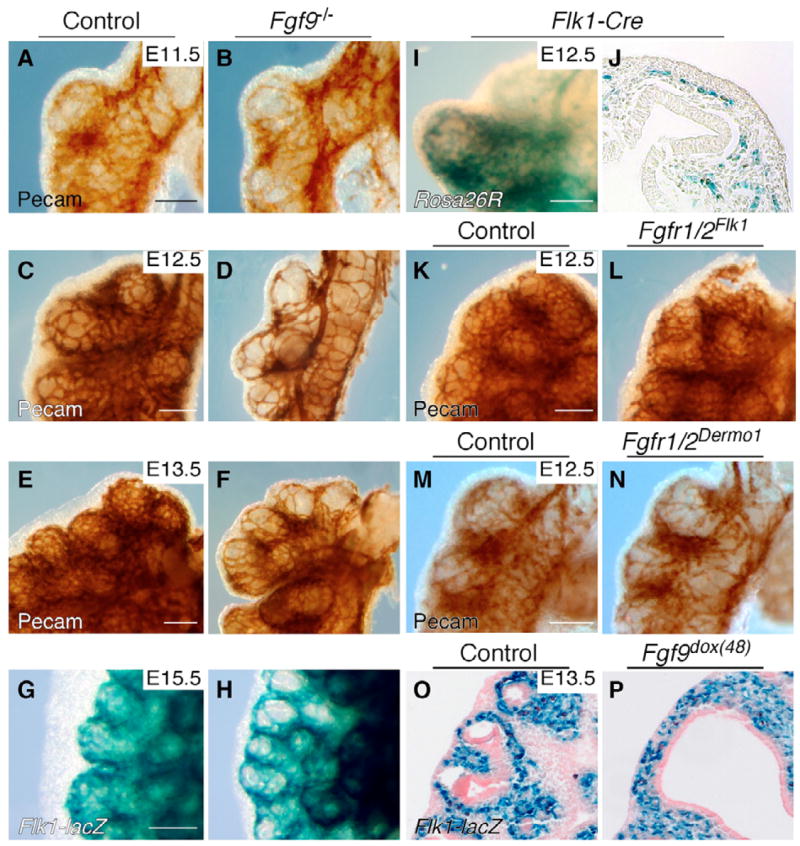
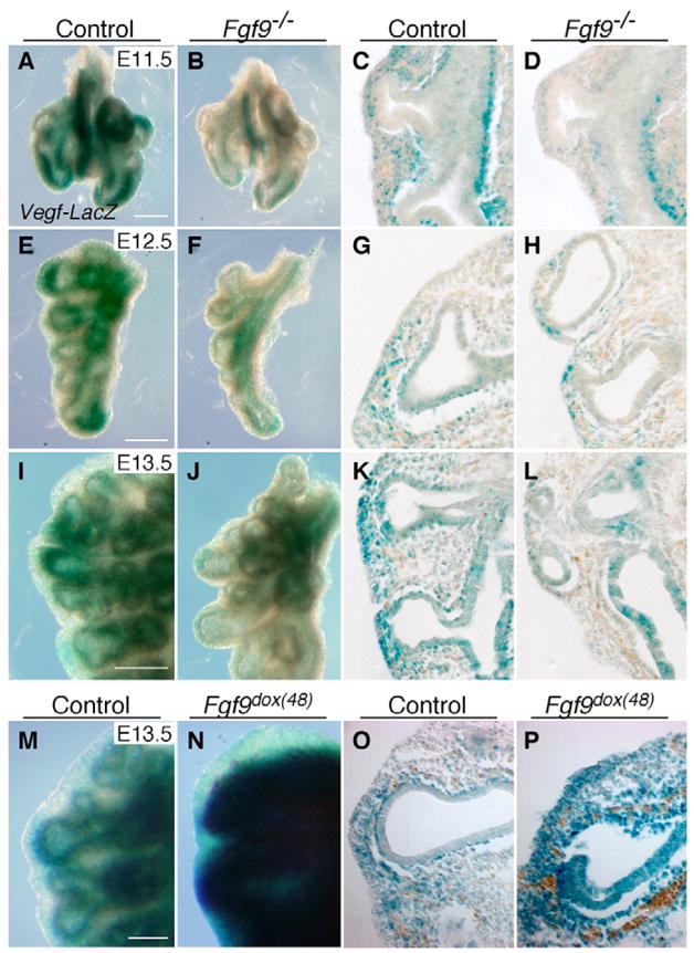
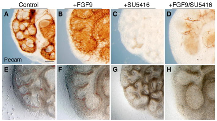
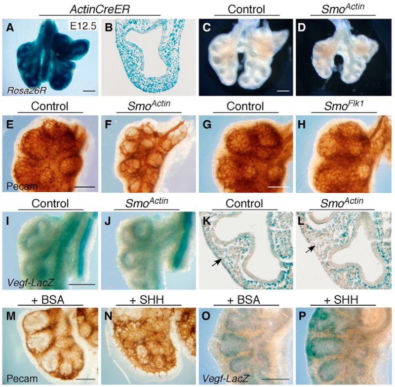
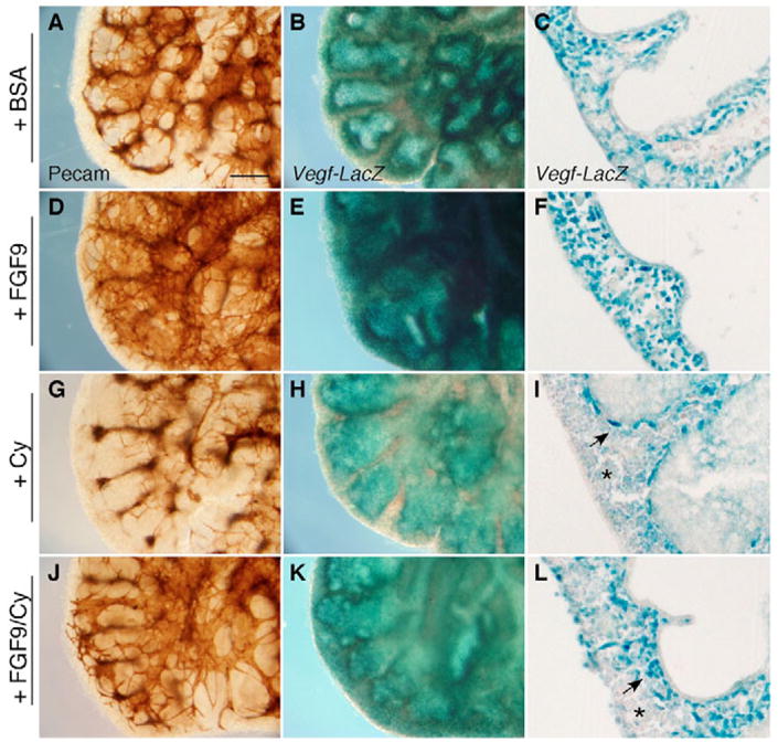
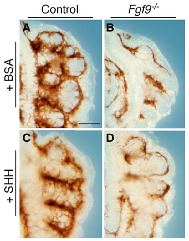
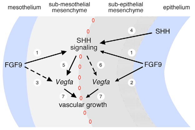
The juxtaposition of a dense capillary network to lung epithelial cells is essential for air-blood gas exchange. Defective lung vascular development can result in bronchopulmonary dysplasia and alveolar capillary dysplasia. Although vascular endothelial growth factor A (Vegfa) is required for formation of the lung capillary network, little is known regarding the factors that regulate the density and location of the distal capillary plexus and the expression pattern of Vegfa. Here, we show that fibroblast growth factor 9 (FGF9) and sonic hedgehog (SHH) signaling to lung mesenchyme, but not to endothelial cells, are each necessary and together sufficient for distal capillary development. Furthermore, both gain- and loss-of-function of FGF9 regulates Vegfa expression in lung mesenchyme, and VEGF signaling is required for FGF9-mediated blood vessel formation. FGF9, however, can only partially rescue the reduction in capillary density found in the absence of SHH signaling, and SHH is unable to rescue the vascular phenotype found in Fgf9(-/-) lungs. Thus, both signaling systems regulate distinct aspects of vascular development in distal lung mesenchyme. These data suggest a molecular mechanism through which FGF9 and SHH signaling coordinately control the growth and patterning of the lung capillary plexus, and regulate the temporal and spatial expression of Vegfa.


Figures

Fig. 1
Fig. 1. FGF9 signaling via mesenchymal FGFR1/2 is necessary and sufficient for distal lung capillary development
(A–F) Whole-mount immunohistochemistry with anti-PECAM antibody showing large gaps between vessels in the distal capillary plexus of Fgf9−/− lungs at E11.5 (B), E12.5 (D) and E13.5 (F) when compared with control lungs (A,C,E). (G,H) Whole-mount lacZ staining with the endothelial cell marker Flk1-lacZ showing a reduction in capillary density around the distal epithelium in Fgf9−/−; Flk1-lacZ+/− lungs at E15.5 (H) compared with control lungs (G). (I,J) Rosa26R-lacZ stain showing endothelial cell-specific Flk1-Cre activity in a pattern consistent with distal lung endothelial cells in whole-mount (I) and frozen (J) sections. (K,L) Fgfr1 and Fgfr2 double conditional knockout using Flk1-Cre (Fgfr1/2Flk; L) showing no difference in distal lung vascular development compared to an Fgfr1/2f/f control (K). (M,N) Fgfr1 and Fgfr2 double conditional knockout using mesenchymal-specific Dermo1-Cre (Fgfr1/2Dermo1), showing reduced distal lung capillary density (N) compared with an Fgfr1/2f/f control (M). (O,P) Induced Fgf9 expression for 48 hours with doxycycline [Fgf9dox(48)] is sufficient to induce Flk1-lacZ expression throughout lung mesenchyme (P), compared with expression only in the sub-epithelial mesenchyme in control lung (O). Histological sections in O and P were photographed through a 20× objective. Scale bars: 50 μm.
Fig. 2
Fig. 2. Fgf9 is necessary and sufficient for mesenchymal Vegfa expression in the lung
(A–L) Fgf9−/−; Vegfa-lacZ lungs show reduced mesenchymal Vegfa-lacZ staining at E11.5 (B,D), E12.5 (F,H) and E13.5 (J,L) compared with control (A,C,E,G,I,K). At E11.5, no Vegfa is observed in the epithelium (C,D). By E13.5, levels of Vegfa-lacZ in the mesenchyme in Fgf9−/− lungs continue to be reduced compared with controls, but epithelial expression is comparable to controls (K,L). (M–P) Fgf9 overexpression results in increased mesenchymal Vegfa in Fgf9dox(48); Vegfa-lacZ lungs at E13.5 (N,P). Left panels, whole-mount β-galactosidase staining; right panels, cryosections of left panels. Histology: 20× objective. Scale bars: 100 μm.
Fig. 3
Fig. 3. FGF9 requires VEGF signaling for the formation and maintenance of the lung capillary network
(A–D) Incubation of E12.5 lung explants for 48 hours with 2.5 ng/μl FGF9 stimulates robust capillary formation (B) compared with untreated lung (A). The VEGFR inhibitor SU5416 (45 μM) eliminates the formation and survival of vessels (C). FGF9 is unable to stimulate growth of new distal vessels in the presence of SU5416 (D) but can partially rescue blood vessel survival in very proximal regions. (E–H) FGF9 stimulates luminal dilation of the epithelial tubules (F). Branching morphogenesis appears comparable to controls in SU5416-treated explants (G). In contrast to capillary development, FGF9-induced luminal dilation is not affected by SU5416 (H). Scale bar: 50 μm.
Fig. 4
Fig. 4. SHH signaling to non-endothelial mesenchyme is necessary for distal lung capillary formation
(A,B) The Rosa26R allele was used to detect Actin-CreER activation throughout the lung mesenchyme and epithelium at E12.5 following tamoxifen injection at E9.5; (A) whole-mount and (B) frozen sections. (C,D) Conditional knockout of Smo, using Actin-CreER (SmoActin), results in lung hypoplasia (D) compared with a Smof/f control (C) at E12.5. (E,F) When assessed by anti-PECAM whole-mount immunohistochemistry, SmoActin lungs showed a reduction in distal capillary network density (F) compared with Smof/f controls (E) at E12.5. (G,H) Distal lung capillary density appeared similar in Smof/f; Flk1-Cre (SmoFlk1; H) and Smof/f control (G) mice, indicating that HH signaling to endothelial cells is not required for development. (I–L) SmoActin lungs (J,L) show a decrease in VegfA-lacZ activity in mesenchyme distal to the sub-epithelial layer (arrow) in whole-lung preparations (I,J) and frozen sections (K,L) compared with controls. (M–P) Lung organ cultures incubated with 500 ng/ml of SHH-N protein show an increase in PECAM-positive cells (N) and VegfA-lacZ staining (P) compared with BSA-incubated controls (M,O) after 24 hours. (B,K,L) Lower left lobe; (EJ) upper left lobe. Histology: 20× objective. Scale bars: 100 μm.
Fig. 5
Fig. 5. FGF9 and SHH signaling are both required for capillary formation
E12.5 lung explants from Vegfa-lacZ mice were incubated for 48 hours with media containing BSA (A–C), FGF9 (D–F), cyclopamine (Cy; G–I) or FGF9 and cyclopamine (J–L). (A,D,G,J) Whole-mount anti-PECAM immunohistochemistry showing increased capillary formation in the presence of 2.5 ng/μl FGF9 (D) and decreased vascular development in the presence of 10 μM cyclopamine (G). FGF9 was able to only partially rescue capillary formation in the presence of cyclopamine (J). (B,E,H,K) Whole-mount preparations and (C,F,I,L) frozen sections stained for lacZ activity. Compared with control explants, Vegfa expression is increased in the presence of FGF9 (E,F) and decreased in the presence of cyclopamine (H,I). FGF9 was able to partially rescue Vegfa expression in the presence of cyclopamine (K,L). Notice that, in the presence of cyclopamine, Vegfa expression is retained only in the inner cell layer of the sub-epithelial mesenchyme (arrow in I), but is significantly reduced in the sub-mesothelial mesenchyme (asterisk in I). FGF9 increases Vegfa expression throughout both mesenchymal layers (F), but in the presence of cyclopamine, Vegfa expression is expanded only in the sub-epithelial layer (arrow in L) and remains low in the sub-mesothelial region (asterisk in L). (AL) Lower left lobe. Histology: 20× objective. Scale bar: 100 μm.
Fig. 6
Fig. 6. SHH is not sufficient to rescue capillary plexus formation in Fgf9−/− lungs
E11.5 explant cultures of wild-type and Fgf9−/− lungs were incubated with BSA (A,B) or SHH (C,D) for 24 hours. Fgf9−/− lungs (B) formed a less dense capillary plexus compared with wild-type controls (A). In contrast to a wild-type lung (A,C), incubation of Fgf9−/− lungs with SHH did not increase capillary plexus density (D). Scale bar: 50 μm.
Fig. 7
Fig. 7. Model for the regulation of Vegfa expression during distal lung vascular development by FGF9 and SHH signaling
(1–3) FGF9 signaling via FGFR1 and FGFR2 stimulates SHH signaling (White et al., 2006) and Vegfa expression both in sub-mesothelial and sub-epithelial mesenchymal layers. In the absence of SHH signaling, FGF9 has a stronger affect on Vegfa expression in the sub-epithelial compartment (2) compared with the sub-mesothelial compartment (3). (4) Epithelial SHH signals to lung mesenchyme (Bellusci et al., 1997a; Weaver et al., 2003). SHH signaling is required for Vegfa expression and subsequent vascular development both in sub-mesothelial mesenchyme (5) and sub-epithelial mesenchyme (6; except for the inner-most cell layer, stippled shading). HH signaling to Flk1-Cre+ endothelial cells is not required for capillary development. (7) VEGF is a primary factor regulating vascular growth in the developing lung. Distal capillaries are denoted by red ovals located between sub-mesothelial and sub-epithelial mesenchyme layers.

Similar articles

Cited by

References

    1. Akeson AL, Greenberg JM, Cameron JE, Thompson FY, Brooks SK, Wiginton D, Whitsett JA. Temporal and spatial regulation of VEGF-A controls vascular patterning in the embryonic lung. Dev Biol. 2003;264:443–455. - PubMed
    1. Asahara T, Bauters C, Zheng LP, Takeshita S, Bunting S, Ferrara N, Symes JF, Isner JM. Synergistic effect of vascular endothelial growth factor and basic fibroblast growth factor on angiogenesis in vivo. Circulation. 1995;92:365–371. - PubMed
    1. Auguste P, Gursel DB, Lemiere S, Reimers D, Cuevas P, Carceller F, Di Santo JP, Bikfalvi A. Inhibition of fibroblast growth factor/fibroblast growth factor receptor activity in glioma cells impedes tumor growth by both angiogenesis-dependent and -independent mechanisms. Cancer Res. 2001;61:1717–1726. - PubMed
    1. Auguste P, Javerzat S, Bikfalvi A. Regulation of vascular development by fibroblast growth factors. Cell Tissue Res. 2003;314:157–166. - PubMed
    1. Bellusci S, Furuta Y, Rush MG, Henderson R, Winnier G, Hogan BL. Involvement of Sonic hedgehog (Shh) in mouse embryonic lung growth and morphogenesis. Development. 1997a;124:53–63. - PubMed

Publication types

MeSH terms

Substances