. 2007 Oct 1;120(Pt 19):3362-71.
doi: 10.1242/jcs.008300.

Specificity of RGS10A as a key component in the RANKL signaling mechanism for osteoclast differentiation

Affiliations

Specificity of RGS10A as a key component in the RANKL signaling mechanism for osteoclast differentiation

Shuying Yang et al. J Cell Sci. .

Abstract

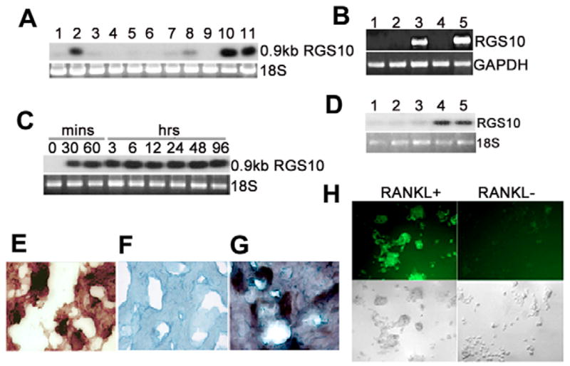
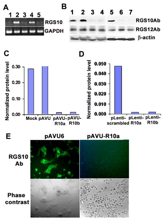
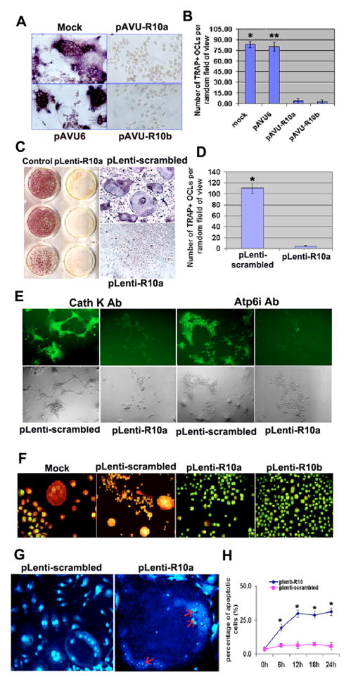
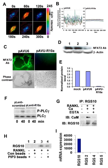
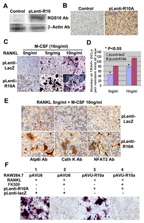
Significant progress has been made in studies of the mechanisms by which RANKL induces terminal osteoclast differentiation. However, many crucial details in the RANKL-evoked signaling pathway for osteoclast differentiation remain to be defined. We characterized genes specifically expressed in osteoclasts by differential screening of a human osteoclastoma cDNA library, and found that the regulator of G-protein signaling 10A (RGS10A), but not the RGS10B isoform, was specifically expressed in human osteoclasts. The expression of RGS10A is also induced by RANKL in osteoclast precursors and is prominently expressed in mouse osteoclast-like cells. RGS10A silencing by RNA interference blocked intracellular [Ca2+]i oscillations, the expression of NFAT2, and osteoclast terminal differentiation in both bone marrow cells and osteoclast precursor cell lines. Reintroduction of RGS10A rescued the impaired osteoclast differentiation. RGS10A silencing also resulted in premature osteoclast apoptosis. RGS10A silencing affected the RANKL-[Ca2+]i oscillation-NFAT2 signaling pathway but not other RANKL-induced responses. Our data demonstrate that target components of RGS10A are distinct from those of RGS12 in the RANKL signaling mechanism. Our results thus show the specificity of RGS10A as a key component in the RANKL-evoked signaling pathway for osteoclast differentiation, which may present a promising target for therapeutic intervention.


Figures

Fig. 1
Fig. 1
RGS10A is prominently expressed in osteoclasts and osteoclast precursors stimulated by RANKL and M-CSF. (A) Northern blot hybridization of mouse RGS10A cDNA to total RNA from mouse tissues and cell lines. Lane 1, calvaria; lane 2, brain; lane 3, long bone; lane 4, heart; lane 5, liver; lane 6, kidney; lane 7, spleen; lane 8, lung; lane 9, muscle; lane 10, RANKL-stimulated RAW264.7 cells; lane 11, RANKL-stimulated MOCP-5 cells. (B) RT-PCR analysis of isoforms RGS10A and RGS10B mRNA in human osteoclasts and brain, showing that hRGS10A is specifically expressed in osteoclasts. Lane 1, negtive control (H2O); lane 2, RNA of isoform A of hRGS10 from brain; lane 3, RNA of isoform B of hRGS10 from brain; lane 4, RNA of isoform B of hRGS10 from osteoclasts; lane 5, RNA of isoform A of hRGS10 from osteoclasts. The primer BOR10-F for RGS10A is located at exon 1 and the primer BOBRR10-R is located at exon 5. The distance between the two exons is 10.8 kb (Gene Access no: NC_000010), so the 669 bp size band is the true RT-PCR product. The RT-PCR fragments from lane 5 were cloned into pBluescript and sequenced. The RT-PCR cDNA fragment from human osteoclast mRNA was 100% homologous to the sequence of the human RGS10A cDNA. (C) Time course of RGS10A mRNA in RANKL-stimulated RAW264.7 cells by northern blotting. Cells were treated with RANKL and M-CSF for the indicated times. RGS10A mRNA was detectable at 30 minutes and continued to increase at 60 minutes, staying at high levels for 96 hours after stimulation with RANKL and M-CSF. (D) Northern blot analysis of RGS10A expression in osteoclasts and BMMs stimulated with M-CSF in the absence of RANKL. No RGS10A expression was detected in Lane 1–3, which are BMMs stimulated with M-CSF for 0, 12, and 24 hours. Lane 4–5, BMMs stimulated with RANKL and M-CSF for 12 and 24 hours. (E–G) In situ hybridization of RGS10A mRNA in a human osteoclastoma. Counterstain, Methyl Green. (E) TRAP+ human osteoclasts. (F) Sense probe as control. (G) Antisense probe. (H) Expression of RGS10A protein in RANKL-stimulated BMMs. Multinucleated osteoclasts were formed only in RANKL-stimulated BMMs, and strongly expressed RGS10A. RGS10A protein expression was absent in cells not stimulated with RANKL.
Fig. 2
Fig. 2
RGS10A silencing blocks RGS10A expression. RAW264.7 cells stably transfected with pAVU-scrambled, pAVU-R10a or pAVU-R10b constructs, and BMMs with pLenti-scrambled or pLenti-R10a were stimulated with RANKL and M-CSF for 96 hours. (A) RT-PCR analysis. Lane 1, negative control (H2O). The expression of RGS10A mRNA was weak or absent in RGS10A-silenced cells (lane 2, pAVU-R10a; lane 4, pAVU-R10b). (B) Western blotting of RGS10A protein and RGS12 protein in control and RGS10A-silenced RAW264.7 cells (lanes 1–4) and BMMs (lanes 5–7) stimulated with RANKL and M-CSF. For RGS10A protein, the signals were strong in control cells (lanes 1, 2, 5) and weak in RGS10A-silenced cells (lanes 3, 4, 6, 7). However, for RGS12 protein, there is no apparent difference in signals between control cells and RGS10A silenced cells. Lane 1, mock; lane 2, pAVU-scrambled shRNA; lane 3, pAVU-R10a; lane 4, pAVU-R10b; lane 5, pLenti-scrambled shRNA; lane 6, pLenti-R10a; lane 7, pLenti-R10b. Ab, antibody. (C,D) Quantification of RGS10A levels by western blotting as in B. RGS10A protein levels in transfected cells were normalized to the loading control β-actin. (E) Immunofluorescence staining. RGS10A expression was silenced in the cells transfected with pAVU-R10a. These are images of phase-contrast as an internal control.
Fig. 3
Fig. 3
RGS10A silencing blocks osteoclast differentiation. (A) TRAP+ MNCs were formed in RANKL-stimulated RAW264.7 cells transfected with mock shRNA or pAVU-scrambled shRNA, but not in the cells transfected with pAVU-R10a or pAVU-R10b. (B) Quantitative analysis of TRAP+ MNCs in (A). TRAP+ MNCs in control groups, mock (*) or pAVU-scrambled (**), are more than 15 times (P<0.05) that of RGS10A-silenced groups (pAVU-R10a or pAVU-R10b). (C) Formation of TRAP+ MNCs in RANKL-stimulated BMMs infected with pLenti-scrambled and the absence of TRAP+ MNCs in the cells infected with pAVU-R10a are shown. (D) Quantitative analysis of TRAP+ MNCs in (C), pLenti-R10a vs the RGS10A-silenced group (*P<0.05). (E) Immunofluorescent staining of cathepsin K and Atp6i in RGS10A-silenced BMMs stimulated with RANKL and M-CSF. RGS10A silencing blocks the expression of cathepsin K and Atp6i as showed in pLenti-R10a groups. (F) Acridine orange staining. Strong orange fluorescence indicates extracellular acidification in the mock or pLenti-scrambled, but not in pLenti-R10a or pLenti-R10b. (G,H) RGS10A silencing-mediated apoptosis in multinucleated osteoclasts with RANKL stimulation. (G) The nuclei of osteoclast cells are condensed and fragmented in the RGS10A-silenced cells as indicated by the arrows, compared with the control cells. (H) Time course of RGS10A silencing-mediated apoptosis. Apoptosis was quantified by counting multinucleated cells with condensed nuclei (each point *P<0.05 vs control).
Fig. 4
Fig. 4
RGS10A signaling is essential for induction of [Ca2+]i oscillations by RANKL and NFAT2 expression. (A) [Ca2+]i oscillations were observed in RANKL-stimulated BMMs or RAW264.7 cells, 48–72 hours after RANKL stimulation. The picture shows an example of successive pseudocolored Ca2+ images of cells treated with RANKL for 48 hours. Oscillations were produced at 2-minute intervals. (B) Ca2+ changes were traced in single RGS10A-silenced or control cells treated with RANKL and M-CSF for 72 hours. Ca2+ changes were estimated as the ratio of fluorescence intensity of the Fluo-4 to Fura Red, plotted at 5-second intervals. Each color indicates a different cell in the same field. [Ca2+]i oscillations are blocked in RGS10A-silenced cells (pAVU-R10a). (C) Immunofluorescence revealed that the expression of NFAT2 was blocked in RGS10A-silenced cells. (D) There were weak signals of NFAT2 protein detected in RGS10A-silenced cells (lane 3) as compared to the controls (lane 1: mock; lane 2: pAVU6). (E) Quantification of the bands in D. NFAT2 signals in the mock or pAVU6, are 4–4.7 times stronger than in pAVU-R10a. (F) Western blot analysis of activation of PLCγ after stimulation with RANKL for 40 minutes. Phosphorylation of PLCγ was impaired in the RGS10A-silenced group (plenty-R10a) following a 40-minute stimulation with RANKL. (G) Co-immunoprecipitation of RGS10A and CaM. No interaction was observed in unstimulated cells. CaM bound to RGS10A in the presence of 1 mM CaCl2. This interaction was blocked by 0.5 mM EGTA. (H) PdIns(3,4,5)P3-bead binding assay. RGS10A was not detected after stimulation with M-CSF alone (lane 1) or with control beads without PdIns(3,4,5)P3 (negative control, lane 2). RGS10A was detected after stimulation with RANKL (positive control, lane 3). RGS10A was detected after stimulation with RANKL in a PdIns(3,4,5)P3 pull-down assay (lane 4). (I) mRNA expression of RGS 2, 4, 5, and 10A in RANKL-stimulated OCLs. RGS10A is predominantly expressed in OCLs, compared with RGS2, RGS4, and RGS5.
Fig. 5
Fig. 5
Overexpression of RGS10A significantly increases the sensitivity to RANKL signaling during osteoclast differentiation. BMMs were infected with pLenti-LacZ or pLenti-R10A. (A) Western blotting. Without RANKL stimulation, the signals of RGS10A were only detectable in cells infected with pLenti-R10A but not in the pLenti-LacZ-transfected cells (control). (B) Immunostaining. 80–90% of the cells express RGS10A in pLenti-R10A; however, there are no stained cells in pLenti-LacZ- infected cells. (C) TRAP+ staining. The cells were treated with 0, 5 and 10 ng/ml of RANKL in the presence of 10 ng/ml of M-CSF for 4 days. Without RANKL stimulation, 8% of precursor cells differentiated into mononuclear TRAP+ cells in pLenti-R10A (left lower panel). In pLenti-R10A cells, there are more TRAP+ cells and mature multinuclear cells compared with the control group in the presence of 5 or 10 ng/ml RANKL together with 10 ng/ml M-CSF (middle and right lower panels). (D) Quantitative analysis of TRAP+ MNCs in (C). The number of TRAP+ MNCs in pLenti-R10A is 1.5 times higher than that of the control group (P<0.05 vs pLenti-LacZ). (E) Immunostaining. With 5 ng/ml RANKL stimulation, pLenti-R10A-infected cells expressed Atp6i, cathepsin K, and NFAT2 at higher levels than the control cells. (F) The effect of FK506 on osteoclastogenesis induced by RGS10A overexpression in RGS10A-silenced RAW264.7 or control cells. FK506 (1 μg/ml) inhibited osteoclast differentiation from RANKL-stimulated RAW264.7 cells infected by pLenti-LacZ (panel 2) as compared with the culture without FK506 (panel 1). FK506 inhibited the rescue effect of RGS10A reintroduction as shown in panel 3 in RGS10A-silenced RAW264.7 cells infected with pLenti-RGS10a (panel 4).

Similar articles

Cited by

References

    1. Alliston T, Derynck R. Medicine: interfering with bone remodelling. Nature. 2002;416:686–687. - PubMed
    1. Boyle WJ, Simonet WS, Lacey DL. Osteoclast differentiation and activation 1. Nature. 2003;423:337–342. - PubMed
    1. Chambers TJ, Fuller K, Darby JA, Pringle JA, Horton MA. Monoclonal antibodies against osteoclasts inhibit bone resorption in vitro. Bone Miner. 1986;1:127–135. - PubMed
    1. Chen W, Li YP. Generation of mouse osteoclastogenic cell lines immortalized with SV40 large T antigen. J Bone Miner Res. 1998;13:1112–1123. - PubMed
    1. Crabtree GR, Olson EN. NFAT signaling: choreographing the social lives of cells. Cell. 2002;109:S67–S79. - PubMed

Publication types

MeSH terms