Comparative Study
. 2007 Sep 19;27(38):10084-93.
doi: 10.1523/JNEUROSCI.2211-07.2007.

Signaling properties of a short-wave cone visual pigment and its role in phototransduction

Affiliations
Comparative Study

Signaling properties of a short-wave cone visual pigment and its role in phototransduction

Guang Shi et al. J Neurosci. .

Abstract

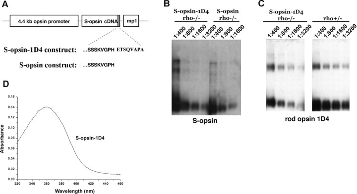
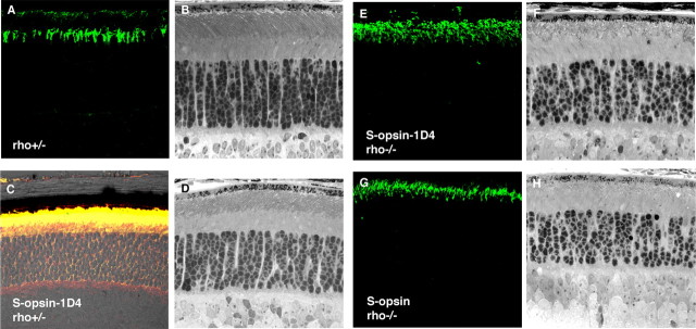
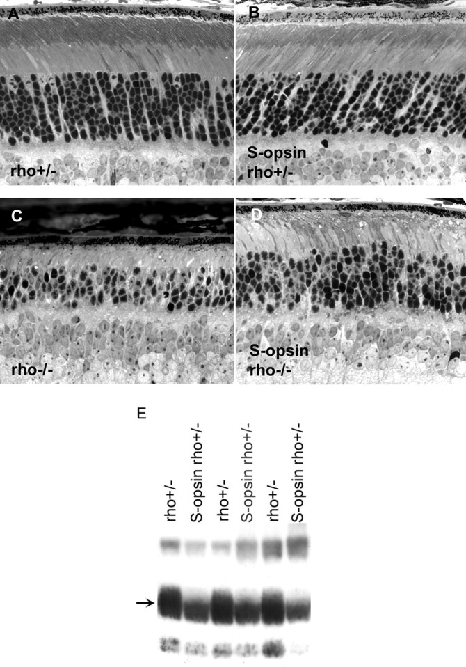
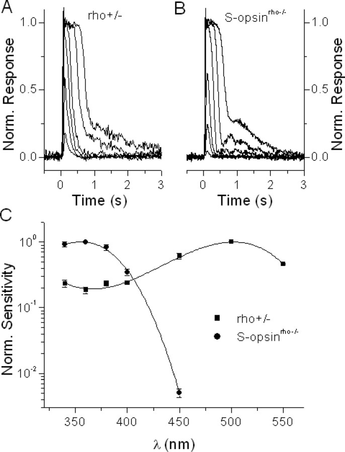
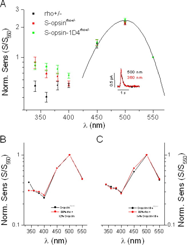
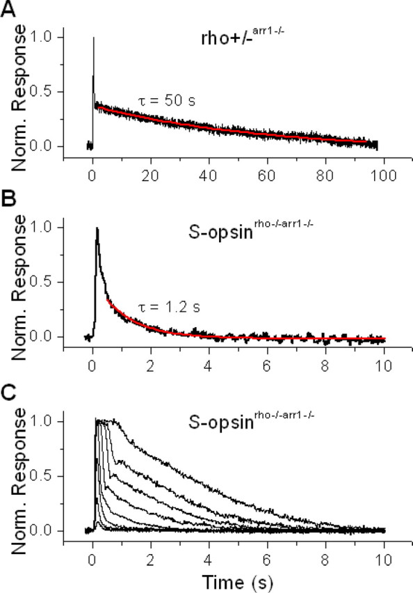
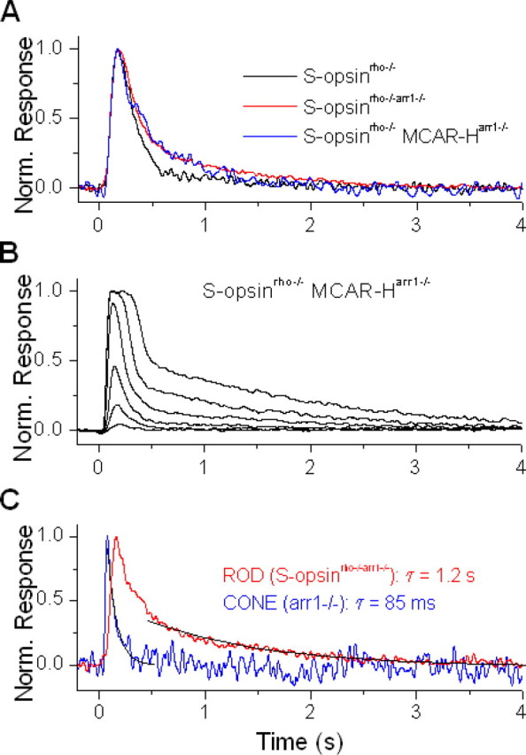
Although visual pigments play key structural and functional roles in photoreceptors, the relationship between the properties of mammalian cone pigments and those of mammalian cones is not well understood. We generated transgenic mice with rods expressing mouse short-wave cone opsin (S-opsin) to test whether cone pigment can substitute for the structural and functional roles of rhodopsin and to investigate how the biophysical and signaling properties of the short-wave cone pigment (S-pigment) contribute to the specialized function of cones. The transgenic S-opsin was targeted to rod outer segments, and formed a pigment with peak absorption at 360 nm. Expression of S-opsin in rods lacking rhodopsin (rho-/-) promoted outer segment growth and cell survival and restored their ability to respond to light while shifting their action spectrum to 355 nm. Using the spectral separation between S-pigment and rhodopsin, we found that the two pigments produced similar photoresponses. Dark noise did not increase in transgenic rods, indicating that thermal activation of S-pigment might not contribute to the low sensitivity of mouse S-cones. Using rod arrestin knock-out animals (arr1-/-), we found that the physiologically active (meta II) state of S-pigment decays 40 times faster than that of rhodopsin. Interestingly, rod arrestin was efficient in deactivating S-pigment in rods, but its deletion did not have any obvious effect on dim-flash response shutoff in cones. Furthermore, transgenic cone arrestin was not able to rescue the slow shutoff of S-pigment dim-flash response in arr1-/- rods. Thus, the connection between rod/cone arrestins and S-pigment shutoff remains unclear.


Figures

Figure 1.
Figure 1.
Expression of S-opsin in transgenic mouse rods. A, The mouse rod opsin (Lem et al., 1991) promoter was used to direct expression of mouse S-opsin in rods. One construct contained the full coding sequence of S-opsin, whereas S-opsin-1D4 contained the terminal eight amino acid residues from rhodopsin (ETSQVAPA) appended to the C terminus of S-opsin. B, Western blot of serially diluted retinal extracts from age-matched S-opsin-1D4rho−/− and S-opsinrho−/− mice probed with an antibody against S-opsin. The ratios indicate the fraction of a retina loaded per lane. C, Western blot of serially diluted retinal extracts from age-matched S-opsin-1D4rho−/− and rho+/− mice probed with the monoclonal antibody 1D4, which recognizes the terminal eight amino acid residues on rhodopsin. The presence of 1D4 on S-opsin-1D4 allows for direct comparison of expression levels between S-opsin and rhodopsin in rho+/− retinas. Ratios indicate the fraction of retina loaded per lane. D, Spectrometric measurement of purified S-opsin-1D4 shows peak absorbance at 360 nm.
Figure 2.
Figure 2.
Localization of S-opsin and retinal morphology in 3-week-old mice. A, C, E, G, Localization of S-opsin (green) visualized by immunofluorescence microscopy of frozen retinal sections. B, D, F, H, Retinal morphology of the corresponding genotype. A, S-opsin reactivity in endogenous cones of the control rho+/− retina. B, Transgenic S-opsin-1D4rho+/− labeled with S-opsin (green) and rhodopsin (red) show colocalization (yellow). Retinal morphology is mostly normal in these genotypes (B, D). S-opsin-1D4 and S-opsin are correctly localized to the outer segments when they are expressed in the rho−/− background (E, G). Disorganized outer segment structures are seen in these mice (F, H).
Figure 3.
Figure 3.
Analysis of retinal morphology and rhodopsin content in 2-month-old mice. A, Retinal morphology is largely normal in rho+/− mice. B, Retinal morphology is unchanged by the expression of S-opsin in this genetic background. C, The outer segment does not form in the absence of rhodopsin, and the number of rods decreases as a function of age. D, The presence of S-opsin delays retinal degeneration in rho−/− mice (compare C, D). E, Rhodopsin content is lowered when S-opsin is expressed; transgene-negative rho+/− littermates show higher rhodopsin content. Individual retinas were homogenized, and an equal fraction (1/3200) from each retina was loaded per lane. The proteins were blotted onto nitrocellulose and probed with 4D2, a monoclonal antibody against rhodopsin (arrow).
Figure 4.
Figure 4.
Transgenic S-opsin expressed in rods produces rod-like photoresponses while inducing S-cone-like spectral sensitivity. A, Family of rhodopsin flash responses from control rho+/− rods. B, Family of S-pigment flash responses from transgenic S-opsinrho−/− rods. Flashes for both figures were delivered at t = 0 s and flash intensity was increased in 0.6 log units. C, Average spectral sensitivities of control rho+/− rods (squares, normalized at 500 nm) and of transgenic S-opsinrho−/− rods (circles, normalized at 360 nm). Error bars indicate SEM. Solid lines represent a third-order polynomial fit to each spectrum.
Figure 5.
Figure 5.
Transgenic expression of S-opsin in rho+/− rods enhances their sensitivity to UV light without affecting their response properties. A, Comparison of spectral sensitivities of control rho+/− rods (black), transgenic S-opsinrho+/− rods (red), and transgenic S-opsin-1D4rho+/− rods (green). Spectral sensitivities are shown normalized at 550 nm, where contribution of S-pigment to the overall action spectrum in the transgenic cells is negligible (Fig. 4C). This point is also supported by the good fit at long wavelengths of both control and transgenic action spectra to the rhodopsin A1 template with λmax = 500 nm (solid line). The inset shows the similar amplitudes and kinetics of the single photon responses from one S-opsinrho−/− rod at 500 nm (black) and 360 nm (red). B, The action spectrum of S-opsinrho+/− rods (black) can be fit by a combination of the action spectra of rho+/− rods (Fig. 5A) and S-opsinrho−/− rods (Fig. 4C) in an 88%:12% ratio (red). C, Similar fit for the S-opsin-1D4rho+/− rod action spectrum (black) with an 86%:14% ratio of rho+/− and S-opsin-1D4rho+/− rod spectra (red). Error bars indicate SEM.
Figure 6.
Figure 6.
The physiologically active (meta II) state of S-pigment decays significantly faster than that of rhodopsin. A, Fitting the slow recovery phase of normalized dim-flash response from rho+/− arr1−/− rod with a single exponential decay function gave time constant of rhodopsin meta II of 50 s. B, Similar fit of the dim-flash response from S-opsinrho−/−arr1−/− rod gave an S-pigment meta II time constant of 1.2 s. C, In contrast to the case of rhodopsin, S-pigment meta II decay is sufficiently fast to allow collection of a complete family of flash responses, from threshold to saturation.
Figure 7.
Figure 7.
Cone arrestin is less efficient than rod arrestin at inactivating transgenic S-pigment, expressed in rods. A, Normalized dim-flash responses from transgenic S-pigment in control rod (S-opsinrho−/−, black), rod lacking rod arrestin (S-opsinrho−/−arr1−/−, red), and rod lacking rod arrestin but expressing cone arrestin (S-opsinrho−/− MCAR-Harr1−/−, blue). Deletion of rod arrestin results in slower recovery of the dim-flash response, which is not reversed by the transgenic expression of cone arrestin. B, Family of flash responses from S-opsinrho−/− MCAR-Harr1−/− rod. Cone arrestin partially reverses the slow decay of the responses to brighter flashes (compare with Fig. 6C). C, Comparison of normalized dim-flash responses from S-opsinrho−/−arr1−/− rod (red) and arr1−/− cone (blue), with recovery fit by a single exponential decay function (black) in each case. The cone-response recovery is significantly faster than the decay of S-opsin meta II, which is rate limiting for the recovery of the rod.

References

    1. Burns ME, Mendez A, Chen J, Baylor DA. Dynamics of cyclic GMP synthesis in retinal rods. Neuron. 2002;36:81–91. - PubMed
    1. Calvert PD, Govardovskii VI, Krasnoperova N, Anderson RE, Lem J, Makino CL. Membrane protein diffusion sets the speed of rod phototransduction. Nature. 2001;411:90–94. - PubMed
    1. Carter-Dawson LD, LaVail MM. Rods and cones in the mouse retina. I. Structural analysis using light and electron microscopy. J Comp Neurol. 1979;188:245–262. - PubMed
    1. Chan S, Rubin WW, Mendez A, Liu X, Song X, Hanson SM, Craft CM, Gurevich VV, Burns ME, Chen J. Functional comparisons of visual arrestins in rod photoreceptors of transgenic mice. Invest Ophthalmol Vis Sci. 2007;48:1968–1975. - PMC - PubMed
    1. Concepcion F, Mendez A, Chen J. The carboxyl-terminal domain is essential for rhodopsin transport in rod photoreceptors. Vision Res. 2002;42:417–426. - PubMed

Publication types