Comparative Study
. 2007 Sep 19;27(38):10094-105.
doi: 10.1523/JNEUROSCI.2459-07.2007.

System x(c)- activity and astrocytes are necessary for interleukin-1 beta-mediated hypoxic neuronal injury

Affiliations
Comparative Study

System x(c)- activity and astrocytes are necessary for interleukin-1 beta-mediated hypoxic neuronal injury

Birgit Fogal et al. J Neurosci. .

Abstract

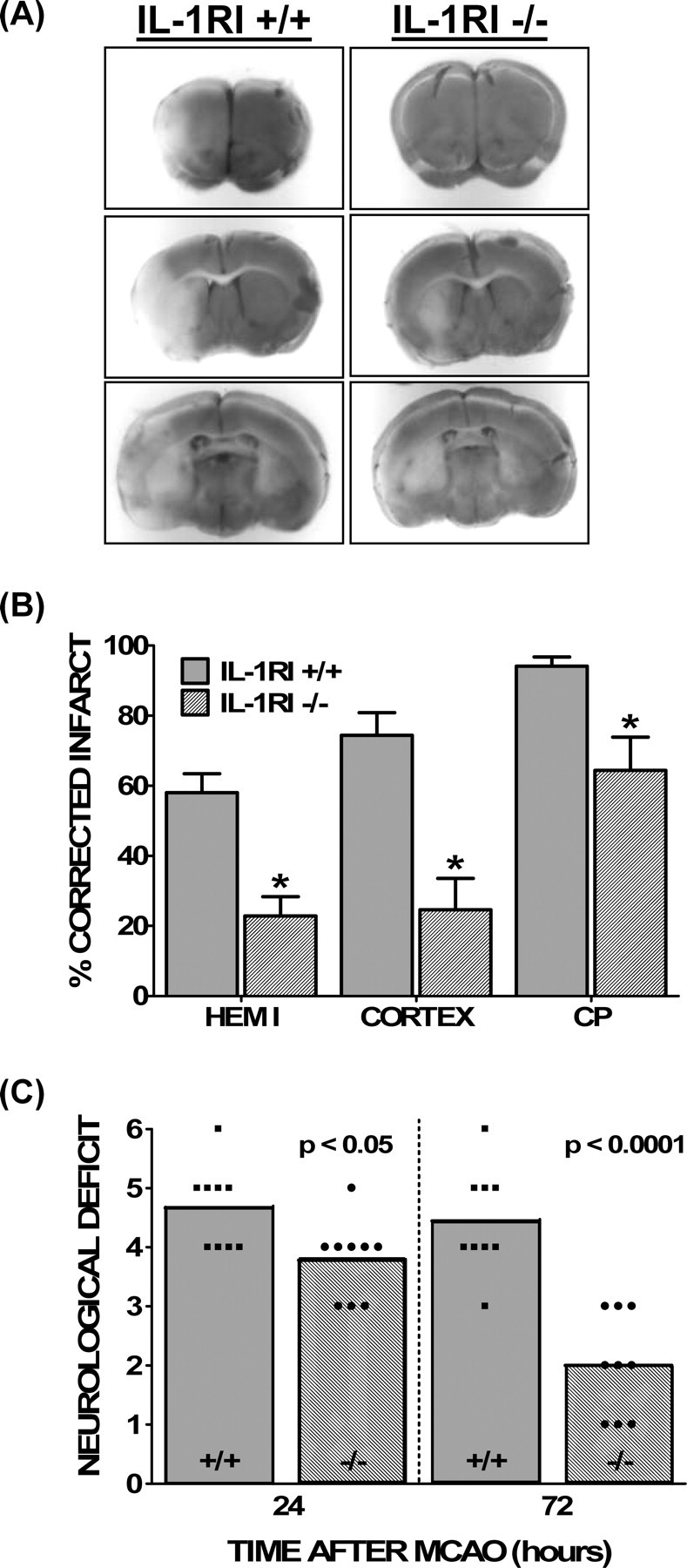
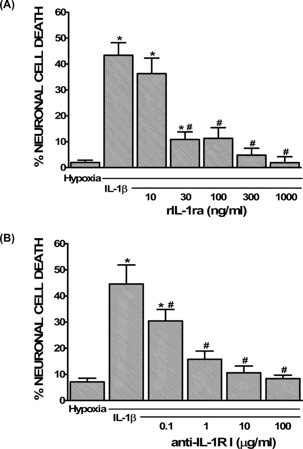
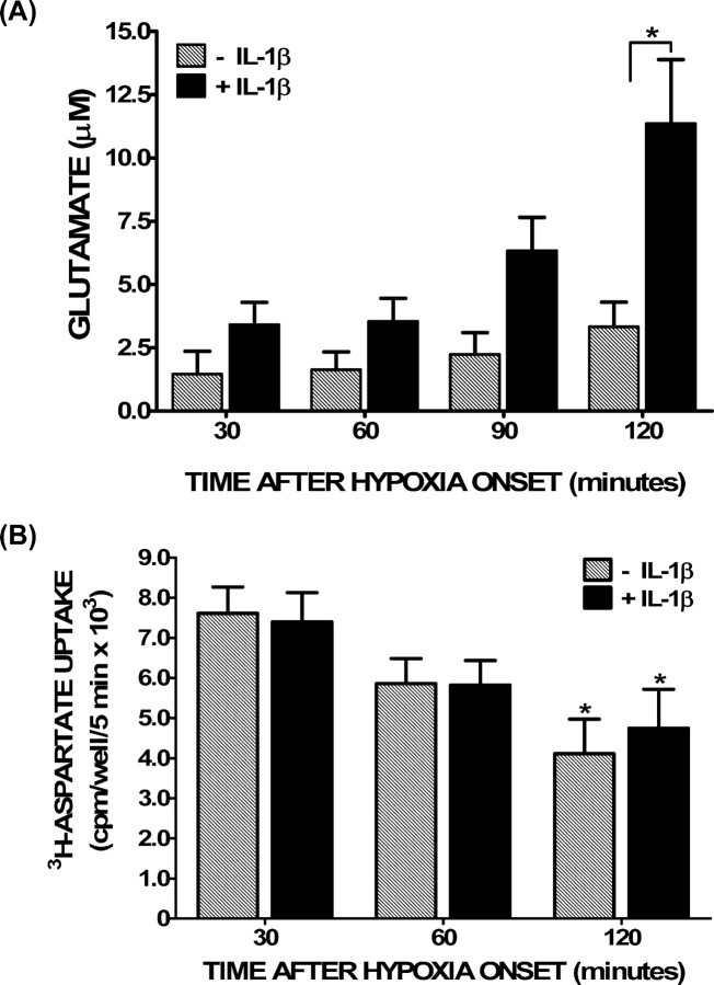
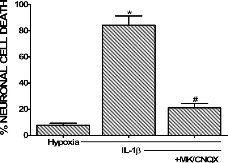
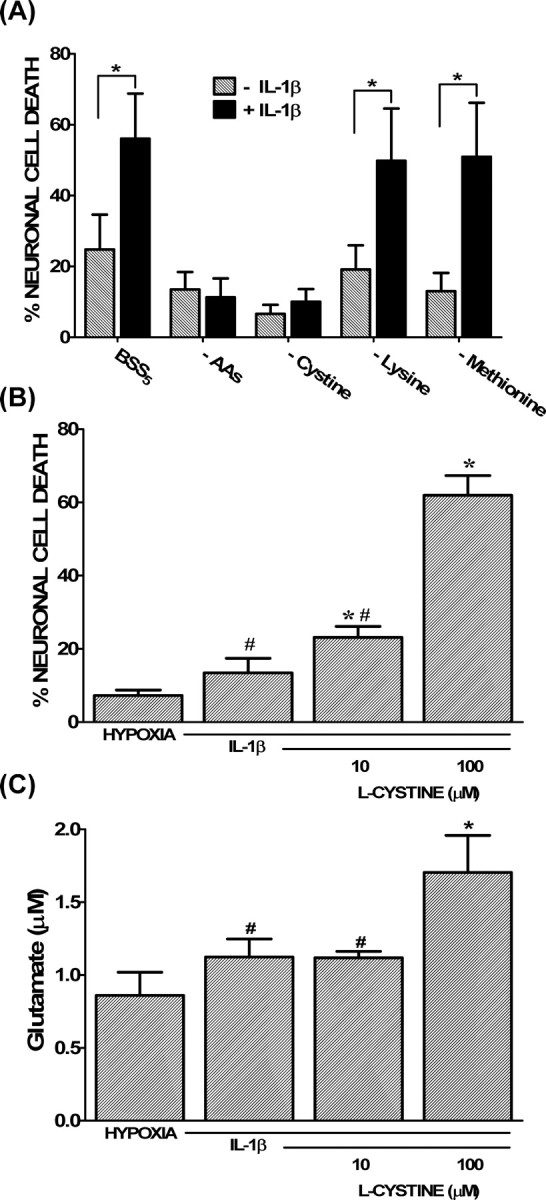
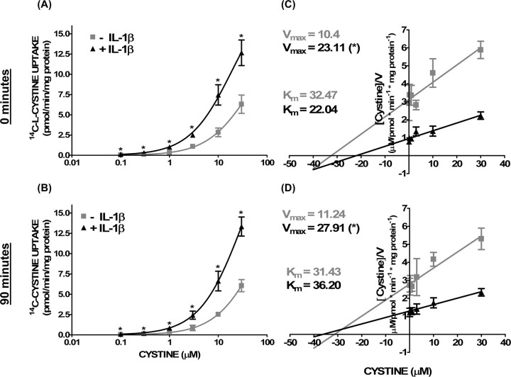
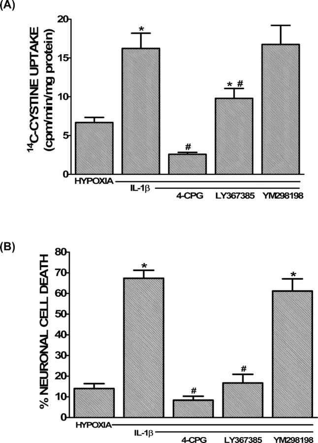
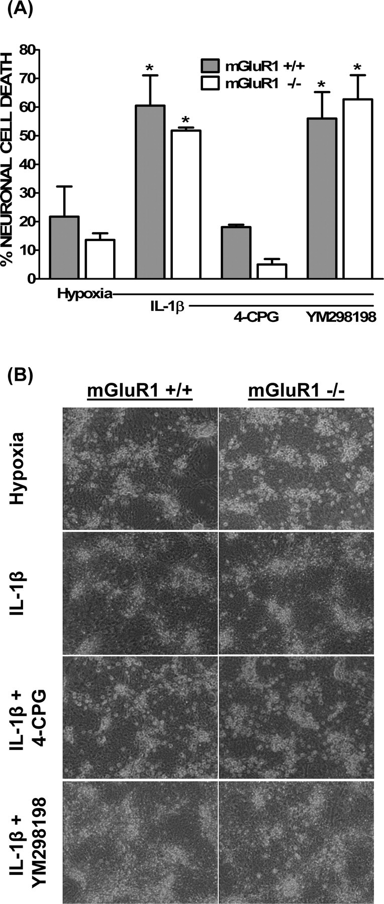
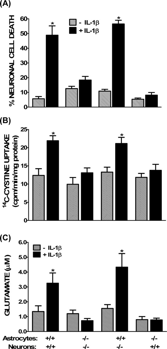
The purpose of this study was to elucidate the cellular/biochemical pathway(s) by which interleukin-1beta (IL-1beta) contributes to the pathogenesis of hypoxic-ischemic brain damage. In vivo, IL-1 receptor type I (IL-1RI)-deficient mice showed smaller infarcts and less neurological deficits than wild-type animals after a 90 min reversible middle cerebral artery occlusion. In vitro, IL-1beta mediated an enhancement of hypoxic neuronal injury in murine cortical cultures that was lacking in cultures derived from IL-1RI null mutant animals and was blocked by the IL-1 receptor antagonist or an IL-1RI blocking antibody. This IL-1beta-mediated potentiation of hypoxic neuronal injury was associated with an increase in both cellular cystine uptake ([cystine]i) and extracellular glutamate levels ([glutamate]e) and was prevented by either ionotropic glutamate receptor antagonism or removal of L-cystine, suggesting a role for the cystine/glutamate antiporter (System x(c)-). Indeed, dual System x(c)-/metabotropic glutamate receptor subunit 1 (mGluR1) antagonism but not selective mGluR1 antagonism prevented neuronal injury. Additionally, cultures derived from mGluR1-deficient mice exhibited the same potentiation in injury after treatment with IL-1beta as wild-type cultures, an effect prevented by System x(c)-/mGluR1 antagonism. Finally, assessment of System x(c)- function and kinetics in IL-1beta-treated cultures revealed an increase in velocity of cystine transport (Vmax), in the absence of a change in affinity (Km). Neither the enhancement in [cystine]i, [glutamate]e, or neuronal injury were observed in chimeric cultures consisting of IL-1RI(+/+) neurons plated on top of IL-1RI(-/-) astrocytes, highlighting the importance of astrocyte-mediated alterations in System x(c)- as a novel contributor to the development and progression of hypoxic neuronal injury.


Figures

Figure 1.
Figure 1.
Comparison of ischemic brain damage between wild-type and IL-1RI null mutant animals. A, Representative TTC staining of coronal brain sections (2 mm) 70.5 h after a 90 min reversible MCAO for wild-type (IL-1RI+/+; left) and IL-1RI-deficient (IL-1RI−/−; right) male mice. The lack of TTC staining indicates an infarcted area. B, Infarct volumes in the hemisphere (hemi), cerebral cortex (cortex), and caudate–putamen (CP) of wild-type (IL-1RI+/+; gray bars) and IL-1RI knock-out (IL-1RI−/−; hatched bars) male mice were measured over five coronal sections. Infarct volumes are reported as the mean ± SEM percentage of non-ischemic hemisphere, corrected for edema. Actual infarct volumes for wild-type versus null mutant animals were as follows: hemisphere, 177.3 ± 18.0 versus 65 ± 16.4 mm3; cortex, 99.8 ± 8.2 versus 29.7 ± 11.3 mm3; caudate–putamen, 31.2 ± 2.0 versus 18.1 ± 2.9 mm3. An asterisk indicates a value significantly different from wild-type animals, as assessed via an unpaired t test (n = 9 animals per group). Significance was assessed at p < 0.05. C, Post-ischemic neurological deficits were scored as described in Materials and Methods at 24 and 72 h after MCAO in wild-type (IL-1RI+/+; squares; gray bars) and IL-1RI null mutant (IL-1RI−/−; circles; hatched bars) male mice. Neurological scores between groups were different at 24 and 72 h after MCAO, as determined by a Mann–Whitney U test (n = 9 animals per group, statistical significance was assessed at p < 0.05).
Figure 2.
Figure 2.
Role of IL-1RI signaling in the potentiation of hypoxic neuronal injury by IL-1β in vitro. A, B, Mixed cortical cell cultures were treated with 1 ng/ml IL-1β for 20–24 h in the presence or absence of rIL-1ra (10–1000 ng/ml; A) or anti-IL-1RI (0.1–100 μg/ml; B), washed, and then deprived of oxygen (5 h). The percentage of total neuronal cell death was determined 20–24 h later. An asterisk indicates values significantly greater than hypoxia alone, whereas # denotes a significant diminution of the IL-1β-mediated increase in injury (IL-1β) as determined by one-way ANOVA followed by a Student–Newman–Keuls t test. Significance was assessed at p < 0.05 (n = 3–9 cultures pooled from 2–3 different experiments).
Figure 3.
Figure 3.
Glutamate accumulation and glutamate transporter function under hypoxic conditions after IL-1β pretreatment. Mixed cortical cell cultures were treated with 1 ng/ml IL-1β for 20–24 h (black bars) or vehicle (hatched bars), washed, and then deprived of oxygen. A, After the period of time indicated, supernatant was collected to measure accumulation of glutamate in the bathing medium via HPLC. Data are expressed as the mean ± SEM glutamate accumulation in micromolar. An asterisk denotes a significant between group difference as assessed by two-way ANOVA followed by a Bonferroni's post hoc test (p < 0.001; n = 8 cultures pooled from 3 different experiments). B, After the times indicated, cells were washed, and [3H]d-aspartate (0.1 μCi) was added in the presence of 50 μm nonradioactive d-aspartate. Uptake was terminated after 5 min, and the amount of [3H]d-aspartate that accumulated into cells was measured via liquid scintillation counting. Data are expressed as mean ± SEM counts per minute per well per 5 min × 103. There was no statistically significant between-group difference in [3H]d-aspartate uptake as determined by two-way ANOVA. An asterisk indicates a significant decrease in [3H]d-aspartate uptake from the 30 min time point within each group as determined by two-way ANOVA followed by a Bonferroni's post hoc test for multiple comparisons. Significance was assessed at p < 0.05 (n = 8 cultures pooled from four different experiments).
Figure 4.
Figure 4.
Effect of glutamate receptor antagonism on IL-1β-mediated potentiation of hypoxic neuronal cell death. Mixed cortical cell cultures were treated with 1 ng/ml IL-1β for 20–24 h, washed, and then deprived of oxygen (5 h) in presence or absence of the ionotropic glutamate receptor antagonists MK-801 [(+)-5-methyl-10,11-dihydro-5H-dibenzo [a,d] cyclohepten-5,10-imine maleate] (10 μm) and CNQX (30 μm). The percentage of total neuronal cell death was determined 20–24 h later. The asterisk indicates values significantly greater than cells deprived of oxygen only (Hypoxia), whereas # denotes a significant decrease in IL-1β-mediated potentiation of hypoxic neuronal cell death as determined by one-way ANOVA followed by a Student–Newman–Keuls t test. Significance was assessed at p < 0.05 (n = 14 cultures pooled from four different experiments).
Figure 5.
Figure 5.
l-Cystine is necessary for the IL-1β-mediated potentiation of hypoxic neuronal cell death. Mixed cortical cell cultures were treated with 1 ng/ml IL-1β or vehicle for 20–24 h, after which cells were moved to the anaerobic chamber. A, Cells were deprived of oxygen (5 h) in a medium that contained MEM amino acids (BSS5), was devoid of MEM amino acids (−AAs), or one that lacked l-cystine (−Cystine), l-lysine (−Lysine), or l-methionine (−Methionine) only. The percentage of total neuronal cell loss was determined 20–24 h later. The asterisk indicates a significant between group difference as determined by two-way ANOVA followed by a Bonferroni's post hoc test (n = 8 cultures pooled from 3 different experiments). B, Cells treated with IL-1β were deprived of oxygen (5 h) in medium devoid or containing l-cystine (10 or 100 μm). The medium in which the control cells (HYPOXIA) were deprived of oxygen contained 100 μm l-cystine. The percentage of total neuronal cell death was determined 20–24 h later. An asterisk indicates a significant increase from hypoxia alone, whereas # denotes a significant difference from 100 μm l-cystine, the standard concentration of this amino acid in BSS5 medium, as determined via one-way ANOVA followed by a Student–Newman–Keuls test (n = 9 cultures pooled from 3 different experiments). C, Cells were treated as described in B, except supernatant was collected after 120 min of oxygen deprivation to measure accumulation of glutamate in the bathing medium via HPLC. Data are expressed as the mean ± SEM glutamate accumulation in micromolar (n = 4–5 cultures pooled from 2 different experiments). The asterisk indicates a value significantly greater than hypoxia alone, whereas # denotes a significant difference from 100 μm l-cystine, the standard concentration of this amino acid in BSS5 medium, as determined by one-way ANOVA followed by a Student–Newman–Keuls test. In all cases, significance was assessed at p < 0.05.
Figure 6.
Figure 6.
Increase in cystine transporter activity follows IL-1β treatment. Mixed cortical cell cultures were treated with 1 ng/ml IL-1β or vehicle for 20–24 h. A–D, Cells were then moved to the anaerobic chamber, washed into deoxygenated BSS5, and deprived of oxygen for 0 min (A, C) or 90 min (B, D). Thereafter, cells were washed with HBSS, and 14C-l-cystine was added for 5 min. A, C, Data are expressed as 14C-l-cystine uptake in picomoles per minute per milligram protein. The asterisk indicates a significant between-group difference as determined by two-way ANOVA followed by a Bonferroni's post hoc test. Significance was assessed at p < 0.05 (n = 6 cultures pooled from 3 different experiments). C, D, Vmax and Km of 14C-l-cystine uptake in cultures were estimated using the Hanes–Woolf plot and are evident as 1/slope and −x-intercept, respectively. An asterisk indicates a significant increase from nontreated cultures (p < 0.05; n = 6 cultures pooled from 3 different experiments).
Figure 7.
Figure 7.
Prevention of the IL-1β-mediated increase in cystine uptake and neuronal injury by System xc/mGluR1 antagonism. Mixed cortical cell cultures were treated with 1 ng/ml IL-1β for 20–24 h or vehicle (hypoxia control), washed, and then deprived of oxygen. A, After 60 min, cells were washed with HBSS, and 14C-l-cystine was added for 5 min in the absence or presence of the System xc/mGluR1 antagonists 4-CPG (50 μm) or LY367385 (50 μm) or the selective mGluR1 antagonist YM298198 (10 μm). Intracellular 14C-l-cystine was measured via liquid scintillation counting as described in Materials and Methods. Data are expressed as 14C-l-cystine uptake in picomoles per minute per milligram protein (n = 6–12 cultures pooled from 2–4 different experiments). B, The percentage of total neuronal cell death in a parallel sister plate was determined 20–24 h later as described in Materials and Methods (n = 9 cultures pooled from 3 different experiments). An asterisk indicates a significant increase from hypoxia alone, whereas # denotes a significant diminution from the IL-1β-mediated enhancement of hypoxic neuronal cell death, as determined by one-way ANOVA followed by a Student–Newman–Keuls test. Significance was assessed at p < 0.05.
Figure 8.
Figure 8.
mGluR1 signaling is not required for the IL-1β-mediated potentiation of hypoxic neuronal cell death. Mixed cortical cell cultures were obtained from mGluR1−/− (white bars) or mGluR1+/+ (gray bars) animals. These cultures were treated with vehicle (hypoxia) or 1 ng/ml IL-1β for 20–24 h washed and then deprived of oxygen for 5 h in the absence or presence of 4-CPG (50 μm) or YM298198 (10 μm). A, The percentage of total neuronal cell death was determined 20–24 h later as described in Materials and Methods. An asterisk indicates a significant increase from control cultures of the respective genotype (hypoxia), whereas # denotes a significant between-group difference, as determined by two-way ANOVA followed by a Bonferroni's post hoc test. Significance was assessed at p < 0.05 (n = 4–6 cultures pooled from 2–3 different experiments). B, Representative phase contrast micrographs (20×) from cultures in A taken 20–24 h after a 5 h oxygen deprivation period.
Figure 9.
Figure 9.
Astrocyte IL-1RI signaling is required for the mediation of IL-1β-induced hypoxic neuronal injury. Chimeric mixed cortical cell cultures were obtained by plating wild-type (IL-1RI+/+) or IL-1RI-deficient (IL-1RI−/−) neurons on either wild-type (IL-1RI+/+) or IL-1RI-null mutant (IL-1RI−/−) astrocytes These cultures were treated with 1 ng/ml IL-1β for 20–24 h (black bars) or vehicle (hatched bars), washed, and then deprived of oxygen. A, The percentage of total neuronal cell death was determined 20–24 h later as described in Materials and Methods (n = 18–24 cultures pooled from 4 different experiments). B, After 60 min, cells were washed with HBSS, and 14C-l-cystine was added for 5 min as described in Materials and Methods. Data are expressed as 14C-l-cystine uptake in picomoles per minute per milligram protein (n = 6–12 cultures pooled from 2–4 different experiments). C, After 120 min of hypoxia, supernatant was collected to measure accumulation of glutamate in the bathing medium via HPLC. Data are expressed as the mean ± SEM glutamate accumulation in micromolar (n = 6 cultures pooled from 3 different experiments). An asterisk indicates a value significantly greater than oxygen deprivation alone (−IL-1β), as determined by two-way ANOVA followed by a Bonferroni's t test for multiple comparisons. Significance was assessed at p < 0.05.

References

    1. Allan SM, Parker LC, Collins B, Davies R, Luheshi GN, Rothwell NJ. Cortical cell death induced by IL-1 is mediated via actions in the hypothalamus of the rat. Proc Natl Acad Sci USA. 2000;97:5580–5585. - PMC - PubMed
    1. Andre R, Moggs JG, Kimber I, Rothwell NJ, Pinteaux E. Gene regulation by IL-1beta independent of IL-1R1 in the mouse brain. Glia. 2006;53:477–483. - PubMed
    1. Araque A, Parpura V, Sanzgiri RP, Haydon PG. Tripartite synapses: glia, the unacknowledged partner. Trends Neurosci. 1999;22:208–215. - PubMed
    1. Arend WP. Interleukin-1 receptor antagonist. Adv Immunol. 1993;54:167–227. - PubMed
    1. Baird AE, Benfield A, Schlaug G, Siewert B, Lovblad KO, Edelman RR, Warach S. Enlargement of human cerebral ischemic lesion volumes measured by diffusion-weighted magnetic resonance imaging. Ann Neurol. 1997;41:581–589. - PubMed

Publication types

MeSH terms

Substances

LinkOut - more resources