. 2007 Sep 25;104(39):15537-42.
doi: 10.1073/pnas.0707484104. Epub 2007 Sep 19.

Fragile X mental retardation protein deficiency leads to excessive mGluR5-dependent internalization of AMPA receptors

Affiliations

Fragile X mental retardation protein deficiency leads to excessive mGluR5-dependent internalization of AMPA receptors

Mika Nakamoto et al. Proc Natl Acad Sci U S A. .

Abstract

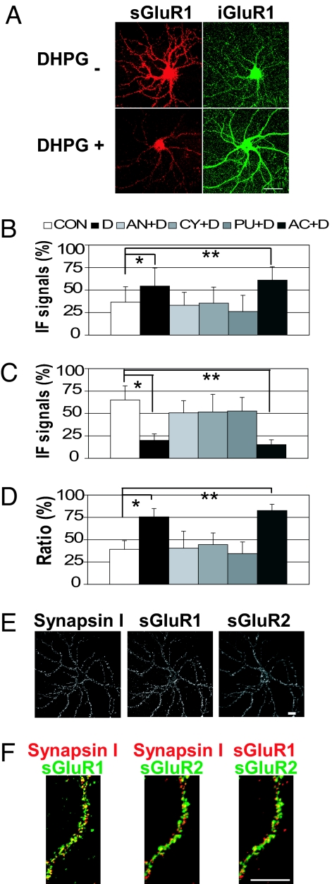
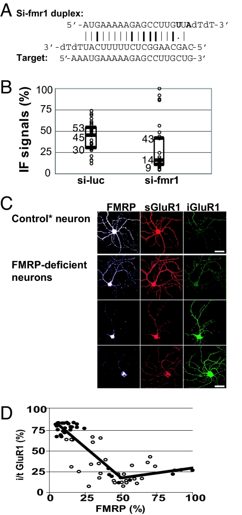
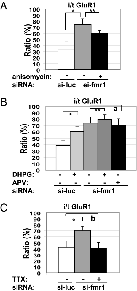
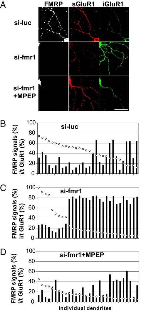
Fragile X syndrome (FXS), a common inherited form of mental retardation, is caused by the functional absence of the fragile X mental retardation protein (FMRP), an RNA-binding protein that regulates the translation of specific mRNAs at synapses. Altered synaptic plasticity has been described in a mouse FXS model. However, the mechanism by which the loss of FMRP alters synaptic function, and subsequently causes the mental impairment, is unknown. Here, in cultured hippocampal neurons, we used siRNAs against Fmr1 to demonstrate that a reduction of FMRP in dendrites leads to an increase in internalization of the alpha-amino-3-hydroxy-5-methyl-4-isoxazole propionic acid receptor (AMPAR) subunit, GluR1, in dendrites. This abnormal AMPAR trafficking was caused by spontaneous action potential-driven network activity without synaptic stimulation by an exogenous agonist and was rescued by 2-methyl-6-phenylethynyl-pyridine (MPEP), an mGluR5-specific inverse agonist. Because AMPAR internalization depends on local protein synthesis after mGluR5 stimulation, FMRP, a negative regulator of translation, may be viewed as a counterbalancing signal, wherein the absence of FMRP leads to an apparent excess of mGluR5 signaling in dendrites. Because AMPAR trafficking is a driving process for synaptic plasticity underlying learning and memory, our data suggest that hypersensitive AMPAR internalization in response to excess mGluR signaling may represent a principal cellular defect in FXS, which may be corrected by using mGluR antagonists.


Conflict of interest statement

The authors declare no conflict of interest.

Figures

Fig. 1.
Fig. 1.
Staining of AMPARs in wild-type live neurons. (A) Representative images of DHPG-induced GluR1 internalization. Remaining surface and internalized GluR1 signals are labeled as sGluR1 and iGluR1, respectively. (Scale bar: 50 μm.) (B–D) Translation inhibitors block DHPG-induced internalization of GluR1. Mean IF signals of internalized GluR1 (B), labeled GluR1 remaining on the surface (C), and ratio of internalized GluR1 in total labeled GluR1 (i/t GluR1) in wild-type dendrites (D) (n = 15 per column). Error bars represent standard deviations. CON, control; D, DHPG, AN, anisomycin; CY, cycloheximide; PU, puromycin; AC, actinomycin D. (B) *, P = 1.3 × 10⁻², **, P = 2.8 × 10⁻⁴. (C) *, P = 6.8 × 10⁻¹¹; **, P = 2.7 × 10⁻¹². (D) *, P = 4.4 × 10⁻¹¹; **, P = 3.9 × 10⁻¹⁴. (E and F) Colocalization of surface GluR1, GluR2, and Synapsin I, a synaptic marker. (Scale bars: 20 μm.) (E) Representative IF images of a wild-type neuron stained for surface GluR1 (sGluR1) and surface GluR2 (sGluR2) under a nonpermeabilized condition followed by Synapsin I staining. (F) Higher magnification images of a dendrite showing colocalization of Synapsin I, sGluR1, and sGluR2 IF signals.
Fig. 2.
Fig. 2.
Reduction of FMRP by siRNA in dendrites correlates with a reduction of labeled GluR1 remaining on the surface and an increase of internalized GluR1. (A) Sequence of the siRNA duplex designed to target Fmr1. Induced mutations are shown in bold letters. G:C and A:U bonds and G:U wobble are indicated by bold lines, plain lines, and a dot, respectively. (B) Box plot of FMRP IF signals in individual dendrites (n = 30) transfected with si-luc or si-fmr1. Median, first quartile, and third quartile are indicated by middle, lower, and upper lines of the boxes and by numbers next to the lines. (C) IF images of representative neurons transfected with si-fmr1. Control neuron was chosen from si-fmr1-treated neurons that showed FMRP levels comparable to si-luc-treated control neurons. (Scale bars: 50 μm.) (D) Piecewise linear regression between FMRP and i/t GluR1 in si-luc- (open circles) and si-fmr1- (filled circles) transfected dendrites (n = 60). Calculated breakpoint is x = 49. Correlation P values are <0.05 if FMRP (%) is below the breakpoint.
Fig. 3.
Fig. 3.
Characterization of aberrant GluR1 internalization in FMRP-deficient dendrites (n = 21 per column). (A) A translation inhibitor partially rescued the aberrant GluR1 internalization. *, P = 1.07 × 10⁻¹⁴; **, P = 8.66 × 10⁻⁸. (B) DHPG-induced additional internalization. *, P = 3.14 × 10⁻¹⁴; **, P = 4.88 × 10⁻² (a = 0.293). (C) Tetrodotoxin rescued the aberrant GluR1 internalization. *, P = 6.81 × 10⁻¹² (b = 0.422).
Fig. 4.
Fig. 4.
MPEP rescues the aberrant internalization of GluR1 after loss of FMRP. (A) Representative IF images of transfected dendrites. (Scale bar: 50 μm.) (B–D) Quantification of IF signals in individual dendrites. Open circles, FMRP IF signals; filled bars, i/t GluR1.

References

    1. Malenka RC, Bear MF. Neuron. 2004;44:5–21. - PubMed
    1. Bredt DS, Nicoll RA. Neuron. 2003;40:361–379. - PubMed
    1. Malenka RC. Ann NY Acad Sci. 2003;1003:1–11. - PubMed
    1. Carroll RC, Beattie EC, von Zastrow M, Malenka RC. Nat Rev Neurosci. 2001;2:315–324. - PubMed
    1. Chang EH, Savage MJ, Flood DG, Thomas JM, Levy RB, Mahadomrongkul V, Shirao T, Aoki C, Huerta PT. Proc Natl Acad Sci USA. 2006;103:3410–3415. - PMC - PubMed

Publication types

MeSH terms

LinkOut - more resources