. 2007 Dec;18(12):4750-61.
doi: 10.1091/mbc.e07-02-0127. Epub 2007 Sep 19.

Myosin VI is required for targeted membrane transport during cytokinesis

Affiliations

Myosin VI is required for targeted membrane transport during cytokinesis

Susan D Arden et al. Mol Biol Cell. 2007 Dec.

Abstract

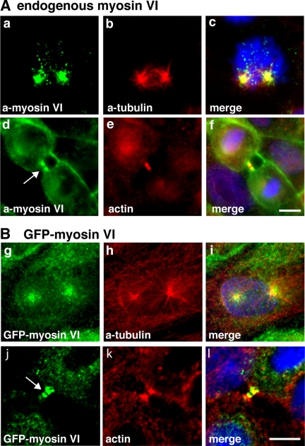
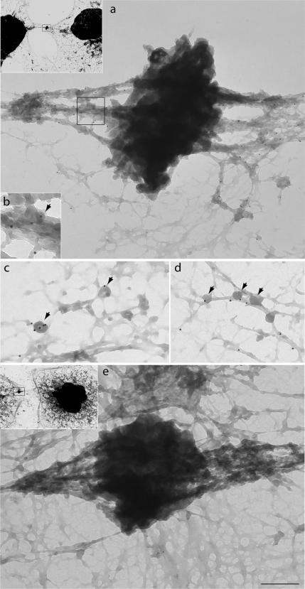
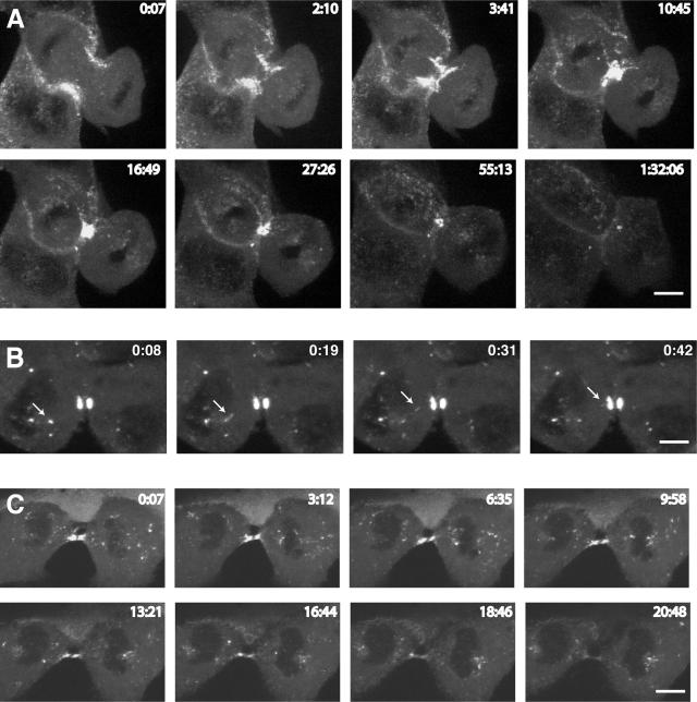
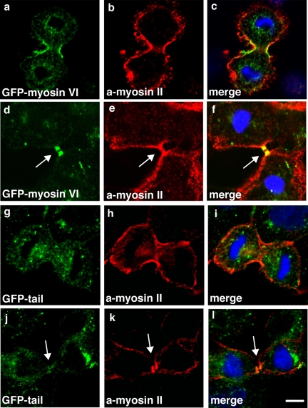
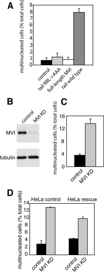
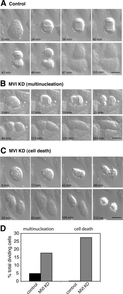
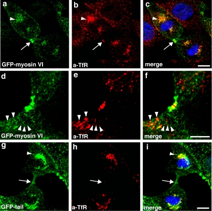
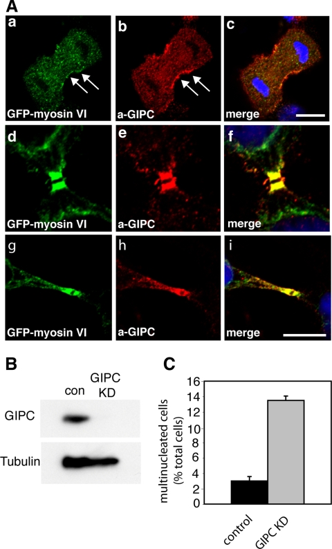
Myosin VI plays important roles in endocytic and exocytic membrane-trafficking pathways in cells. Because recent work has highlighted the importance of targeted membrane transport during cytokinesis, we investigated whether myosin VI plays a role in this process during cell division. In dividing cells, myosin VI undergoes dramatic changes in localization: in prophase, myosin VI is recruited to the spindle poles; and in cytokinesis, myosin VI is targeted to the walls of the ingressing cleavage furrow, with a dramatic concentration in the midbody region. Furthermore, myosin VI is present on vesicles moving into and out of the cytoplasmic bridge connecting the two daughter cells. Inhibition of myosin VI activity by small interfering RNA (siRNA)-mediated knockdown or by overexpression of dominant-negative myosin VI tail leads to a delay in metaphase progression and a defect in cytokinesis. GAIP-interacting protein COOH terminus (GIPC), a myosin VI binding partner, is associated with the function(s) of myosin VI in dividing cells. Loss of GIPC in siRNA knockdown cells results in a more than fourfold increase in the number of multinucleated cells. Our results suggest that myosin VI has novel functions in mitosis and that it plays an essential role in targeted membrane transport during cytokinesis.


Figures

Figure 1.
Figure 1.
In mitotic cells myosin VI is associated with spindle poles in prophase and with the cleavage furrow during cytokinesis. (A) Endogenous myosin VI in MDCK cells was localized using a polyclonal myosin VI tail antibody (a and d). Double labeling with a monoclonal α-tubulin antibody (b) reveals that myosin VI is recruited to spindle poles in prophase and labeling F-actin with Rhodamine-phalloidin (e) highlights the presence of myosin VI in the cytoplasmic bridge between two daughter cells during cytokinesis (d) (arrow). Stable MDCK cells expressing full-length GFP-tagged myosin VI were either stained with GFP-antibody (g and j) or with α-tubulin antibody (h) or for F-actin with Rhodamine-phalloidin (k). GFP-myosin VI is recruited to the spindle poles in prophase (g) and to the midbody region in late cytokinesis (j) (arrow). The merged images together with the DNA stain in blue are shown in (c, f, i, and l). Bars, 10 μm.
Figure 2.
Figure 2.
Immunoelectron microscopy localizes myosin VI to the intercellular bridge on either side of the midbody. The localization of myosin VI at the ultrastructural level was visualized in HeLa cells grown on grids that were saponin extracted and then fixed and stained for immunoelectron microscopy with our polyclonal antibodies to myosin VI tail followed by protein A 10-nm gold. Myosin VI is concentrated along cytoskeletal elements on either side of the dark midbody (a) and is associated with fibrous structures and vesicles of ∼50 nm (arrows highlight vesicles in b–d). A control picture in which grids were incubated with protein A gold only is shown in e. Insets show lower magnification pictures (1000× of original) of cells from which regions were enlarged in a and e. Bar (in e) represents 375 nm (a), 150 nm (b), 200 nm (c), 300 nm (d), and 375 nm (e).
Figure 3.
Figure 3.
Spatial and temporal dynamics of GFP-myosin VI during cytokinesis. To visualize the dynamic behavior of myosin VI during mitosis, MDCK cells stably expressing GFP-myosin VI were imaged using time-lapse microscopy. (A) Gallery of still images of a cell progressing through cytokinesis (see Supplemental Movie 1). (B) A cell in late cytokinesis is shown, highlighting the presence of myosin VI on a vesicle (arrow) moving into the cleavage furrow (see Supplemental Movie 2). (C) Still images of a cell in late cytokinesis show that myosin VI-containing vesicles concentrate on either side of the midbody region and in the polar region around the spindle poles (see Supplemental Movie 3).
Figure 4.
Figure 4.
The myosin VI dominant-negative tail is not recruited into the cleavage furrow or the midbody region. To compare the targeting of full-length myosin VI or only the tail domain during cytokinesis, stable MDCK cells expressing full-length GFP-myosin VI (a and d) or the GFP-tail (g and j) were double labeled with a GFP-mAb and a nonmuscle myosin II polyclonal antibody (b, e, h, and k). The merged images are shown (in c, f, i, and l), and they clearly indicate that the myosin VI tail is not recruited to the walls of the ingressing cleavage furrow or the midbody region. In contrast, in both of these regions full-length myosin VI shows very good colocalization with myosin II. Arrows indicate midbody region. Bars, 10 μm.
Figure 5.
Figure 5.
Loss of Myosin VI causes defects in late cytokinesis. To investigate the role of myosin VI during cytokinesis, wild-type MDCK cells (control) and stable MDCK cell lines expressing either GFP-myosin VI (full-length MVI), GFP-tail (tail wild type), or the GFP-tail with the RRL->AAA mutation (aa 1107–1109) (tail RRL->AAA) were fixed and stained with anti-tubulin antibodies and Hoechst DNA dye. To score the number of multinucleated cells, >2000 interphase cells for each condition were counted using a fluorescence microscope, and the results were plotted as a percentage of the total number of cells (A). Cytokinesis defects in myosin VI KD cells were quantified in HeLa cells that were either mock transfected or transfected with a siRNA SMARTpool specific for myosin VI. After two successive knockdowns, myosin VI protein expression was down to <10% as shown by immunoblotting (B). To quantify cytokinesis defects KD and control cells were fixed and stained with anti-tubulin antibodies and Hoechst DNA dye. The number of multinucleated cells was expressed as a percentage of total cells (C). More than 4000 cells were counted for each condition. (D) For siRNA rescue experiments knockdowns were performed in a HeLa cell line expressing an siRNA-resistant version of myosin VI (HeLa rescue). Numbers of multinucleated cells were quantified as described in C, and they were compared with control HeLa cells. Whereas in wild-type HeLa cells the absence of myosin VI leads to a more than fourfold increase in multinucleation (3–13%), the cell line expressing siRNA-resistant myosin VI only shows a twofold increase in multinucleation (from 4.5 to 9.5%), indicating a 50% rescue resulting from myosin VI expression. More than 2000 cells were counted for each condition.
Figure 6.
Figure 6.
DIC live cell microscopy of myosin VI KD cells characterizes defects in cytokinesis. To visualize how HeLa cells lacking myosin VI are progressing through cytokinesis, control and myosin VI KD cells were imaged using time-lapse DIC microscopy. A gallery of still images of a mock-transfected control cell progressing through cytokinesis is shown in A (see Supplemental Movie 6). (B) Representative series of images of a myosin VI KD cell forming a cleavage furrow that later on regresses to form a binucleated cell (see Supplemental Movie 7). (C) Gallery of images following a cell through cytokinesis; the cell fails to complete abscission and forms two apoptotic daughter cells connected by a thin cytoplasmic bridge (see Supplemental Movie 8). To quantify these defects, the number of multinucleated and dead cells was counted and expressed as a percentage of the total number of dividing cells (D). In total, 41 control and 62 myosin VI KD cells were analyzed and counted. Bars, 20 μm.
Figure 7.
Figure 7.
Overexpression of the dominant-negative myosin VI tail inhibits transport of TfR into the midbody region. Stable MDCK cells expressing full-length GFP-myosin VI (a and d) or only the GFP-tail domain (g) were double labeled in indirect immunofluorescence with a mAb to the TfR (b, e, and h). Full-length GFP-myosin VI colocalizes with the TfR that is concentrated in the polar region (a–c, arrowhead) around the spindle pole and in the midbody region (a–c, arrow). Arrowheads mark single TfR-positive vesicles (d–f) that contain myosin VI. In cells overexpressing the myosin VI tail, the TfR is still present in the polar region, and it is not present in the midbody region (arrow, g–i). Bars, 10 μm.
Figure 8.
Figure 8.
Myosin VI colocalizes with GIPC in dividing cells, and the loss of GIPC causes defects in cytokinesis. (A) MDCK cells stably expressing GFP-myosin VI were labeled with a GFP-antibody (a, d, and g) and with a GIPC antibody (b, e, and h). The merged images together with the DNA stain in blue are shown in c, f, and i. Myosin VI and GIPC are both recruited to the ingressing cleavage furrow in late anaphase/telophase (arrows in a and b), and they show perfect colocalization in the midbody region in late cytokinesis (d–i). Bars, 10 μm. (B) Western blot showing GIPC expression levels in mock-treated control cells and GIPC KD cells transfected with a SMARTpool of siRNA specific for GIPC. An α-Tubulin blot is shown as a loading control. After two knockdowns (72 h), no GIPC can be detected by immunoblotting. (C) HeLa grown on coverslips were transfected twice with SMARTpool siRNA specific for GIPC. Twenty-four hours after the second transfection, the cells were fixed and stained with Hoechst DNA dye. The number of multinucleated cells was counted and expressed as a percentage of the total number of cells. More than 2500 cells were counted for each condition.

References

    1. Albertson R., Riggs B., Sullivan W. Membrane traffic: a driving force in cytokinesis. Trends Cell Biol. 2005;15:92–101. - PubMed
    1. Aschenbrenner L., Lee T., Hasson T. Myo6 facilitates the translocation of endocytic vesicles from cell peripheries. Mol. Biol. Cell. 2003;14:2728–2743. - PMC - PubMed
    1. Au J., Puri C., Ihrke G., Kendrick-Jones J., Buss F. Myosin VI is required for sorting of AP-1B dependent cargo to the basolateral domain in polarised MDCK cells. J. Cell Biol. 2007;177:103–114. - PMC - PubMed
    1. Bunn R. C., Jensen M. A., Reed B. C. Protein interactions with the glucose transporter binding protein GLUT1CBP that provide a link between GLUT1 and the cytoskeleton. Mol. Biol. Cell. 1999;10:819–832. - PMC - PubMed
    1. Buss F., Arden S. D., Lindsay M., Luzio J. P., Kendrick-Jones J. Myosin VI isoform localized to clathrin-coated vesicles with a role in clathrin-mediated endocytosis. EMBO J. 2001;20:3676–3684. - PMC - PubMed

Publication types

LinkOut - more resources