. 2007 Dec;18(12):4780-93.
doi: 10.1091/mbc.e06-12-1144. Epub 2007 Sep 19.

Interaction of ezrin with the novel guanine nucleotide exchange factor PLEKHG6 promotes RhoG-dependent apical cytoskeleton rearrangements in epithelial cells

Affiliations

Interaction of ezrin with the novel guanine nucleotide exchange factor PLEKHG6 promotes RhoG-dependent apical cytoskeleton rearrangements in epithelial cells

Romina D'Angelo et al. Mol Biol Cell. 2007 Dec.

Abstract

The mechanisms underlying functional interactions between ERM (ezrin, radixin, moesin) proteins and Rho GTPases are not well understood. Here we characterized the interaction between ezrin and a novel Rho guanine nucleotide exchange factor, PLEKHG6. We show that ezrin recruits PLEKHG6 to the apical pole of epithelial cells where PLEKHG6 induces the formation of microvilli and membrane ruffles. These morphological changes are inhibited by dominant negative forms of RhoG. Indeed, we found that PLEKHG6 activates RhoG and to a much lesser extent Rac1. In addition we show that ezrin forms a complex with PLEKHG6 and RhoG. Furthermore, we detected a ternary complex between ezrin, PLEKHG6, and the RhoG effector ELMO. We demonstrate that PLEKHG6 and ezrin are both required in macropinocytosis. After down-regulation of either PLEKHG6 or ezrin expression, we observed an inhibition of dextran uptake in EGF-stimulated A431 cells. Altogether, our data indicate that ezrin allows the local activation of RhoG at the apical pole of epithelial cells by recruiting upstream and downstream regulators of RhoG and that both PLEKHG6 and ezrin are required for efficient macropinocytosis.


Figures
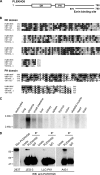
Figure 1.
Figure 1.
Structural organization and expression pattern of human PLEKHG6. (A) PLEKHG6 contains a Dbl homology domain (DH) followed by a plekstrin homology (PH) domain. The bar represents the longest cDNA isolated in the yeast two-hybrid screen that encodes the ezrin binding site (aa 579-790). (B) Sequence comparison of DH and PH domains from PLEKHG6, human GEF720 (Acc. no BAA34440), D. melanogaster CG7323 (Acc. no NP_649182), and C. elegans T08H4.1 (Acc. no NP_494723). Black and gray letters indicate identical and conserved amino acids, respectively. (C) Northern blot analysis of PLEKHG6 mRNA expression in human tissues. A single band at ∼3 kb was detected. (D) PLEKHG6 is expressed in JEG-3, LLC-PK1, and A431 cells. Cells lysates from JEG-3, LLC-PK1, and A431 cells were immunoprecipitated with an anti-PLEKHG6 antibody and immunoblotting on total cell lysates, and immunoprecipitates was performed with the same antibody. Lane 1, lysate from 293T cells transfected with Myc-PLEKHG6 used as control.
Figure 2.
Figure 2.
The C-terminal region of PLEKHG6 interacts with the N-terminal domain of ezrin. (A) Schematic representation of the domain structures of ezrin and PLEKHG6 and of the different forms used in the experiments. Ezrin contains a globular amino-terminal domain (FERM domain) followed by an α-helical region and a F-actin–binding site. (B) S35Met-labeled PLEKHG6-CT produced by in vitro transcription/translation was incubated with either GST or GST-fused ezrin constructs. The translation product corresponding to PLEKHG6-CT used in each reaction (input) or bound to GST-ezrin constructs was visualized by autoradiography. (C) PLEKHG6-wt and -ΔNT interact only with ezrin-NT. GST or GST-fused ezrin constructs immobilized on glutathione beads were incubated with lysates from 293T cells expressing Myc-tagged PLEKHG6-wt, -ΔNT, and -ΔCT. Bound PLEKHG6 proteins were detected by Western blot with an anti-Myc antibody. (D) PLEKHG6 binding site on ezrin is cryptic. 293T cell lysates expressing Myc-tagged PLEKHG6 were incubated with GST-ezrin-NT in the presence or not of ezrin-CT. Left panel, ponceau staining of GST-ezrin-NT and ezrin-CT. Right panel, Western blot with anti-Myc antibody. Addition of the purified ezrin-CT in the pulldown assay prevents the association of PLEKHG6 with GST-ezrin-NT. (E) PLEKHG6 coimmunoprecipitates with ezrin T567D. LLC-PK1 cells stably transfected with the vector (−), the cDNAs coding for wild-type ezrin (wt) or for ezrin T567D (T/D) were transiently transfected with the plasmids encoding either Myc-tagged PLEKHG6-wt or -ΔCT. Immunoprecipitation was performed with an anti-VSVG antibody (top panel). Western blot was performed with anti-Myc or anti-VSVG antibodies on immunoprecipitated proteins (top panel) and on cell lysates (bottom panels). (F) Endogenous PLEKHG6 immunoprecipitates ezrin T567D. Immunoprecipitation with an anti-PLEKHG6 antibody was performed in LLC-PK1 cells stably expressing VSVG-tagged ezrin wt or T567D. Western blot was performed with anti-VSVG and anti-PLEKHG6 on immunoprecipitated proteins (top panel) and cell lysates (bottom panel). Endogenous PLEKHG6 is detectable only after immunoprecipitation.
Figure 3.
Figure 3.
PLEKHG6 displays a guanine nucleotide exchange activity preferentially toward RhoG. (A) Morphogenic effects induced by PLEKHG6 in NIH3T3 cells. NIH3T3 cells transiently expressing Myc-tagged PLEKHG6 were stained with an anti-Myc antibody (green) and rhodamine-phalloidin (F-actin). PLEKHG6 induces dorsal microvilli and ruffles. A loss of actin stress fibers is observed in cells expressing PLEKHG6. Scale bar, 20 μm. (B) PLEKHG6 guanine nucleotide exchange activity in yeast. β-galactosidase activity reflecting the exchange activity of indicated PLEKHG6 forms toward RhoG (left panel) or Rac (right panel) is shown. PLEKHG6 displays an exchange activity toward RhoG and to a much lesser extent toward Rac. Trio-GEFD1 and βPix were used as positive and negative controls, respectively. The different PLEKHG6 proteins were produced at similar levels in yeast (data not shown). (C) PLEKHG6 stimulates GTP-loading on RhoG in vivo. Left panel: lysates of LLC-PK1 cells expressing HA-tagged RhoG and Myc-tagged PLEKHG6 were incubated with GST-ELMO immobilized on beads. GTP-RhoG (top) and total RhoG (middle) were detected with an anti-HA antibody. PLEKHG6 proteins were detected in the lysates with an anti-Myc antibody (bottom). As a control, lysates of cells expressing HA-RhoGQ61L were incubated with GST-ELMO. Right panel: lysates of LLC-PK1 cells expressing PLEKHG6 proteins were incubated with GST-CRIB immobilized on beads. GTP-Rac1 or GTP-Cdc42 (top), and total Rac1 or Cdc42 (middle) was detected with anti-Rac1 and Cdc42 antibodies, respectively. PLEKHG6 were detected with an anti-Myc mantibody (bottom). As a control, lysates of cells expressing Myc-Rac1Q61L or Myc-Cdc42Q61L were incubated with GST-CRIB. Densitometric quantification: Rac activity was calculated from the amount of GTP-bound Rac1 normalized to the amount of total Rac in cell lysates. An arbitrary unit of 1 was given for Rac1 activity in cells that do not produce PLEKHG6. Rac activity is induced 2.3-fold in cells producing PLEKHG6-wt and 1.6-fold in cells producing PLEKHG6-ΔCT. Error bars, the SD between three independent experiments. (D) RhoG is the main target of PLEKHG6 in vivo. 293T cells were cotransfected with plasmids encoding Myc-PLEKHG6 and GFP-RhoGTN17 or GFP-RacT17N. Cell lysates were immunoprecipitated with an anti-GFP antibody and immunoblotting was performed with an anti-Myc antibody to detect associated PLEKHG6 proteins. Western blots were performed on cell lysates with the indicated antibodies (bottom panel).
Figure 4.
Figure 4.
PLEKHG6 induces apical microvilli and ruffles in epithelial cells. (A) LLC-PK1 cells transfected with Myc-tagged PLEKHG6-wt or PLEKHG6-ΔCT were immunostained with a Myc antibody. F-actin was stained with rhodamine-phalloidin. Scale bar, 20 μm. (B) Double immunofluorescence was performed with an anti-Myc and an anti-ezrin antibody. Endogenous ezrin colocalizes with Myc-PLEKHG6 in the apical microvilli and ruffles. PLEKHG6-ΔCT lacking the ezrin binding site induces extensive lamellipodia. Scale bar, 20 μm.
Figure 5.
Figure 5.
Ezrin targets PLEKHG6 to the apical surface of epithelial cells. (A) LLC-PK1 cells expressing Myc-tagged PLEKHG6-N351A or PLEKHG6-N351A-ΔCT were immunostained with a monoclonal anti-Myc antibody (red) and a polyclonal anti-ezrin antibody (green). PLEKHG6-N351A but not PLEKHG6-N351A-ΔCT colocalizes with ezrin to the apical microvilli (merged and inset). (B) PLEKHG6-induced morphogenic effects are impaired in presence of RhoGT17N or RhoGF37A. Immunofluorescence of LLC-PK1 cells expressing Myc-tagged PLEKHG6-wt and GFP-RhoGT17N or GFP-RhoGF37A was performed with an anti-Myc antibody. Scale bar, 20 μm.
Figure 6.
Figure 6.
Ezrin forms a ternary complex with PLEKHG6 and RhoG. 293T cells were cotransfected with plasmids encoding Myc-PLEKHG6-wt or Myc-PLEKHG6-ΔCT, GFP-RhoGT17N and VSVG-ezrin-Δ29 as indicated. Immunoprecipitations were performed with an anti-VSVG antibody followed by immunoblotting with anti-Myc and anti-GFP antibodies. Western blots were performed on cell lysates with the indicated antibodies (bottom panel).
Figure 7.
Figure 7.
Ezrin and PLEKHG6 interact with ELMO. (A) PLEKHG6, ezrin, and ELMO form a ternary complex. 293T cells were cotransfected with the indicated plasmids. Immunoprecipitations were performed with anti-GFP (top panel) or anti-VSVG (middle panel) antibodies. Western blots on the immunoprecipitated proteins and cell lysates were performed with the indicated antibodies. (B) PLEKHG6 interacts with ELMO. Lysates of 293T cells producing various Myc-tagged forms of PLEKHG6 were incubated with GST-ELMO. Proteins were detected by Western blot with an anti-Myc antibody.
Figure 8.
Figure 8.
PLEKHG6 and ezrin are required for macropinocytosis in EGF-stimulated A431 cells. (A) Inhibition of PLEKHG6 expression by shRNAs. Top panel, JEG-3 cells were cotransfected with PLEKHG6 or scramble psiRNAs and cDNA encoding Myc-PLEKHG6. The levels of Myc-PLEKHG6 in cells producing three different shRNAs were analyzed by Western blot with an anti-Myc antibody. Anti-tubulin antibody was used as loading control. Bottom panel, semiquantitative RT-PCR was performed with total RNA extracted from A431 cells transfected with PLEKHG6 or scramble psiRNAs. GADPH was used as control for PCR. (B) Top panel, Western blot performed with anti-ezrin and anti-tubulin antibodies on A431 cell extracts after 3-d transfection with ezrin or scramble psiRNAs. Bottom panel, fluorescent staining with anti-ezrin antibody and Alexa Fluor 350 phalloidin was performed on cells transfected with scramble or ezrin psiRNAs. Transfected cells express GFP. No ezrin staining was detected in cells transfected with either psiEz1 or psiEz2 (not shown). (C) Ezrin knockdown inhibits PLEKHG6-induced dorsal ruffles. After 2-d transfection with ezrin or scramble psiRNAs, A431 cells were transfected with the plasmid encoding Myc-PLEKHG6-wt. Fluorescent staining was performed with anti-myc antibody and Alexa Fluor 350 phalloidin. The merged image represents F-actin staining in red, and the GFP is indicative of cells transfected with the psiRNA. (D) Fluorescent-dextran uptake in EGF-treated A431 cells. Three days after transfection of A431 cells with psiRNAs, the uptake of TRITC-dextran (TRDx) was performed in presence of EGF. Dextran uptake was scored in transfected cells producing GFP. For each psiRNA and in three independent experiments, at least 50 cells expressing GFP were examined for dextran uptake. As shown in the bar diagram, TRITC-dextran uptake was observed in ~65% of cells producing scramble shRNA, whereas only ~23 and 27% of cells producing PLEKHG6 and ezrin shRNAs, respectively, were positive. Error bars, SD. Scale bar, 20 μm.

References

    1. Aghazadeh B., Zhu K., Kubiseski T. J., Liu G. A., Pawson T., Zheng Y., Rosen M. K. Structure and mutagenesis of the Dbl homology domain. Nat. Struct. Biol. 1998;5:1098–1107. - PubMed
    1. Barret C., Roy C., Montcourrier P., Mangeat P., Niggli V. Mutagenesis of the phosphatidylinositol 4,5-biphosphate (PIP2) binding site in the NH2-terminal domain of ezrin correlates with its altered cellular distribution. J. Cell Biol. 2000;151:1067–1079. - PMC - PubMed
    1. Berryman M., Franck Z., Bretscher A. Ezrin is concentrated in the apical microvilli of a wide variety of epithelial cells whereas moesin is found primarily in endothelial cells. J. Cell Sci. 1993;105:1025–1043. - PubMed
    1. Berryman M., Gary R., Bretscher A. Ezrin oligomers are major cytoskeletal components of placental microvilli: a proposal for their involvement in cortical morphogenesis. J. Cell Biol. 1995;131:1231–1242. - PMC - PubMed
    1. Blangy A., Vignal E., Schmidt S., Debant A., Gauthier-Rouviere C., Fort P. TrioGEF1 controls Rac- and Cdc42-dependent cell structures through the direct activation of rhoG. J. Cell Sci. 2000;113(Pt 4):729–739. - PubMed

Publication types

MeSH terms

LinkOut - more resources