. 2007 Dec;18(12):4847-58.
doi: 10.1091/mbc.e06-06-0539. Epub 2007 Sep 19.

Distinct sequence elements of cyclin B1 promote localization to chromatin, centrosomes, and kinetochores during mitosis

Affiliations

Distinct sequence elements of cyclin B1 promote localization to chromatin, centrosomes, and kinetochores during mitosis

Anna M Bentley et al. Mol Biol Cell. 2007 Dec.

Abstract

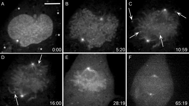
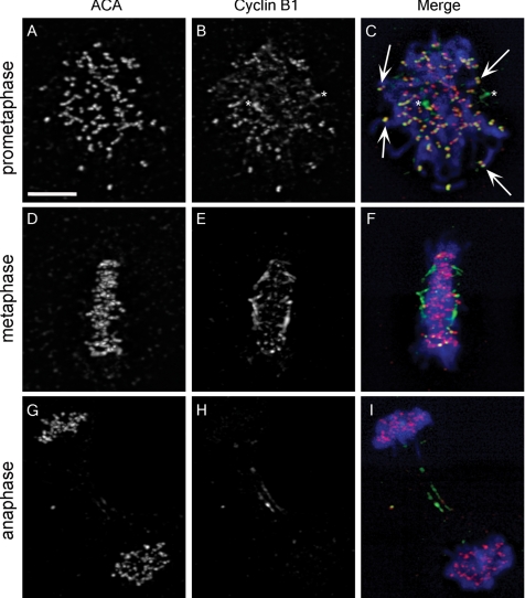
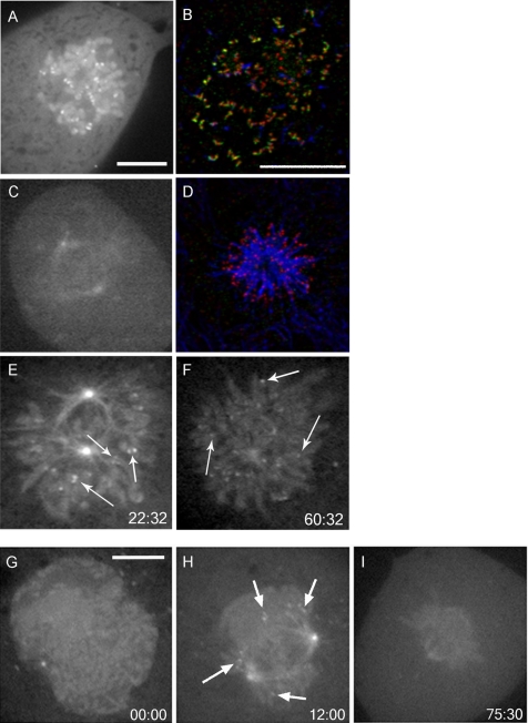
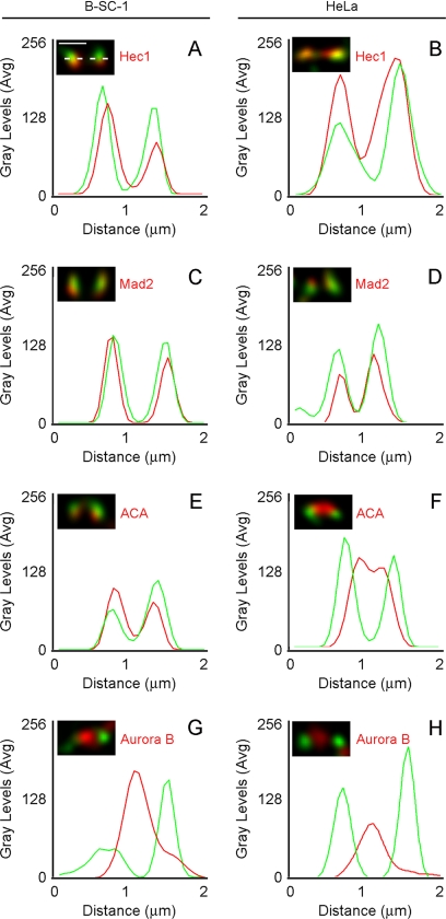
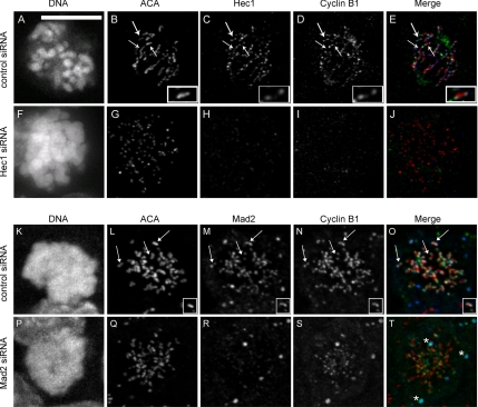
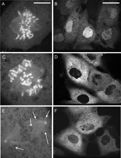
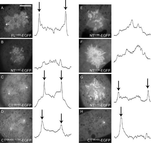
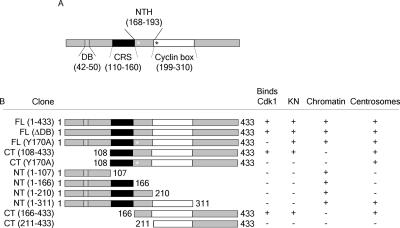
The mitotic cyclins promote cell division by binding and activating cyclin-dependent kinases (CDKs). Each cyclin has a unique pattern of subcellular localization that plays a vital role in regulating cell division. During mitosis, cyclin B1 is known to localize to centrosomes, microtubules, and chromatin. To determine the mechanisms of cyclin B1 localization in M phase, we imaged full-length and mutant versions of human cyclin B1-enhanced green fluorescent protein in live cells by using spinning disk confocal microscopy. In addition to centrosome, microtubule, and chromatin localization, we found that cyclin B1 also localizes to unattached kinetochores after nuclear envelope breakdown. Kinetochore recruitment of cyclin B1 required the kinetochore proteins Hec1 and Mad2, and it was stimulated by microtubule destabilization. Mutagenesis studies revealed that cyclin B1 is recruited to kinetochores through both CDK1-dependent and -independent mechanisms. In contrast, localization of cyclin B1 to chromatin and centrosomes is independent of CDK1 binding. The N-terminal domain of cyclin B1 is necessary and sufficient for chromatin association, whereas centrosome recruitment relies on sequences within the cyclin box. Our data support a role for cyclin B1 function at unattached kinetochores, and they demonstrate that separable and distinct sequence elements target cyclin B1 to kinetochores, chromatin, and centrosomes during mitosis.


Figures

Figure 1.
Figure 1.
FL1-433 localizes to unattached kinetochores during mitosis. (A–E) BS-C-1 cells expressing FL1-433 were imaged using time-lapse, spinning disk confocal microscopy from prophase through metaphase completion. Elapsed time (minutes:seconds) from the first acquisition is shown in the lower right corner. (A) FL1-433 localizes to the nucleus and centrosomes in prophase. Asterisks indicate punctate cytoplasmic signal. (B) After NEB, FL1-433 associates with chromatin and centrosomes, and punctate kinetochore signal becomes apparent. (C) Later in prometaphase, kinetochore signal is prominent at outlying chromosomes (arrows) and along spindle microtubules. (D) As chromosomes congress to the metaphase plate, kinetochore-associated signal is diminished, but kinetochores adjacent to centrosomes continue to show strong kinetochore signal (arrows). (E) By metaphase, kinetochore signal is not observed, but chromatin and centrosome signal remain. (F) Before its degradation, FL1-433 signal is lost from chromatin. Bar, 10 μm (A). A movie of this cell is shown in Supplemental Video 1.
Figure 2.
Figure 2.
Endogenous cyclin B1 localizes to kinetochores. (A, D, and G) ACA recognize kinetochores during prometaphase, metaphase, and anaphase. (B) An antibody that reacts with an N-terminal epitope in cyclin B1 has signal at kinetochores, but only during prometaphase. Arrows in C indicate colocalization of cyclin B1 and ACA. Asterisks in B and C designate centrosomes. (E and F) Cyclin B1 localization is depleted at kinetochores during metaphase, whereas ACA signal is robust (D and F). In anaphase, ACA remains at kinetochores (G and I), but cyclin B1 signal is depleted except for a reproducible population at the midzone (H and I). Merged images show DNA staining in blue, ACA staining in red, and cyclin B1 staining in green (C, F, and I). Bar, 10 μm (A).
Figure 3.
Figure 3.
Kinetochore localization of FL1-433 is sensitive to microtubule attachment but not tension. FL1-433–expressing BS-C-1 cells were treated with 0.5 μM nocodazole (A and B), 0.5 μM taxol (C and D), or monastrol (100 μg/ml) (E and F). (A) After nocodazole treatment, FL1-433 localizes to all kinetochores. (C) Taxol causes collapse of the spindle but no accumulation of FL1-433 at kinetochores. Cells treated with nocodazole (B) or taxol (D) were stained for cyclin B1 (green), tubulin (blue), and ACA (red), and recapitulate cyclin B1 dynamics was observed in live cells. (E and F) Cells were treated with monastrol and imaged using time-lapse, spinning disk confocal microscopy. Elapsed time (minutes:seconds) from the addition of monastrol and first acquisition is shown in the bottom right corner. (E) Early in monastrol treatment, FL1-433 localizes to centrosomes, microtubules and chromatin. FL1-433 is also present at paired kinetochores (arrows). (F) After prolonged monastrol arrest, FL1-433 is retained at centrosomes, microtubules, and chromatin. Paired FL1-433 evolves into singlets of kinetochore signal (arrows). This cell is shown in Supplemental Video 2. (G–I) Cells expressing FLΔDB were imaged using time-lapse, spinning disk confocal microscopy. Elapsed time (minutes:seconds) from the first acquisition is shown in the bottom right corner. (G) FLΔDB is enriched in the nucleus at prophase and localizes to centrosomes, spindle microtubules, chromatin, and kinetochores during mitosis (H). (I) FLΔDB is delocalized from kinetochores upon metaphase completion. Bar, 10 μm (A) for A, C, E and F; for B and D (B), and for G–I (G).
Figure 4.
Figure 4.
Cyclin B1 colocalizes with Hec1 and Mad2 to the outer kinetochore. (A–H) Images of fixed B-SC-1 and HeLa cells were acquired by spinning disk confocal microscopy at 0.3-μm Z-intervals and deconvolved. (A) Kinetochore-associated signal was measured by line scan (dotted line) across the length of a kinetochore pair. Average gray levels were plotted against calibrated distance for each line scan. (A–H) Cyclin B1 signal (green) was compared with the signal from Hec1, Mad2, ACA, or Aurora B antibodies (red). The relative distributions of cyclin B1 were similar in B-SC-1 and HeLa cells. At least five kinetochore pairs from each of five separate cells were assessed to select representative pairs for this analysis. Bar, 1 μm (A).
Figure 5.
Figure 5.
Cyclin B1 does not localize to kinetochores after Hec1 or Mad2 depletion. HeLa cells were transfected with control (A–E, and K–O), Hec1 (F–J), or Mad2-directed siRNA (P–T) for 48 h and then processed for immunofluorescence. In control siRNA-transfected cells, cyclin B1 (D and N) localized with Hec1 (C) or Mad2 (M) at kinetochores (B and L, arrows) in mitotic cells. A single kinetochore pair is enlarged to better show colocalization (B–E and L–O, insets). In cells depleted for Hec1 (F–J), chromatin is disorganized within the spindle (F). ACA (G) is present on kinetochores that are depleted for Hec1 (H), whereas cyclin B1 is absent from kinetochores (I). In the merged images (E and J), ACA (red), Hec1 (blue), and cyclin B1 (green) are shown. In cells depleted for Mad2 (P–T), ACA (Q) is present on kinetochores that are depleted for Mad2 (R), whereas cyclin B1 is reduced (S). In the merged images (O and T), ACA (red), Mad2 (blue), and cyclin B1 (green) are shown. Asterisks in T represent nonkinetochore signal specific to the primary antibody. Scale, 10 μm (A).
Figure 6.
Figure 6.
The N-terminal domain of cyclin B1 is necessary and sufficient for localization to chromatin. (A) During mitosis, NT1-107 localizes predominantly to chromatin. Centrosome signal is severely reduced and kinetochore signal is absent. (B) NT1-107 is constitutively nuclear during interphase. NT1-166 localizes to chromatin but not to kinetochores during mitosis (C) and is cytoplasmic during interphase (D). (E) CT108-433 localizes to centrosomes, spindle microtubules, and kinetochores (arrows) during prometaphase but not to chromatin (E). (F) CT108-433 is cytoplasmic during interphase. Bar, 10 μm (A), which also applies to C and E. Bars, 10 μm (A; same scale for C and E) or 35 μm (B; same scale for D and F).
Figure 7.
Figure 7.
Cyclin B1 localizes to kinetochores by CDK-1–dependent and – independent mechanisms. Lysates from adenovirally infected BS-C-1 cells were immunoprecipitated with an anti-GFP antibody. (A) Control BS-C-1 lysates (C, lane 1), uninfected BS-C-1 (UN), FL1-433 (FL), NT1-107 (NT), FLY170A (Y170A), CT108-433 (CT), and CT108-433, Y170A (CT-Y170A) immunoprecipitates were immunoblotted for GFP and CDK1. An immunoglobulin G-reactive band is present in each sample (α-GFP). CDK1 associates with FL1-433 and CT108-433 but not with NT1-107, FLY170A or CT108-433, Y170A (α-CDK1). By spinning disk confocal microscopy, FL1-433 (B) and FLY170A (C) localize to chromatin, centrosomes, spindle microtubules, and kinetochores. Arrows in C indicate kinetochore localization. (D) CT108-433 localizes to centrosomes, spindle microtubules, and kinetochores as shown in Figure 6F. (E) CT108-433, Y170A does not localize to kinetochores. (F) Quantitation of normalized integrated signal intensity at kinetochores of FL1-433–, FLY170A–, and CT108-433–expressing cells in untreated conditions and after treatment with 100 nM nocodazole. Error bars indicate 95% confidence levels. An asterisk represents a statistically significant difference in integrated intensity at kinetochores in the presence of 100 nM nocodazole versus untreated. Bar, 10 μm (A).
Figure 8.
Figure 8.
A complete cyclin box is required for cyclin B1 localization to centrosomes. (A–H) Centrosome signal was measured by line scan across the length of the spindle. Representative examples of B-SC-1 mitotic cells expressing FL1-433 (A), NT1-107 (B), CT108-433 (C), CT108-433, Y170A (D), NT1-166 (E), NT1-210 (F), NT1-311 (G), and CT166-433 (H) are shown. Peaks in integrated intensity (arrows) correspond with positive centrosome signal. The presence of one centrosome in the plane of focus in H is reflected by a single peak in the corresponding line scan. Bar, 10 μm (A).
Figure 9.
Figure 9.
A summary of localization determinants of cyclin B1. (A) Schematic of significant sequence domains of cyclin B1. The Destruction box (DB), CRS, N-terminal helix (NTH), and cyclin box are shown. (A) Residues are indicated for each domain in parentheses. A white asterisk represents the Y170A point mutation, and a black asterisk represents the MRAIL residues in the beginning of the cyclin box. (B) The name and schematic for each cyclin B1 construct is indicated. The + and − indicate whether that particular construct binds CDK1, localizes to kinetochores (KN), chromatin or centrosomes.

Similar articles

Cited by

References

    1. Acquaviva C., Herzog F., Kraft C., Pines J. The anaphase promoting complex/cyclosome is recruited to centromeres by the spindle assembly checkpoint. Nat. Cell Biol. 2004;6:892–898. - PubMed
    1. Arnaoutov A., Dasso M. The Ran GTPase regulates kinetochore function. Dev. Cell. 2003;5:99–111. - PubMed
    1. Bailly E., Cabantous S., Sondaz D., Bernadac A., Simon M. N. Differential cellular localization among mitotic cyclins from Saccharomyces cerevisiae: a new role for the axial budding protein Bud3 in targeting Clb2 to the mother-bud neck. J. Cell Sci. 2003;116:4119–4130. - PubMed
    1. Brown N. R., Lowe E. D., Petri E., Skamnaki V., Antrobus R., Johnson L. N. Cyclin B and cyclin a confer different substrate recognition properties on CDK2. Cell Cycle. 2007;6:1350–1359. - PubMed
    1. Brown N. R., Noble M. E., Endicott J. A., Johnson L. N. The structural basis for specificity of substrate and recruitment peptides for cyclin-dependent kinases. Nat. Cell Biol. 1999;1:438–443. - PubMed

Publication types

MeSH terms