. 2007 Sep 20;449(7160):346-50.
doi: 10.1038/nature06129.

Generation of functional multipotent adult stem cells from GPR125+ germline progenitors

Affiliations

Generation of functional multipotent adult stem cells from GPR125+ germline progenitors

Marco Seandel et al. Nature. .

Abstract

Adult mammalian testis is a source of pluripotent stem cells. However, the lack of specific surface markers has hampered identification and tracking of the unrecognized subset of germ cells that gives rise to multipotent cells. Although embryonic-like cells can be derived from adult testis cultures after only several weeks in vitro, it is not known whether adult self-renewing spermatogonia in long-term culture can generate such stem cells as well. Here, we show that highly proliferative adult spermatogonial progenitor cells (SPCs) can be efficiently obtained by cultivation on mitotically inactivated testicular feeders containing CD34+ stromal cells. SPCs exhibit testicular repopulating activity in vivo and maintain the ability in long-term culture to give rise to multipotent adult spermatogonial-derived stem cells (MASCs). Furthermore, both SPCs and MASCs express GPR125, an orphan adhesion-type G-protein-coupled receptor. In knock-in mice bearing a GPR125-beta-galactosidase (LacZ) fusion protein under control of the native Gpr125 promoter (GPR125-LacZ), expression in the testis was detected exclusively in spermatogonia and not in differentiated germ cells. Primary GPR125-LacZ SPC lines retained GPR125 expression, underwent clonal expansion, maintained the phenotype of germline stem cells, and reconstituted spermatogenesis in busulphan-treated mice. Long-term cultures of GPR125+ SPCs (GSPCs) also converted into GPR125+ MASC colonies. GPR125+ MASCs generated derivatives of the three germ layers and contributed to chimaeric embryos, with concomitant downregulation of GPR125 during differentiation into GPR125- cells. MASCs also differentiated into contractile cardiac tissue in vitro and formed functional blood vessels in vivo. Molecular bookmarking by GPR125 in the adult mouse and, ultimately, in the human testis could enrich for a population of SPCs for derivation of GPR125+ MASCs, which may be employed for genetic manipulation, tissue regeneration and revascularization of ischaemic organs.


Figures
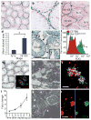
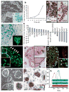
Figure 1
Figure 1. Restricted GPR125 expression in adult mouse testis and derivation of multipotent cells from spermatogonial progenitor cells (SPCs)
a–c, X-gal staining of adult GPR125–LacZ mouse testis. d, Quantification of X-gal staining (arbitrary units) in tubules grouped as stages IV–V (0.98 ± 0.11 (mean ± s.e.m.); n = 30 tubules) versus stages VII–VIII (3.84 ± 0.49; n = 28; *P <0.001 by Wilcoxon test). e, Anti-GPR125 staining (brown, arrows) of adult mouse testis. f, Flow cytometry on freshly dissociated adult Gpr125lacZ/lacZ testis. Roman numerals in ce denote approximate stages of the seminiferous tubules. g, Anti-CD34 staining (brown) of peritubular/interstitial mouse cells, which remain CD34+ (inset, green staining) following in vitro expansion. h, i, Highly proliferative SPC colonies (h) that express PLZF (i, green staining) after expansion on inactivated CD34+MTS. j, The number (mean ± s.d.) of SPCs doubled every ~2 days. k, l, The appearance of MASCs derived from Gt(Rosa26)Sor-lacZ SPCs following transfer to MEF for expansion and antibody staining, revealing OCT4 expression in the nucleus (green, right panel in l). Scale bars, 50 μm. Nuclei are shown in red (ac, i, l) or blue (e, g).
Figure 2
Figure 2. Characterization and multipotent derivatives of Gpr125lacZ/lacZ SPC lines
a, Morphology of SPC colonies and expression of GPR125 by X-gal staining (blue, inset). b, Proliferation of GPR125+ SPCs (GSPCs) in culture. c, Immunolabelling by germ-cell markers GCNA (brown, left panel), and anti-DAZL (brown, right panel). Absent staining in feeders is denoted by asterisks. d, Expression of GPR125–LacZ in cloned GSPCs (tracked by GFP (green, inset), using lentivirus). e, Quantitative PCR of Gpr125lacZ/lacZ GSPCs compared to Gpr125lacZ/lacZ total testis. Bars depict fold change (± scaled s.d.) of gene expression (compared with total testis) of genes associated with GSPCs or differentiating spermatogenic cells. fh, Engraftment of Gpr125lacZ/lacZ GSPCs microinjected into busulphan-treated testes. f, Confocal slices (~1 μm, inset) distinguishing areas with GFPbright spermatogonia along the basement membrane (arrows) from centrally located areas containing smaller, round GFPdim differentiating cells, in the projection of 32 slices. g, GPR125 expression by X-gal staining (blue, arrowheads) present in engrafted cells along the basement membrane. h, Differentiation of donor-derived GFP+ cells and GFP non-engrafted tubules (arrowheads, GFP+ spermatids; asterisk, non-engrafted tubule). i, Derivation of GPR125+MASCs (blue staining in inset, X-gal) from GSPCs. j, Nuclear labelling by anti-OCT4 (brown). k, Flow cytometry for GPR125 expression in Gpr125lacZ/lacZ MASCs or GSPCs by FDG staining (mean fluorescence intensity: 22.1 or 18.2, respectively, versus 2.2 in wild-type GSPC control). Scale bars, 50 μm.
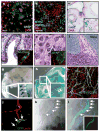
Figure 3
Figure 3. GPR125–LacZ MASCs exhibit multipotency and can form functional vessels
a, b, Embryoid bodies differentiated in vitro and immunolabelled for neuroectoderm (anti-GFAP, green; a); mesoderm (anti-myosin heavy chain, myosin HC, blue; b); and endoderm or ectoderm (using anti-HNF3β, green; b). c, X-gal-stained GPR125–LacZ MASC teratoma formed in NOD-SCID mice. df, Teratoma histology showing endodermal (d), ectodermal (e), and mesodermal (f) elements. Immunofluorescence (green) in insets: d, anti-Muc5ac; e, anti-GFAP. g, h, Whole-mount embryo X-gal staining. g, E13.5 GPR125–LacZ MASC chimaera formed by blastocyst injection. h, E14.5 full heterozygous Gpr125+/lacZ embryo. g, h, Arrowheads denote putative ossification centres. i, GPR125–LacZ MASCs differentiated in vitro (22 days) and stained with anti-VE-cadherin/CDH5 (green). jl, Cloned MASCs that were previously transduced in vitro with a VE-cadherin (Cdh5) promoter fragment driving GFP (green) in a lentiviral vector, form functional teratoma vessels in vivo, demonstrated by perfusion with mouse endothelial specific lectin (red in j) or by the presence of blood in GFP+ vessels (black in k, l; inset in l shows GFP alone). Arrows denote donor-derived vessels. Nucleic acid counter stain in ac, d (inset), e (inset), and i is red. Scale bars, 50 μm.
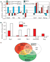
Figure 4
Figure 4. Gpr125lacZ/lacZ MASCs have an expression profile different from mouse embryonic stem cells
a, b, Quantitative PCR comparing expression of relevant genes in vitro in Gpr125lacZ/lacZ MASCs versus wild-type ESCs, Gpr125lacZ/lacZ GSPCs, and MEFs. c, Venn diagram illustrating transcripts unique or common to GSPCs, MASCs and ESCs.

References

    1. Guan K, et al. Pluripotency of spermatogonial stem cells from adult mouse testis. Nature. 2006;440:1199–1203. - PubMed
    1. Kanatsu-Shinohara M, Shinohara T. The germ of pluripotency. Nature Biotechnol. 2006;24:663–664. - PubMed
    1. Valenzuela DM, et al. High-throughput engineering of the mouse genome coupled with high-resolution expression analysis. Nature Biotechnol. 2003;21:652–659. - PubMed
    1. Oakberg EF. A description of spermiogenesis in the mouse and its use in analysis of the cycle of the seminiferous epithelium and germ cell renewal. Am J Anat. 1956;99:391–413. - PubMed
    1. Fiering SN, et al. Improved FACS-Gal: flow cytometric analysis and sorting of viable eukaryotic cells expressing reporter gene constructs. Cytometry. 1991;12:291–301. - PubMed

Publication types

MeSH terms