

Comparison of morphology of human macular and peripheral Bruch's membrane in older eyes
- PMID: 17882712
- PMCID: PMC2562033
- DOI: 10.1080/02713680701550660
Comparison of morphology of human macular and peripheral Bruch's membrane in older eyes
Abstract
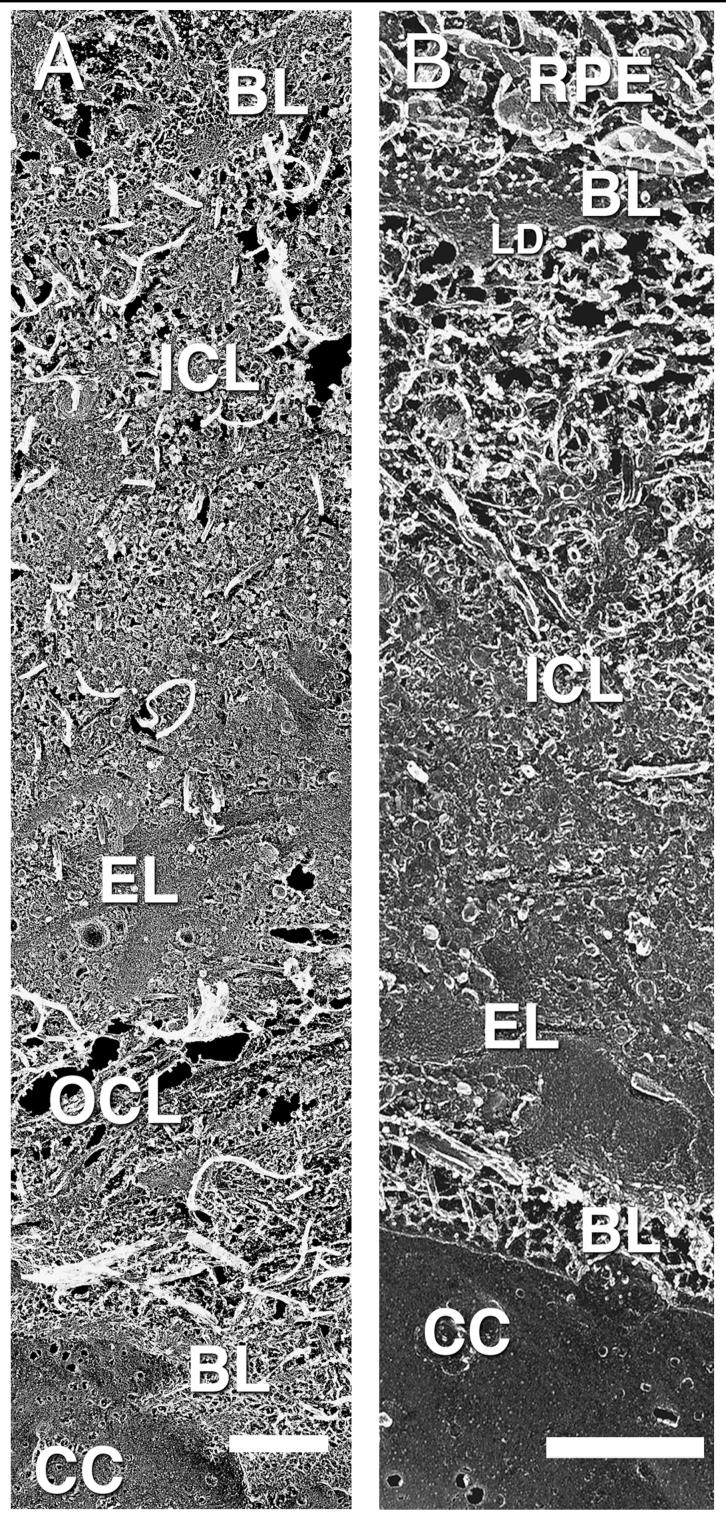
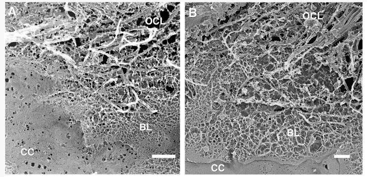
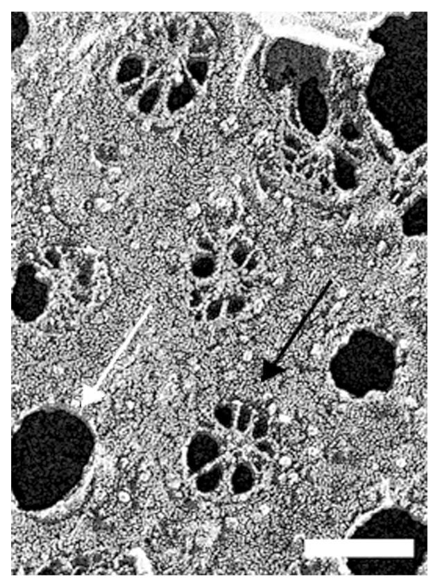
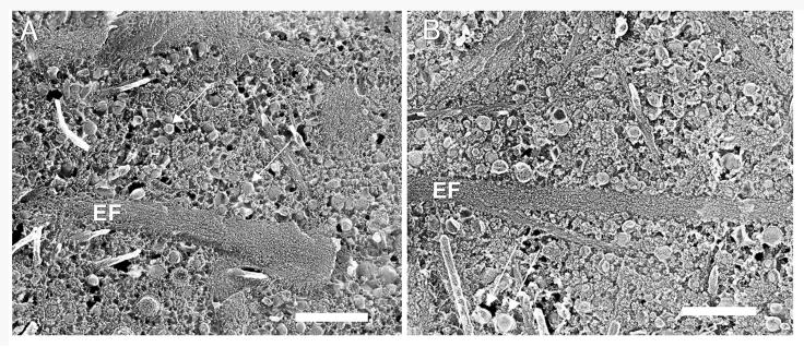
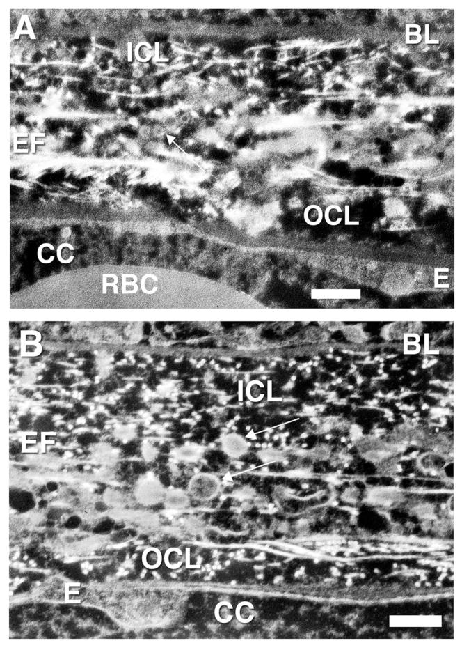
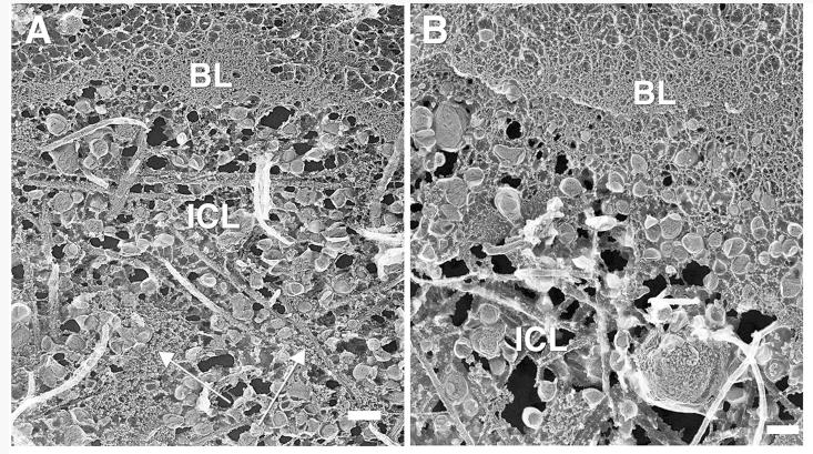
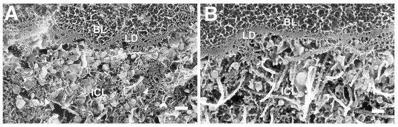
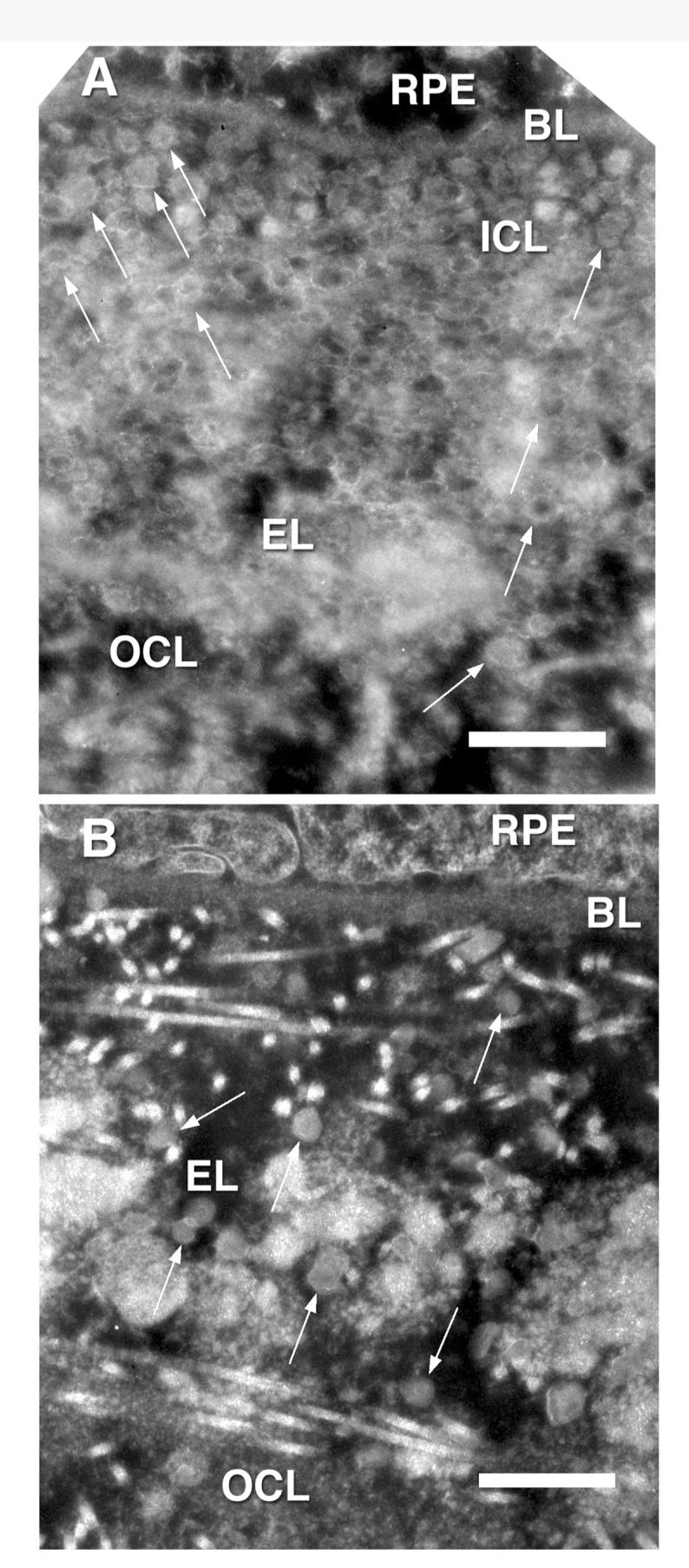
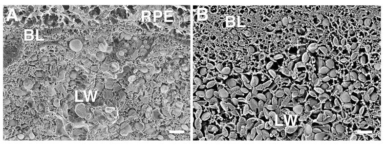
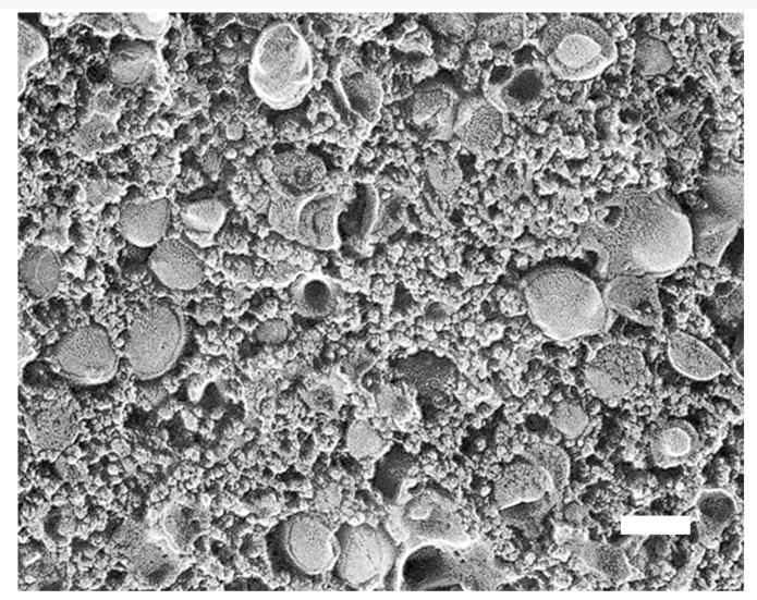
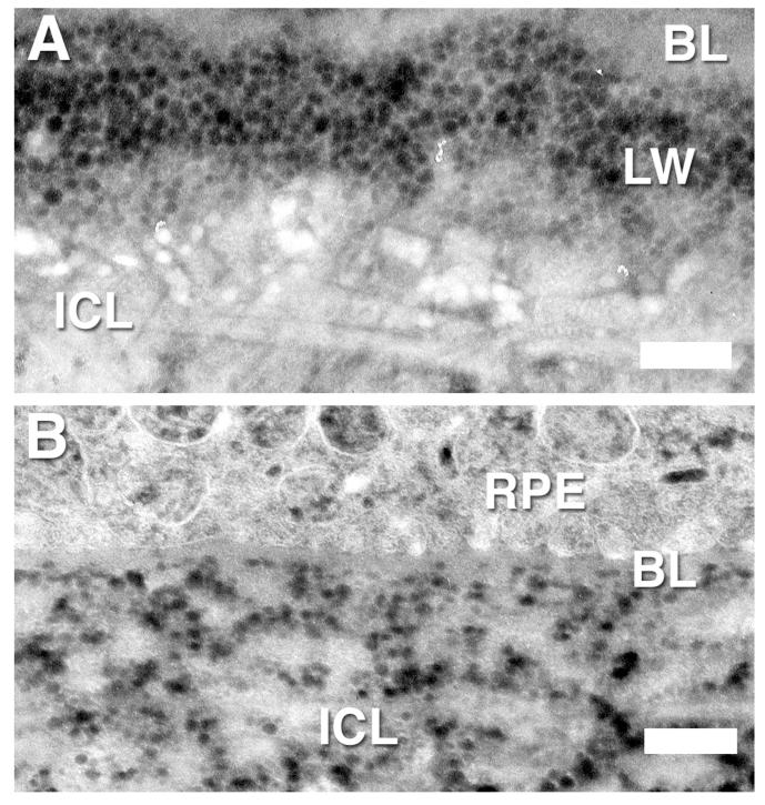
Deposits in macular human Bruch's membrane (BrM) increase with age and have been postulated to be associated with age-related maculopathy. We used two ultrastructural methods to compare these deposits by electron microscopy in macular and peripheral BrM of eight eyes from donors 63-86 years of age. Quick-freeze/deep-etch (QFDE) was used to prepare replicas that showed the ultrastructure of deposits, and osmium-tannic acid-paraphenylenediamine (OTAP) was used to preserve small extracellular lipid particles. We found that an accumulation of lipoprotein-like particles (LLPs) occurred in the peripheral BrM just as it does in the macular region, but with perhaps a somewhat slower time course. The "lipid wall," reported in macular BrM, was also found occasionally in the peripheral regions. The same processes that lead to age-related accumulation of LLPs in macular BrM appear to also occur in the peripheral regions.
Figures
Similar articles
-
Age-related changes in human macular Bruch's membrane as seen by quick-freeze/deep-etch.Exp Eye Res. 2007 Aug;85(2):202-18. doi: 10.1016/j.exer.2007.03.011. Epub 2007 May 3. Exp Eye Res. 2007. PMID: 17586493 Free PMC article.
-
Morphometric analysis of lipoprotein-like particle accumulation in aging human macular Bruch's membrane.Invest Ophthalmol Vis Sci. 2008 Jun;49(6):2721-7. doi: 10.1167/iovs.07-1196. Epub 2008 Feb 22. Invest Ophthalmol Vis Sci. 2008. PMID: 18296655 Free PMC article.
-
Quick-freeze/deep-etch visualization of age-related lipid accumulation in Bruch's membrane.Invest Ophthalmol Vis Sci. 2003 Apr;44(4):1753-9. doi: 10.1167/iovs.02-0496. Invest Ophthalmol Vis Sci. 2003. PMID: 12657618
-
The oil spill in ageing Bruch membrane.Br J Ophthalmol. 2011 Dec;95(12):1638-45. doi: 10.1136/bjophthalmol-2011-300344. Epub 2011 Sep 2. Br J Ophthalmol. 2011. PMID: 21890786 Free PMC article. Review.
-
[Drusen in Bruch's membrane. Their significance for the pathogenesis and therapy of age-associated macular degeneration].Ophthalmologe. 1992 Oct;89(5):363-86. Ophthalmologe. 1992. PMID: 1304217 Review. German.
Cited by
-
Expression of human complement factor H prevents age-related macular degeneration-like retina damage and kidney abnormalities in aged Cfh knockout mice.Am J Pathol. 2015 Jan;185(1):29-42. doi: 10.1016/j.ajpath.2014.08.026. Epub 2014 Nov 1. Am J Pathol. 2015. PMID: 25447048 Free PMC article.
-
Visualizing lipid behind the retina in aging and age-related macular degeneration, via indocyanine green angiography (ASHS-LIA).Eye (Lond). 2022 Sep;36(9):1735-1746. doi: 10.1038/s41433-022-02016-3. Epub 2022 Mar 21. Eye (Lond). 2022. PMID: 35314773 Free PMC article. Review.
-
Location of Retinal Pigment Epithelial Cells in the Eye Is Critical to Their Function.Am J Pathol. 2023 Nov;193(11):1691-1693. doi: 10.1016/j.ajpath.2023.04.005. Epub 2023 May 7. Am J Pathol. 2023. PMID: 37160188 Free PMC article. No abstract available.
-
Aging, age-related macular degeneration, and the response-to-retention of apolipoprotein B-containing lipoproteins.Prog Retin Eye Res. 2009 Nov;28(6):393-422. doi: 10.1016/j.preteyeres.2009.08.001. Epub 2009 Aug 19. Prog Retin Eye Res. 2009. PMID: 19698799 Free PMC article. Review.
-
LXRs regulate features of age-related macular degeneration and may be a potential therapeutic target.JCI Insight. 2020 Jan 16;5(1):e131928. doi: 10.1172/jci.insight.131928. JCI Insight. 2020. PMID: 31829999 Free PMC article.
References
-
- Lewis H, Straatsma BR, Foos RY. Chorioretinal juncture. Multiple extramacular drusen. Ophthalmology. 1986;93:1098–1112. - PubMed
-
- Sheraidah G, Steinmetz R, Maguire J, Pauleikhoff D, Marshall J, Bird AC. Correlation between lipids extracted from Bruch’s membrane and age. Ophthalmology. 1993;100:47–51. - PubMed
-
- Holz FG, Sheraidah G, Pauleikhoff D, Bird AC. Analysis of lipid deposits extracted from human macular and peripheral Bruch’s membrane. Arch Ophthalmol. 1994;112:402–406. - PubMed
-
- Curcio CA, Millican CL, Bailey T, Kruth HS. Accumulation of cholesterol with age in human Bruch’s membrane. Invest Ophthalmol Vis Sci. 2001;42:265–274. - PubMed
Publication types
MeSH terms
Grants and funding
LinkOut - more resources
Full Text Sources
Medical