

Changes in the oviducal epithelium during the estrous cycle in the marsupial Monodelphis domestica
- PMID: 17883438
- PMCID: PMC2375823
- DOI: 10.1111/j.1469-7580.2007.00794.x
Changes in the oviducal epithelium during the estrous cycle in the marsupial Monodelphis domestica
Abstract
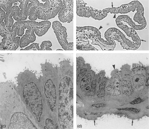
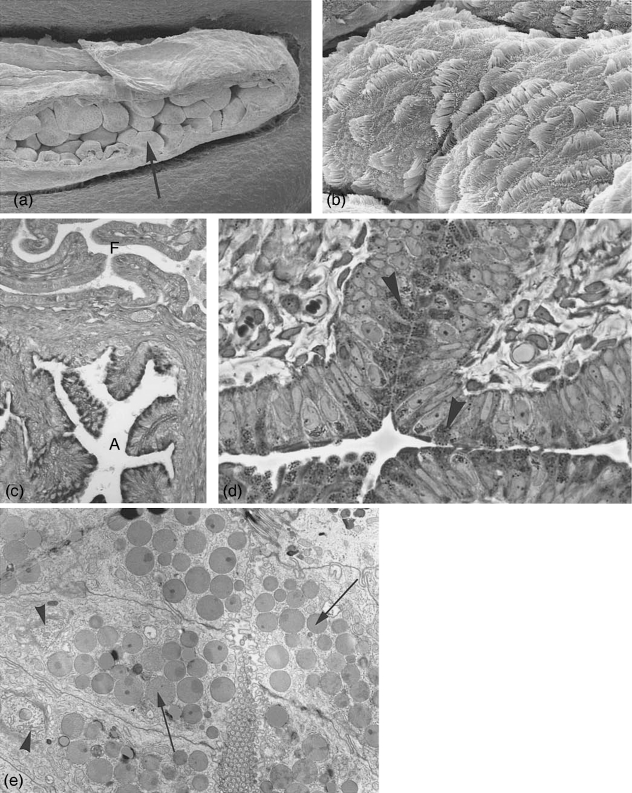
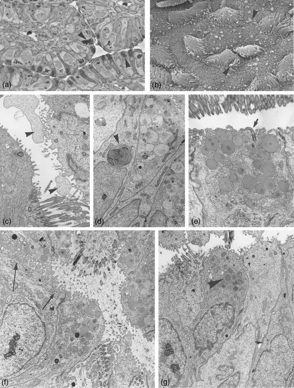
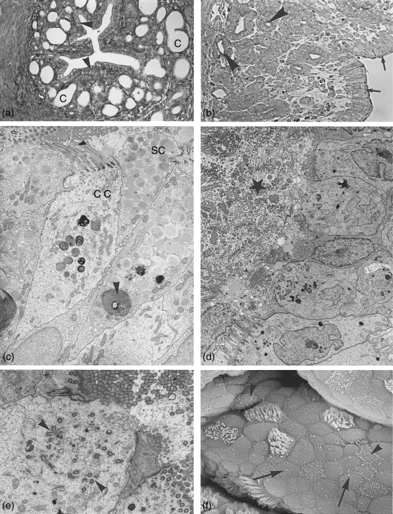
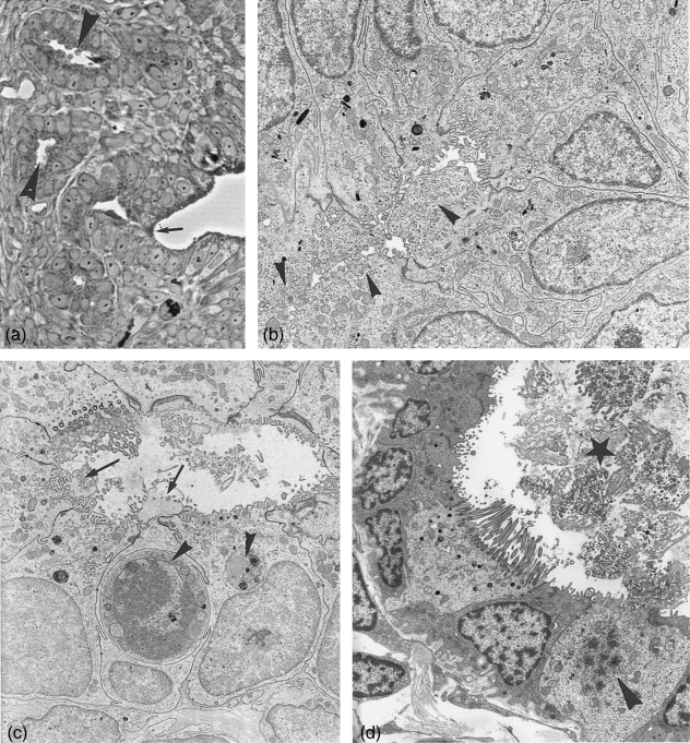
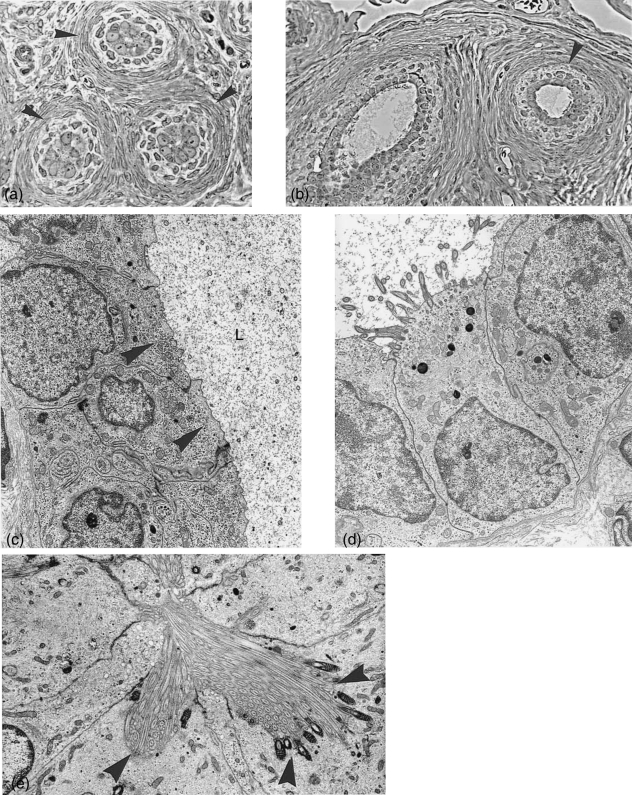
The Monodelphis oviduct can be divided into four anatomical segments: preampulla (comprising fimbriae and infundibulum), ampulla, isthmus with crypts and uterotubal junction. Ovaries are enclosed in a periovarial sac, the bursa, and in some specimens tubules of an epoophoron could be identified. In both structures non-ciliated cells develop small translucent vesicles, which accumulate in the cell apices and presumably produce fluid as often seen in the bursa and in the tubules of the epooophoron. These vesicles do not stain with Alcian blue or PAS. The same applies also to the non-ciliated cells of the fimbriae. The oviducal epithelium of ampulla and the surface epithelium of the isthmus consisting of ciliated and non-ciliated, secretory cells undergo considerable changes during the estrous cycle. Proestrus shows low numbers of ciliated cells, some are in the process of neo-ciliogenesis, non-ciliated cells carry solitary cilia and few remnant secretory granules from the previous cycle may be found. At estrus the amount of ciliated cells in ampulla and isthmus has increased, most non-cililated cells lost the solitary cilia, developed longer microvilli and formed numerous secretory granules in their cell apices. At postestrus secretory products, often surrounded by membranes, are extruded into the oviducal lumen and contribute towards egg coat formation. First signs of deciliation processes are apparent. Solitary cilia reappear. At metestrus only few secretory cells are left with some secretory material. The lumen is often filled with shed cilia and cell apices. Proliferation of basal bodies within non-secretory cells indicate the formation of new ciliated cells. The non-ciliated epithelial cells of the isthmic crypts form no secretory granules but accumulate a great number of translucent vesicles, which in contrast to the secretory granules do not stain with Alcian blue or PAS.
Figures
Similar articles
-
Morphometric and ultrastructural features of the mare oviduct epithelium during oestrus.Theriogenology. 2011 Mar 1;75(4):671-8. doi: 10.1016/j.theriogenology.2010.10.007. Epub 2010 Dec 15. Theriogenology. 2011. PMID: 21111474
-
Cyclic ultrastructural changes in ewe uterine tube (oviduct) infundibular epithelium.Am J Vet Res. 1976 Aug;37(8):923-33. Am J Vet Res. 1976. PMID: 986123
-
Changes in the epithelium of the vaginal complex during the estrous cycle of the marsupial Monodelphis domestica. 2. Scanning electron microscopy study.Cells Tissues Organs. 2004;178(1):48-59. doi: 10.1159/000081092. Cells Tissues Organs. 2004. PMID: 15550759
-
Ultrastructural changes in the cervical epithelium during the estrous cycle of the marsupial Monodelphis domestica (grey short-tailed opossum).Cells Tissues Organs. 2002;171(2-3):162-76. doi: 10.1159/000063710. Cells Tissues Organs. 2002. PMID: 12097839
-
Ultrastructural changes in the uterine luminal and glandular epithelium during the oestrous cycle of the marsupial Monodelphis domestica (grey short-tailed opossum).Cells Tissues Organs. 2002;170(2-3):111-31. doi: 10.1159/000046185. Cells Tissues Organs. 2002. PMID: 11731700
Cited by
-
Fallopian tube rheology regulates epithelial cell differentiation and function to enhance cilia formation and coordination.Nat Commun. 2024 Aug 28;15(1):7411. doi: 10.1038/s41467-024-51481-9. Nat Commun. 2024. PMID: 39198453 Free PMC article.
-
Molecular Evolution of CatSper in Mammals and Function of Sperm Hyperactivation in Gray Short-Tailed Opossum.Cells. 2021 Apr 29;10(5):1047. doi: 10.3390/cells10051047. Cells. 2021. PMID: 33946695 Free PMC article.
References
-
- Abe H. Regional variations in the ultrastructural features of secretory cells in the rat oviductal epithelium. Anat Rec. 1994;240:77–85. - PubMed
-
- Andersen DH. Comparative anatomy of the tubo-uterine junction. Histology and physiology in the sow. Am J Anat. 1928;42:255–305.
-
- Armati-Gulson P, Lowe J. Histology and scanning electron microscopy of the development of the reproductive tract of the common ringtail possum Pseudocheirus peregrinus Pseudocheiridae: Marsupialia. Aust Mammalogy. 1985;8:97–109.
-
- Arnold R, Shorey CD. Structure of the oviducal epithelium of the brushtailed possum Trichosurus vulpecula. J Reprod Fert. 1985;73:9–19. - PubMed
-
- Baggot LM, Moore HD. Early embryonic development of the grey short-tailed opossum, Monodelphis domestica, in vivo and in vitro. J Zool Lond. 1990;222:623–639.
Publication types
MeSH terms
LinkOut - more resources
Full Text Sources
