. 2007 Dec;36(4):462-71.
doi: 10.1016/j.mcn.2007.08.003. Epub 2007 Aug 15.

Mis-expression of L1 on pre-crossing spinal commissural axons disrupts pathfinding at the ventral midline

Affiliations

Mis-expression of L1 on pre-crossing spinal commissural axons disrupts pathfinding at the ventral midline

Ralph Imondi et al. Mol Cell Neurosci. 2007 Dec.

Abstract

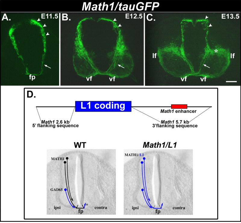
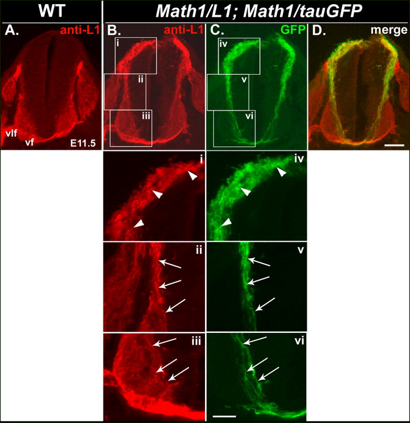
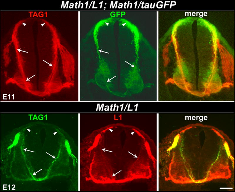
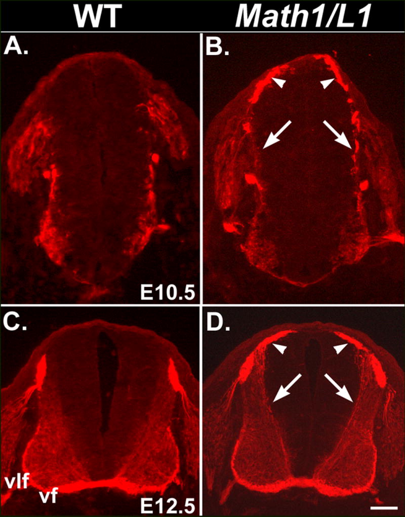
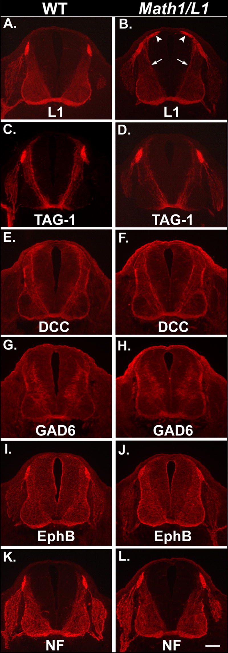
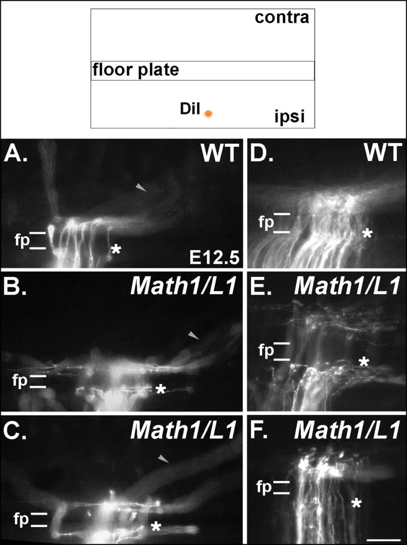
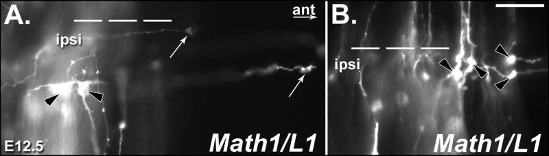
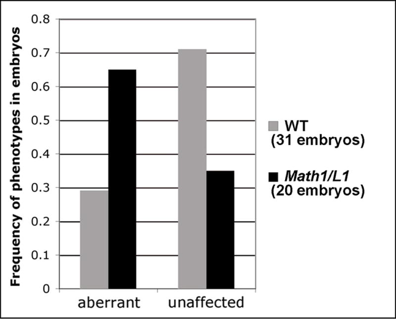
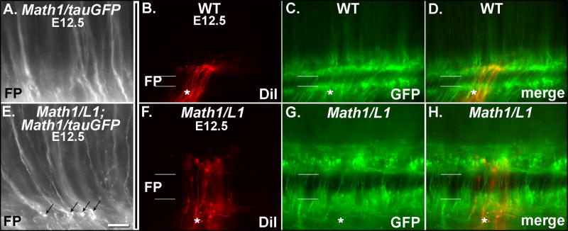
In vertebrates, spinal commissural axons project along a transverse path toward and across the floor plate (FP). Post-crossing commissural axons alter their responsiveness to FP-associated guidance cues and turn to project longitudinally in a fasciculated manner prior to extending away from the midline. The upregulation of the neural cell adhesion molecule L1 on crossed commissural axon segments has been proposed to facilitate pathfinding on the contralateral side of the FP. To explore this possibility in vivo, we used Math1 regulatory sequences to target L1 to commissural axons before they cross the ventral midline. L1 mis-expression did not alter the distribution of commissural axon-associated markers or the ventral extension of commissural axons toward the midline. However, commissural axons often stalled or inappropriately projected into the longitudinal plane at the ipsilateral FP margin. These observations suggest that L1-mediated pathfinding decisions are normally delayed until axons have crossed the ventral midline (VM).


Figures

Figure 1
Figure 1. Math1 regulatory sequences drive transgene expression in commissural neurons/axons
(A-C) Transverse cryosections obtained from E11.5 (A), E12.5 (B) and E13.5 (C) Math1/tauGFP embryos. (A, B) At E11.5 and E12.5, tauGFP is expressed by dorsally-located cell bodies of Math1 progenitors, the commissural neurons derived from these progenitors (arrowheads), and the corresponding axons (arrows) that extend ventrally toward and across the floor plate (fp) and assemble into the longitudinally projecting ventral funiculus (vf). (C) Expression of tauGFP in commissural neurons/axons persists in E13.5 embryos. A large, ventrally located population of neurons (asterisk) that appears to extend axons directly into the lateral funiculus (lf) also expresses the transgene at this age. (D, top panel) Construct used to generate Math1/L1 transgenic mice. Genomic sequences flanking the Math1 coding region (black line), including the Math1 enhancer (red), were used to flank the mouse L1 coding sequence (blue). (D, lower panels) In wild-type (WT) embryos, L1 is expressed on axons emanating from Math1-positive dorsal commissural axons only after they cross through the FP (asterisk). A more ventral population of GAD-65-positive commissural neurons extends L1-positive axons prior to reaching the FP. In Math1/L1 mice, the L1 transgene should be expressed on pre-crossing segments of axons emanating from dorsal commissural neurons. Scale bar in (C), 100 μm (A-C). ipsi, ipsilateral; contra, contralateral.
Figure 2
Figure 2. L1 is mis-expressed on pre-crossing segments of commissural axons in Math1/L1 mice
(A-D) Transverse cryosections obtained from an E11.5 wild-type embryo (A) or an E11.5 embryo derived from a cross between Math1/L1 and Math1/tauGFP mice (B-D) that were labeled with anti-L1. In Math1/L1;Math1/tauGFP (B-D), but not in wild-type (A), embryos, anti-L1 labels dorsally located cell bodies of Math1 progenitors, the commissural neurons derived from these progenitors, and the corresponding axons of these cells, which project ventrally toward the FP and express the tauGFP transgene (C, D). Panel (D) represents a merge of panels (B) and (C). Ventral funiculus (vf), ventrolateral funiculus (vlf). Scale bar in (D), 100 μm (A-D). (i-vi) Higher magnification views of boxed regions in panels (B) and (C). Arrowheads (i, iv) indicate L1- and GFP-expressing cell bodies of Math1 progenitors and post-mitotic dI1 commissural neurons, as well as the most proximal portions of their pre-crossing axons. Arrows identify pre-crossing segments of L1- and GFP-expressing commissural axons in an intermediate (ii, v) and a ventral (iii, vi) region of the spinal cord. Scale bar in (vi), 50 μm (i-vi).
Figure 3
Figure 3. The distribution of TAG-1 overlaps with the expression of tauGFP and L1 in Math1/L1; Math1/tauGFP and Math1/L1 mice, respectively
(Top panels) Transverse cryosection obtained from an E11 Math1/L1; Math1/tauGFP embryo labeled with anti-TAG-1. TAG-1 (left) and GFP (middle) expression overlap on the dorsally located cells bodies of Math1 progenitors and post-mitotic commissural neurons (arrowheads), as well as on the pre-crossing segments of their commissural axons (arrows). (Bottom panels) Transverse cryosection obtained from an E12 Math1/L1 embryo labeled with anti-TAG-1 and anti-L1. TAG-1 (left) and L1 (middle) expression overlap on the dorsally located cells bodies of Math1 progenitors and post-mitotic commissural neurons (arrowheads), as well as on the pre-crossing segments of their commissural axons (arrows). In each case, the right panel represents a merge of the left and middle panels. Scale bar in lower right panel, 100 μm (all panels).
Figure 4
Figure 4. Mis-expression of L1 is detectable at E10.5 and E12.5 in Math1/L1 mice
(A-D) Transverse cryosections were obtained from E10.5 and E12.5 wild-type (A, C) or Math1/L1 (B, D) embryos and labeled with anti-L1. In Math1/L1 embryos, ectopic L1 expression on the cell bodies of Math1 progenitors and the commissural neurons derived from these progenitors (arrowheads), as well as on their ventrally projecting axons (arrows), is detected at both E10.5 (B) and E12.5 (D). The punctate anti-L1 labeling of axons at E10.5 (arrows in B) reflects the relatively poor morphology characteristic of cryosections obtained from these young embryos. Ventral funiculus (vf), ventrolateral funiculus (vlf). Scale bar in (D), 100 μm (C, D); 65 μm (A, B).
Figure 5
Figure 5. L1 mis-expression does not affect the ventral extension of commissural axons or the distribution of commissural axon markers
(A-L) Semi-serial transverse cryosections obtained from E12.5 wild-type (left column) or Math1/L1 embryos (right column) that were labeled with a variety of commissural axon markers. In Math1/L1 embryos, L1 is mis-expressed on dorsally located cell bodies of Math1 progenitors and the commissural neurons derived from these progenitors (white arrowheads), as well as on the corresponding axons (arrows) that extend ventrally toward and across the floor plate (B). The distribution of other commissural axon markers is unaffected in Math1/L1 embryos (D, F, H, J, L). Scale bar in (L), 100 μm (A-L).
Figure 6
Figure 6. Mis-expression of L1 results in commissural axon projection errors in the immediate vicinity of the FP
(Top) The pathfinding behavior of commissural axons was visualized through the application of DiI into a dorsal region of fixed, open-book spinal cord preparations taken from E12.5 embryos. (A-F) Photomicrographs of DiI-labeled axons in open book preparations derived from wild type (WT) and Math1/L1 embryos. (A, D) In WT embryos, commissural axons project directly through the FP in a well-organized fashion before turning in a predominantly rostral direction on the contralateral side of the spinal cord. (B, C, E) In Math1/L1 embryos, commissural axons “pile-up” and stall at the ipsilateral margin of the FP, or project along this boundary before ultimately crossing the FP. The arrowheads in A, B and C denote out of focus axons that are projecting along appropriately shaped contralateral trajectories in WT and Math1/L1 embryos. (F) On rare occasions, some commissural axons were observed to stall at the contralateral margin of the FP. In A-F, asterisks demarcate the position of the ipsilateral FP margin. In each panel, rostral is to the right. Scale bar in (F), 95 μm (A-C); 50 μm (D-F).
Figure 7
Figure 7. Many of the commissural axon-associated growth cones in Math1/L1 embryos display a swollen/bulbous morphology typically associated with a stalling behavior
(A, B) Photomicrographs of DiI-labeled axons in open book preparations derived from E12.5 Math1/L1 embryos. (A) This particular open-book preparation contains examples of both swollen/bulbous, stalled growth cones (black arrowheads) and commissural axons tipped by wild type-like growth cones (white arrows) that inappropriately turn into the longitudinal plane on the ipsilateral side of the FP. (B) In this open-book preparation, there are at least four examples of presumably stalled growth cones (black arrowheads). In each panel, the dashed lines demarcate the position of the FP. Ipsi, ipsilateral; ant, anterior direction. Scale bar in (B), 25 μm (A, B).
Figure 8
Figure 8. Quantification of pathfinding errors in wild-type (WT) and Math1/L1 embryos
Frequency histogram summarizing the phenotypes (unaffected and aberrant) observed in Math1/L1 versus WT embryos (see text for scoring criteria and details).
Figure 9
Figure 9. GFP-labeled commissural axons exhibit pathfinding defects in Math1/L1;Math/tauGFP (Math1/L1) embryos
(A, E) Photomicrographs of GFP-labeled commissural axons in open book preparations derived from an E12.5 Math1/tauGFP (A) and a Math1/L1;Math1/tauGFP (E) embryo. Several examples of stalled growth cones (black arrows in E) are observed in the open-book preparation derived from the Math1/L1;Math1/tauGFP, but not the Math1/tauGFP, embryo. The FP is located at the bottom of each panel. (B-D, F-H). Photomicrographs of DiI- or GFP-labeled commissural axons in open book preparations derived from a Math1/tauGFP (WT; B-D) and a Math1/L1;Math1/tauGFP (Math1/L1; F-H) embryo. (B, F) DiI labeling. (C, G) GFP expression. (D, H) Merge of DiI labeling and GFP expression. In B-D and F-H, the asterisk demarcates the ipsilateral side of the FP. In each panel, rostral is to the right. Scale bar in (E), 25 μm (A, E); 45 μm (B-D, F-H).

Similar articles

Cited by

References

    1. Bovolenta P, Dodd J. Guidance of commissural growth cones at the floor plate in embryonic rat spinal cord. Development. 1990;109:435–447. - PubMed
    1. Brittis PA, Lu Q, Flanagan JG. Axonal protein synthesis provides a mechanism for localized regulation at an intermediate target. Cell. 2002;110:223–235. - PubMed
    1. Brummendorf T, Kenwrick S, Rathjen FG. Neural cell recognition molecule L1: from cell biology to human hereditary brain malformations. Curr Opin Neurobiol. 1998;8:87–97. - PubMed
    1. Butner KA, Kirschner MW. Tau protein binds to microtubules through a flexible array of distributed weak sites. J Cell Biol. 1991;5:717–730. - PMC - PubMed
    1. Caspary T, Anderson KV. Patterning cell types in the dorsal spinal cord: what the mouse mutants say. Nat Rev Neurosci. 2003;4:289–297. - PubMed

Publication types

MeSH terms

Substances