. 2007 Nov 30;282(48):34605-10.
doi: 10.1074/jbc.M705100200. Epub 2007 Sep 20.

IL-21 is produced by Th17 cells and drives IL-17 production in a STAT3-dependent manner

Affiliations

IL-21 is produced by Th17 cells and drives IL-17 production in a STAT3-dependent manner

Lai Wei et al. J Biol Chem. .

Abstract

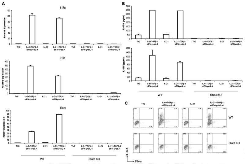
CD4(+) helper T cells can differentiate into several possible fates including: Th1, Th2, T regulatory, and Th17 cells. Although, cytokine production by non-T cells is an important factor in helper T cell differentiation, a characteristic feature of both Th1 and Th2 lineages is their ability to secrete cytokines that promote their respective differentiation. However, cytokines produced by T cells that help to sustain Th17 cells have not yet been identified. Here we show that IL-21 is a product of Th17 cells, which is induced in a Stat3-dependent manner. Additionally, Stat3 can directly bind the Il21 promoter. IL-21 also induces IL-17 production and expression of the transcription factor, RORgammat. Furthermore, generation of Th17 cells in the conventional manner is attenuated by blocking IL-21. IL-21 is known to activate Stat3 and its ability to induce Th17 differentiation is abrogated in the absence of Stat3. These data argue that IL-21 serves as an autocrine factor secreted by Th17 cells that promotes or sustains Th17 lineage commitment.


Figures

Figure 1
Figure 1. IL-21 is selectively produced by Th17 cells and is regulated by Stat3
(A) Total RNA was isolated from wild-type naïve CD4+ T cells cultured under Th0, Th1, Th2, or Th17 conditions. IL-21 mRNA level was detected by quantitative real-time PCR using Taqman assay kit from ABI. Gene expression was normalized to actin mRNA level in each sample. (B) ELISA was performed to detect IL-21 production in supernatants from wild-type naïve CD4+ T cells cultured under Th0, Th1, Th2, or Th17 conditions. (C) Total RNA was isolated from wild-type (WT) or CD4-Cre Stat3fl/fl (Stat3 KO) naïve CD4+ T cells cultured under Th0 or Th17 conditions. IL-21 mRNA levels were detected by quantitative real-time PCR using Taqman assay kit from ABI. (D) ELISA was performed to detect IL-21 production in supernatants from wild-type (WT) or CD4-Cre Stat3fl/f (Stat3 KO) naïve CD4+ T cells cultured under Th0 or Th17 conditions. (E) Peripheral CD4+ T cells were polarized under Th0 or Th17 conditions for 72 h. ChIP assays were performed using anti-Stat3 antibody.
Figure 2
Figure 2. IL-21 can replace IL-6 to promote Th17 differentiation
Peripheral naïve CD4+ T cells from wild-type mice (WT) or CD4-Cre Stat3fl/fl (Stat3 KO) mice were cultured under the indicated conditions for 72 h. (A) Levels of mRNA for IL-17A, IL-17F, and Rorc were measured by quantitative real-time PCR using Taqman assay kits from ABI. Gene expression was normalized to Actin mRNA level in each sample. (B) ELISA was performed to detect IL-17A and IL-17F production in cell culture supernatants. (C) Proportions of IL-17A- and IFN-γ-producing cells in Stat3fl/f (WT) and MMTV-Cre Stat3fl/fl (Stat3 KO) cell cultures were determined by intracellular cytokine staining and flow cytometry.
Figure 3
Figure 3. Conventional Th17 differentiation is dependent upon IL-21
Peripheral naïve CD4+ T cells from wild type mice were cultured in neutral (Th0), optimal Th17, or Th17 conditions with anti-IL-21 antibody for 72 h. (A) Levels of mRNA for IL-17A and IL-17F were assayed by quantitative real-time PCR using Taqman assay kits from ABI. Gene expression was normalized to Actin mRNA level in each sample. (B) ELISA was performed to detect IL-17A and IL-17F production in cell culture supernatants. (C) Proportions of IL-17A- and IFN-γ-producing cells were determined by intracellular cytokine staining and flow cytometry.

References

    1. O’Garra A, Vieira P. Nat Med. 2004;10(8):801–805. - PubMed
    1. Murphy KM, Ouyang W, Farrar JD, Yang J, Ranganath S, Asnagli H, Afkarian M, Murphy TL. Annu Rev Immunol. 2000;18:451–494. - PubMed
    1. Szabo SJ, Sullivan BM, Peng SL, Glimcher LH. Annu Rev Immunol. 2003;21:713–758. - PubMed
    1. Ansel KM, Djuretic I, Tanasa B, Rao A. Annu Rev Immunol. 2006;24:607–656. - PubMed
    1. Weaver CT, Harrington LE, Mangan PR, Gavrieli M, Murphy KM. Immunity. 2006;24(6):677–688. - PubMed

Publication types

MeSH terms