

Estrogen and progesterone are critical regulators of Stat5a expression in the mouse mammary gland
- PMID: 17884938
- PMCID: PMC2194608
- DOI: 10.1210/en.2007-0594
Estrogen and progesterone are critical regulators of Stat5a expression in the mouse mammary gland
Abstract
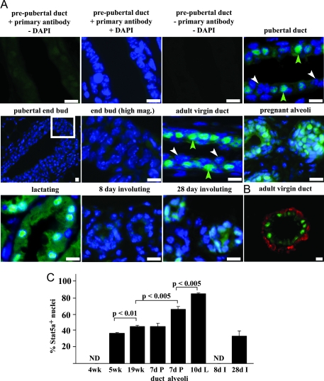
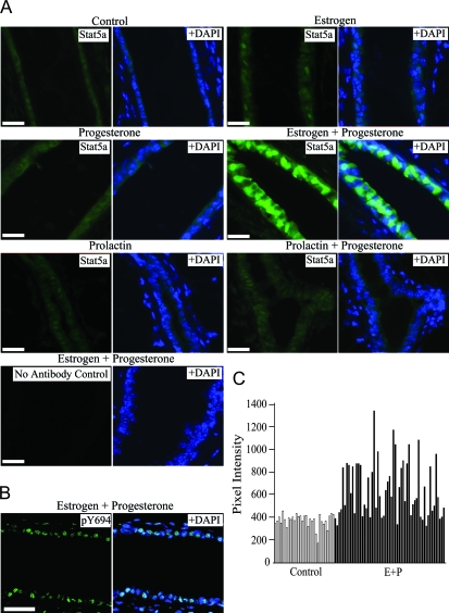
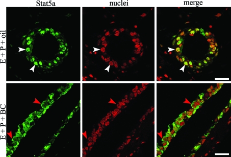
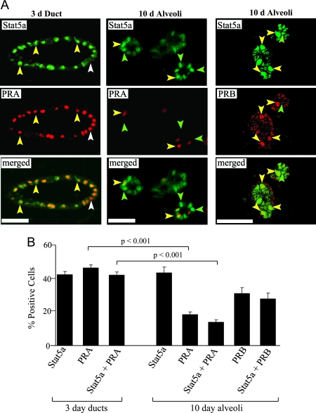
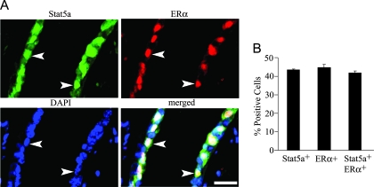
Signal transducer and activator of transcription (Stat)5a is a well-established regulator of mammary gland development. Several pathways for activating Stat5a have been identified, but little is known about the mechanisms that regulate its expression in this tissue. In this report, we used immunofluorescent staining to examine Stat5a expression in mammary epithelial cells during normal development and in response to treatment with the ovarian hormones estrogen (E) and progesterone (P). Stat5a was present at very low levels in the prepubertal gland and was highly induced in a subset of luminal epithelial cells during puberty. The percentage of positive cells increased in adult virgin, pregnant, and lactating animals, dropped dramatically during involution, and then increased again after weaning. Ovariectomy ablated Stat5a expression in virgin animals, and treatment with both E and P was necessary to restore it. Double-labeling experiments in animals treated with E plus P for 3 d demonstrated that Stat5a was localized exclusively to cells containing both E and P receptors. Together, these results identify a novel role for E and P in inducing Stat5a expression in the virgin mammary gland and suggest that these hormones act at the cellular level through their cognate receptors.
Figures
Similar articles
-
Signal transducer and activator of transcription 5a mediates mammary ductal branching and proliferation in the nulliparous mouse.Endocrinology. 2010 Jun;151(6):2876-85. doi: 10.1210/en.2009-1282. Epub 2010 Apr 14. Endocrinology. 2010. PMID: 20392833 Free PMC article.
-
Regulation of gene expression in the bovine mammary gland by ovarian steroids.J Dairy Sci. 2007 Jun;90 Suppl 1:E55-65. doi: 10.3168/jds.2006-466. J Dairy Sci. 2007. PMID: 17517752 Review.
-
Activation of Stat5a and Stat5b by tyrosine phosphorylation is tightly linked to mammary gland differentiation.Mol Endocrinol. 1996 Dec;10(12):1496-506. doi: 10.1210/mend.10.12.8961260. Mol Endocrinol. 1996. PMID: 8961260
-
Fibronectin and the alpha(5)beta(1) integrin are under developmental and ovarian steroid regulation in the normal mouse mammary gland.Endocrinology. 2001 Jul;142(7):3214-22. doi: 10.1210/endo.142.7.8273. Endocrinology. 2001. PMID: 11416044
-
The hyperplastic phenotype in PR-A and PR-B transgenic mice: lessons on the role of estrogen and progesterone receptors in the mouse mammary gland and breast cancer.Vitam Horm. 2013;93:185-201. doi: 10.1016/B978-0-12-416673-8.00012-5. Vitam Horm. 2013. PMID: 23810007 Review.
Cited by
-
Introduction: hormonal regulation of mammary development and milk protein gene expression at the whole animal and molecular levels.J Mammary Gland Biol Neoplasia. 2009 Sep;14(3):317-9. doi: 10.1007/s10911-009-9146-4. J Mammary Gland Biol Neoplasia. 2009. PMID: 19657596 No abstract available.
-
Strain-specific differences in the mechanisms of progesterone regulation of murine mammary gland development.Endocrinology. 2009 Mar;150(3):1485-94. doi: 10.1210/en.2008-1459. Epub 2008 Nov 6. Endocrinology. 2009. PMID: 18988671 Free PMC article.
-
17β-Estradiol and ICI182,780 Differentially Regulate STAT5 Isoforms in Female Mammary Epithelium, With Distinct Outcomes.J Endocr Soc. 2018 Feb 26;2(3):293-309. doi: 10.1210/js.2017-00399. eCollection 2018 Mar 1. J Endocr Soc. 2018. PMID: 29594259 Free PMC article.
-
Hormone-sensing cells require Wip1 for paracrine stimulation in normal and premalignant mammary epithelium.Breast Cancer Res. 2013 Jan 31;15(1):R10. doi: 10.1186/bcr3381. Breast Cancer Res. 2013. PMID: 23369183 Free PMC article.
-
Genistein Directly Represses the Phosphorylation of STAT5 in Lactating Mammary Epithelial Cells.ACS Omega. 2021 Aug 25;6(35):22765-22772. doi: 10.1021/acsomega.1c03107. eCollection 2021 Sep 7. ACS Omega. 2021. PMID: 34514247 Free PMC article.
References
-
- Leonard WJ, O’Shea JJ 1998 Jaks and STATs: biological implications. Annu Rev Immunol 16:293–322 - PubMed
-
- Akira S 1999 Functional roles of STAT family proteins: lessons from knockout mice. Stem Cells 17:138–146 - PubMed
-
- Buitenhuis M, Coffer PJ, Koenderman L 2004 Signal transducer and activator of transcription 5 (STAT5). Int J Biochem Cell Biol 36:2120–2124 - PubMed
-
- Darnell Jr JE 1997 STATs and gene regulation. Science 277:1630–1635 - PubMed
Publication types
MeSH terms
Substances
Grants and funding
LinkOut - more resources
Full Text Sources
Other Literature Sources
Miscellaneous
