. 2007 Oct 2;104(40):15693-8.
doi: 10.1073/pnas.0703080104. Epub 2007 Sep 20.

Solution structure of the A4 domain of factor XI sheds light on the mechanism of zymogen activation

Affiliations

Solution structure of the A4 domain of factor XI sheds light on the mechanism of zymogen activation

Dharmaraj Samuel et al. Proc Natl Acad Sci U S A. .

Abstract

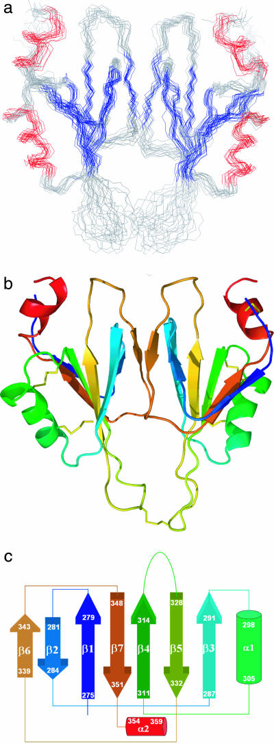
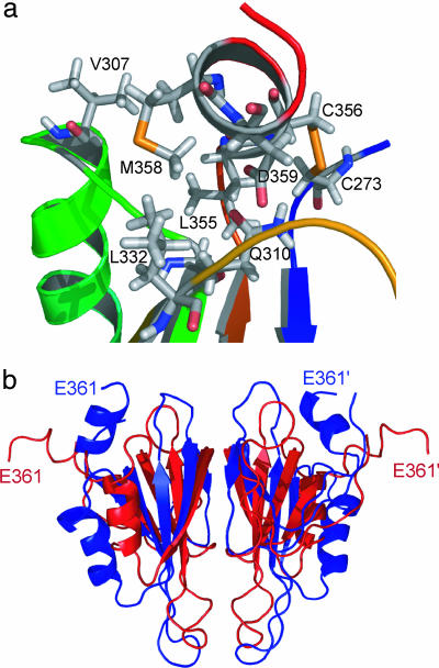
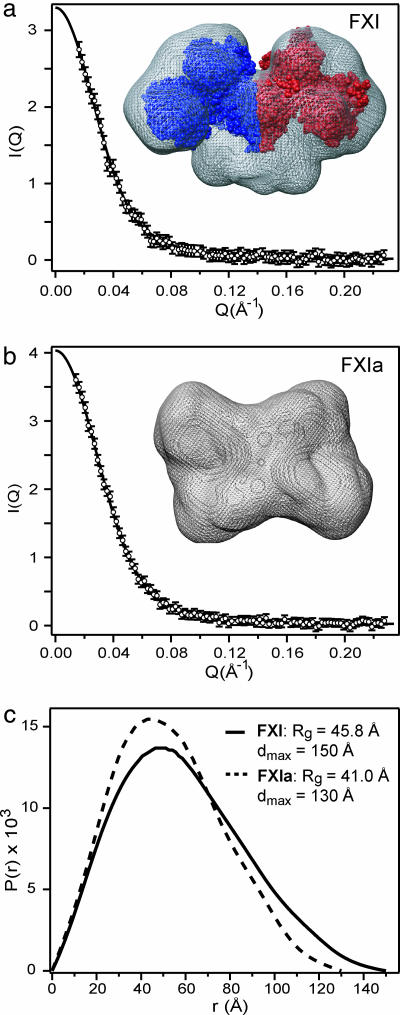
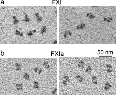
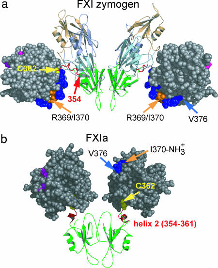
Factor XI (FXI) is a homodimeric blood coagulation protein. Each monomer comprises four tandem apple-domain repeats (A1-A4) and a serine protease domain. We report here the NMR solution structure of the A4 domain (residues 272-361), which mediates formation of the disulfide-linked FXI dimer. A4 exhibits characteristic features of the plasminogen apple nematode domain family, including a five-stranded beta-sheet flanked by an alpha-helix on one side and a two-stranded beta-sheet on the other. In addition, the solution structure reveals a second alpha-helix at the C terminus. Comparison with a recent crystal structure of full-length FXI, combined with molecular modeling, suggests that the C-terminal helix is formed only upon proteolytic activation. The newly formed helix disrupts interdomain contacts and reorients the catalytic domains, bringing the active sites into close proximity. This hypothesis is supported by small-angle x-ray scattering and electron microscopy data, which indicate that FXI activation is accompanied by a major change in shape. The results are consistent with biochemical evidence that activated FXI cleaves its substrate at two positions without release of an intermediate.


Conflict of interest statement

The authors declare no conflict of interest.

Figures

Fig. 1.
Fig. 1.
Solution structure of the A4 dimer. (a) The backbone overlay of 14 low-energy structures of FXI A4. Colored segments indicate β-sheet (blue) and helices (red). (b) Ribbon representation of the two domains of the dimer rendered in colors ranging from blue (N-terminal) to red (C-terminal). (c) Topology diagram of the NMR structure of the FXI A4 domain. The color scheme follows that in b.
Fig. 2.
Fig. 2.
C-terminal helix. (a) Local environment of the C-terminal helix (α2) in the NMR structure of A4. Residues involved in interactions between the C-terminal helix and other parts of the protein are shown. (b) Overlay of a representative NMR solution structure (blue) and crystal structure (red) of the A4 dimer. The C-terminal segment (354–361), which is part of an extended connecting loop in the crystal structure, assumes an α-helical conformation in the NMR structure, resulting in an ≈20 Å displacement of the C-terminal Glu-361.
Fig. 3.
Fig. 3.
Conformational changes associated with activation of FXI monitored by SAXS. (a and b) The scattering intensity, I(Q), is plotted vs. scattering vector, Q, for FXI (a) and FXIa (b). (c) The lines represent the predicted scattering profiles corresponding to the P(r) functions. Low-resolution structures derived from the SAXS profiles by ab initio simulated annealing calculations (25) for FXI and FXIa are shown in a and b, respectively, using mesh surface rendering. For comparison, a space-filling representation of FXI based on the crystal structure (7) is included in a.
Fig. 4.
Fig. 4.
Electron micrographs showing domain rearrangements associated with FXI zymogen activation. Representative electron micrographs are shown for samples of FXI (a) and FXIa (b) prepared by using rotary shadowing with tungsten.
Fig. 5.
Fig. 5.
Changes in domain structure upon zymogen activation. (a) Domain arrangement of the FXI zymogen, based on the crystal structure of Papagrigoriou et al. (7). The A4 domain (green) mediating dimerization and the three other apple domains are shown in ribbon representation. The two catalytic domains (gray, space-filling) are connected to the A4 domain via an extended loop (red, residues 354–362). (b) A model representing one of the possible conformations of the catalytic and A4 domains in FXIa triggered by formation of a second α-helix at the C terminus of the A4 domain (red). Upon cleavage of the R369-I370 bond (orange), the 362–482 disulfide bond (yellow) is the only covalent link between the pair of catalytic domains and A4 domains. Residues in the active-site cleft are colored magenta.

References

    1. Fujikawa K, Chung DW, Hendrickson LE, Davie EW. Biochemistry. 1986;25:2417–2424. - PubMed
    1. Walsh PN, Gailani D. In: Hemostasis and Thrombosis: Basic Principles and Clinical Practice. Colman RW, Hirsh J, Marder VJ, Clowes AW, George JN, editors. Philadelphia: Lippincott; 2006. Chap 12.
    1. von dem Borne PA, Meijers JC, Bouma BN. Blood. 1995;86:3035–3042. - PubMed
    1. McMullen BA, Fujikawa K, Davie EW. Biochemistry. 1991;30:2056–2060. - PubMed
    1. Brown PJ, Gill AC, Nugent PG, McVey JH, Tomley FM. FEBS Lett. 2001;497:31–38. - PubMed

Publication types

LinkOut - more resources