. 2007 Nov;145(3):863-74.
doi: 10.1104/pp.107.102657. Epub 2007 Sep 20.

A nonclassical arabinogalactan protein gene highly expressed in vascular tissues, AGP31, is transcriptionally repressed by methyl jasmonic acid in Arabidopsis

Affiliations

A nonclassical arabinogalactan protein gene highly expressed in vascular tissues, AGP31, is transcriptionally repressed by methyl jasmonic acid in Arabidopsis

Chenggang Liu et al. Plant Physiol. 2007 Nov.

Abstract

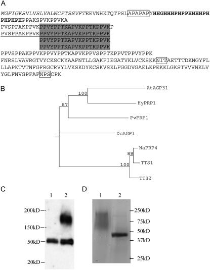
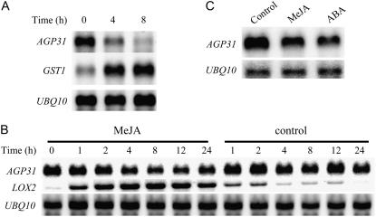
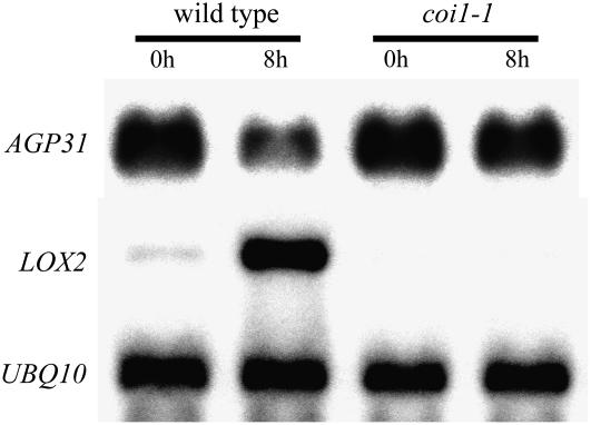
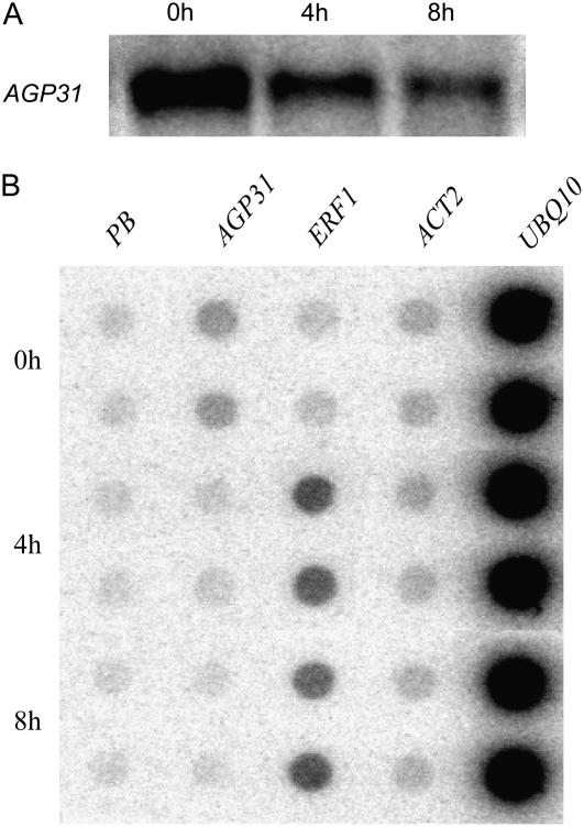
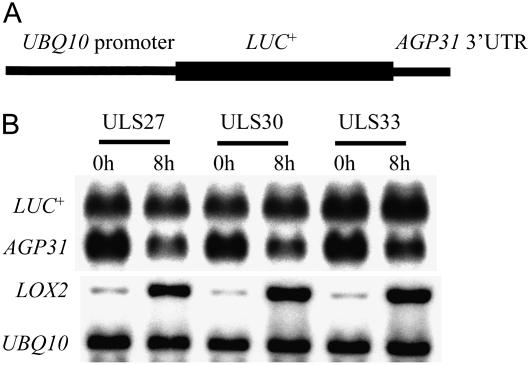
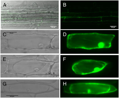
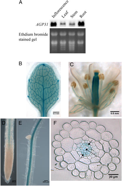
In response to wounding and pathogens, jasmonate (JA) serves as a signal molecule for both induction and repression of gene expression. To examine defense-regulated gene repression in Arabidopsis (Arabidopsis thaliana), we have identified a nonclassical arabinogalactan protein (AGP) gene, AGP31, and show that its mRNA decreased to about 30% of its original level within 8 h in response to methyl JA (MeJA) treatment of whole 7-d-old seedlings. Wounding and abscisic acid treatment had similar effects. MeJA suppression primarily depends on the action of the JA-signaling protein, COI1, as shown by much lower MeJA suppression in coi1-1 mutant plants. The main mechanism of mRNA suppression by MeJA is repression of transcription, as shown by nuclear run-on experiments. The AGP31 protein shares features with several known and putative nonclassical AGPs from other species: a putative signal peptide, a histidine-rich region near the N terminus followed by a repetitive proline-rich domain, and a cysteine-rich C-terminal PAC (for proline-rich protein and AGP, containing cysteine) domain. Positive Yariv reagent interaction demonstrated that the protein is an AGP. Monosaccharide analysis of purified AGP31 indicated it is a galactose-rich AGP. Expression of an AGP31-enhanced green fluorescent protein fusion protein in transgenic cells revealed that the AGP31 protein was localized to the cell wall. AGP31 promoter-beta-glucuronidase reporter gene analysis showed expression in the vascular bundle throughout the plant, except in the flower. In the flower, beta-glucuronidase staining occurred throughout the pistil, except in the stigma. The strong preferential expression in vascular tissues suggests that AGP31 may be involved in vascular tissue function during both the defense response and development.


Figures

Figure 1.
Figure 1.
Characterization of AGP31. A, Structure of AGP31. Shaded regions are four exact repeat units. Underlined regions are two longer repeat units. The smallest repeat unit is PPXX. The italicized region is the predicted signal peptide; the bold region is the His-rich region. Boxed regions are the predicted APAPAP module for arabinogalactan attachment and N-glycosylation sites. B, Neighbor-joining tree generated from the alignment of full-length amino acid sequences of the homologs of AGP31. Numbers indicate the percentage of 1,000 bootstrap replicates. C, Western blot showing that AGP31 exists in AGP precipitation of cell wall protein. Lane 1, AGP fraction from wild-type plants; lane 2, AGP fraction from transgenic plants expressing 35S∷AGP31∷myc. D, Purified AGP31. AGP31 was isolated from high-salt eluted cell wall protein by a Ni-NTA column and deglycosylated by TFMS. Approximately 2 μg of native and deglycosylated AGP31 were separated by SDS-PAGE on a 15% gel and stained by Coomassie Blue. Lane 1, Native AGP31; lane 2, deglycosylated AGP31.
Figure 2.
Figure 2.
Effects of wounding, MeJA, and ABA treatments on AGP31 and reference mRNA levels. A, Northern-blot analysis of RNA from wounded leaves of 4-week-old plants. About one-third of the leaves were pressed with pliers and wounded leaves were harvested for RNA at given time points. The same blot hybridized with the AGP31 probe was washed and rehybridized with GST1 and then again with the UBQ10 probe to show equal loading. B, Northern-blot analysis of RNA from MeJA-treated 7-d-old plants. Seedlings were grown in petri dishes, sprayed with 500 μm MeJA in 0.1% ethanol, 0.01% Tween 20, or solvent control (0.1% ethanol, 0.01% Tween 20), and whole seedlings were harvested at given time points. The same blot hybridized with the AGP31 probe was washed and rehybridized with LOX2 and then again with the UBQ10 probe. C, Northern-blot analysis of RNA from ABA-treated 7-d-old plants. Seedlings were grown in petri dishes, transferred to assay plates containing 0.1% ethanol solvent control, 50 μm MeJA, and 10 μm ABA, and whole seedlings were harvested at 8 h. The same blot hybridized with the AGP31 probe was washed and rehybridized with the UBQ10 probe.
Figure 3.
Figure 3.
Effects of MeJA treatment on AGP31, LOX2, and UBQ10 mRNA levels in wild-type and coi1-1 plants. Northern-blot analysis of RNA from 7-d-old plants, either wild type or coi1-1, transferred to plates containing 50 μm MeJA and whole seedlings were harvested at 8 h. The same blot hybridized with the AGP31 probe was washed and rehybridized with LOX2 and then again with the UBQ10 probe.
Figure 4.
Figure 4.
AGP31 mRNA steady-state levels and transcription rates of different genes in Arabidopsis cultured cells treated with MeJA. A, Northern-blot analysis of AGP31 mRNA in MeJA-treated (50 μm) cultured cells harvested at the given time points. B, Nuclei were isolated from the same batch of 1-week cultured Arabidopsis suspension cells as shown in A at the given time points. After incubation with 32P-UTP to produce radiolabeled run-off transcripts, RNA was purified from nuclei and hybridized to duplicate spots for each linearized plasmid DNA: PB, pBluescript vector; ACT2, ACTIN2.
Figure 5.
Figure 5.
Effect of MeJA on the levels of LUC+ reporter mRNA containing the 3′-UTR of AGP31 in transgenic seedlings. A, The construct structure of UBQ10 promoter∷LUC+AGP31 3′-UTR gene. B, Northern-blot analysis of RNA from transgenic plants carrying a UBQ10 promoter∷LUC+AGP31 3′-UTR construct. Seven-day-old seedlings of three single-insertion lines were sprayed with 500 μm MeJA and harvested at the given times. The same blot was sequentially hybridized to the following probes: 3′-UTR of AGP31 (to detect LUC+ and endogenous AGP31 mRNA), LOX2, and UBQ10.
Figure 6.
Figure 6.
Subcellular localization of AGP31-GFP fusion protein. eGFP was fused with the C terminus of AGP31 and stably transformed into Arabidopsis (A and B) or introduced biolistically into onion epidermal cells (G and H). Control is eGFP alone introduced into onion epidermal cells biolistically (C–F). C and D, Unplasmolyzed cell. E to H, Plasmolyzed cells. The indicated bar scales are used for A and B, C to F, and G and H. A, Overlay of fluorescence image of B on bright-field image. C, E, and G, Bright-field image. B, Confocal image excited by 488-nm light. D, F, and H, Fluorescence images excited by blue light.
Figure 7.
Figure 7.
Localization AGP31 gene in different tissues. An approximately 1.4-kb genomic fragment upstream of the AGP31 ATG start codon was fused with GUS reporter gene and transformed into wild-type Arabidopsis. A, Northern blot of AGP31 mRNA in different plant parts of 4-week-old plants. B, Leaf of 10-d-old plant. C, Flower in stage 15 to 16. D and E, Roots of 7-d-old seedling. F, Cross section of root. Arrow, Phloem; diamond arrow, xylem.

Similar articles

Cited by

References

    1. Ahn JH (2000) RNA extraction for northern blot and RT-PCR. In D Weigel, J Glazebrook, eds, Arabidopsis: A Laboratory Manual. Cold Spring Harbor Laboratory Press, Cold Spring Harbor, NY, pp 172–173
    1. Arnim Av (2002) Subcellular localization of GUS- and GFP-tagged protein in onion epidermal cells. In D Weigel, J Glazebrook, eds, Arabidopsis: A Laboratory Manual. Cold Spring Harbor Laboratory Press, Cold Spring Harbor, NY, pp 252–257
    1. Baldwin TC, Domingo C, Schindler T, Seetharaman G, Stacey N, Roberts K (2001) DcAGP1, a secreted arabinogalactan protein, is related to a family of basic proline-rich proteins. Plant Mol Biol 45 421–435 - PubMed
    1. Bevilacqua A, Ceriani MC, Capaccioli S, Nicolin A (2003) Post-transcriptional regulation of gene expression by degradation of messenger RNAs. J Cell Physiol 195 356–372 - PubMed
    1. Bomblies K (2000) Whole mount GUS staining. In D Weigel, J Glazebrook, eds, Arabidopsis: A Laboratory Manual. Cold Spring Harbor Laboratory Press, Cold Spring Harbor, NY, pp 243–245

Publication types

MeSH terms