. 2007 Oct;28(9):1700-5.
doi: 10.3174/ajnr.A0659. Epub 2007 Sep 20.

CT and MR characteristics of cerebral sparganosis

Affiliations

CT and MR characteristics of cerebral sparganosis

T Song et al. AJNR Am J Neuroradiol. 2007 Oct.

Abstract

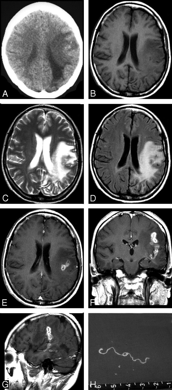
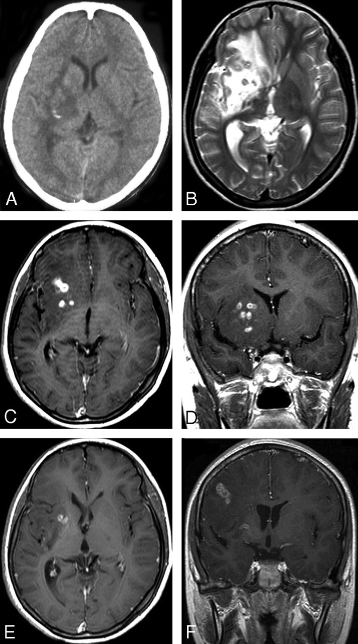
Background and purpose: Sparganosis is a rare parasitic infection in humans by a larval cestode of the genus Spirometra. Preoperative diagnosis of cerebral sparganosis in the past has been very difficult. Our objective was to evaluate the CT and MR features of cerebral sparganosis in order to make a definite diagnosis.

Materials and methods: We retrospectively reviewed 25 patients (13 male and 12 female; age range, 9-83 years) who proved to have cerebral sparganosis. Fifteen patients underwent MR imaging: 2 patients had CT scanning, and the remaining 8 had both CT and MR scanning. We focused on evaluating the imaging features on CT and MR.

Results: All patients showed edema and degeneration of cerebral white matter. All but 1 had a unilateral lesion. Twenty-two patients had ipsilateral ventricular dilation. The new finding was a tunnel sign, approximately 4 cm in length and 0.8 cm in width, column or fusiform shaped on postcontrast coronal and sagittal MR images (n = 10). Thirteen patients showed bead-like enhancement, but solitary ring enhancement was common on the CT images (n = 2). The wall of the ring and tunnel appeared isointense or slightly hyperintense on T2-weighted images. Punctate calcifications were seen in 6 patients on CT images but only in 3 patients on the MR images. Hemorrhage was seen in 4 patients on the MR images. An intact whitish, stringlike, living worm was found (n = 5).

Conclusion: The most characteristic finding was a tunnel sign on postcontrast MR images. The most common finding was bead-shaped enhancement. MR is superior to CT in demonstrating the extent and number of lesions, except punctate calcifications. Combined with clinical data and enzyme-linked immunosorbent assay, the preoperative diagnosis of cerebral sparganosis could be established on MR imaging.


Figures

Fig 1.
Fig 1.
Case 1. Images of the brain of an 80-year-old man with a history of headache, seizure, and left hemiparesis for 2 years. A, Precontrast CT scan shows patchy area of hypoattenuation in the white matter of the right parietal lobe with a punctate calcification located centrally. B, Axial T2-weighted MR image of the same day shows hyperintense area and cortical atrophy in the right parietal lobe. However, calcification seen on CT image cannot be found on MR images. C–D, Sagittal and coronal postcontrast images show tunnel-shaped enhancement representing inflammatory granuloma. No ipsilateral ventricular dilation is seen. E, Postoperative gross photograph of resected specimen shows a degenerated worm of Spirometra mansoni surrounded by inflammatory granulomatous tissues. F, Photomicrograph of histologic specimen shows a removed degenerated worm (W) surrounded by collagen capsule (C) and peripheral inflammatory cells and gliosis (G) (H&E stain × 40). G–H, Sagittal and coronal postcontrast images 1 year after a craniotomy in the same patient show lesions excised, with edematous area in the right parietal lobe.
Fig 2.
Fig 2.
Case 2. Images of the brain of a 45-year-old woman with a 6-year history of severe headache, intermittent seizures, and right hemiparesis. A, Precontrast CT scan reveals unilateral extensive area of low attenuation in the white matter of the left parietal lobe, with ipsilateral ventricular dilation. Small, punctate calcifications are seen in the left parietal lobe. B–D, Axial T1-weighted (A), T2-weighted (B), and FLAIR images (C) of the same section show a wide area of hypointensity on T1-weighted image (B), heterogeneous hyperintensity on T2-weighted (C) and FLAIR images (D), with a small central area of isointensity and slight hyperintensity on T2-weighted image (C), corresponding to isointensity or hypointensity on FLAIR image (D), representing encephalomalacia. E–G, Postcontrast axial (E), coronal (F), and sagittal (G) T1-weighted images show a tunnel about 5 cm in length and 1.5 cm in width, appearing as a hollow tube located in the left temporal and parietal lobe. H, Intraoperative photograph shows a whitish, wrinkled, threadlike live worm approximately 6 cm in length with slow peristalsis.
Fig 3.
Fig 3.
Case 6. Images of the brain of a 14-year-old boy with a 4-year history of seizures and left hemiparesis. A, Precontrast CT scan shows an extensive area of low attenuation in the right basal ganglia with a punctate calcification centrally. B, Axial T2-weighted image of the same section as the CT image in (A) shows hyperintense area in the right basal ganglia. However, calcification seen on CT image cannot be shown clearly on MR image. C–D, Postcontrast axial (C) and coronal (D) T1-weighted images show bead-shaped enhancement in the right basal ganglia. E–F, After 4 months, postcontrast axial (E) and coronal (F) T1-weighted images of the same patient show the tunnel-shaped enhancement in the right parietal lobe with small amounts of residual bead-shaped enhancement in the right basal ganglia, representing the migration of the worm and lesions shifting from the right basal ganglia to the right parietal lobe. Preoperative ELISA on serum and on CSF revealed strongly positive against Spirometra mansoni. A live worm was found at craniotomy.

References

    1. Mueller JF. The biology of Spirometra. J Parasitol 1974;60:3–14 - PubMed
    1. Muller JF, Hart EP, Walsh WP. Human sparganosis in the United States. J Parasitol 1963;49:294–96
    1. Dunn IJ, Palmer PE. Sparganosis. Semin Roentgenol 1998;33:86–88 - PubMed
    1. Chang KH, Cho SY, Chi JG, et al. Cerebral sparganosis: CT characteristics. Radiology 1987;165:505–10 - PubMed
    1. Fan KJ, Pezeshkpour GH. Cerebral sparganosis. Neurology 1986;36:1249–51 - PubMed

LinkOut - more resources